Microenvironmental forces regulate notch signaling through integr
Lực vi môi trường điều hòa tín hiệu Notch qua sự tương tác protein. Khám phá cơ chế phân tử quan trọng trong sinh học tế bào.
Năm xuất bản
Số trang
150
Thời gian đọc
23 phút
Lượt xem
0
Lượt tải
0
Phí lưu trữ
40 Point
Tổng quan nhanh
- Chủ đề:
- Lực Vi Môi Trường Điều Hòa Tín Hiệu Notch Tế Bào
- Số trang:
- 150 trang
- Trường:
- Boise State University
- Chuyên ngành:
- Biology
- Tác giả:
- Michael Allen Detweiler
- Năm:
- 2018
Tóm tắt nội dung luận án
I.Lực Vi Môi Trường Điều Hòa Tín Hiệu Notch Tế Bào
Môi trường ngoại bào tác động đáng kể đến chức năng và hành vi của tế bào. Tương tác tế bào-tế bào rất quan trọng. Tương tác tế bào-chất nền cũng ảnh hưởng lớn. Các lực vật lý từ môi trường ngoại bào thay đổi hành vi tế bào. Tế bào tiếp nhận các tín hiệu và lực này. Các kênh và thụ thể màng truyền tín hiệu. Tín hiệu đi từ bên ngoài vào bên trong tế bào. Tín hiệu Notch cổ điển được kích hoạt khi có tương tác phối tử với các tế bào lân cận. Tuy nhiên, bằng chứng gần đây cho thấy sự đa dạng. Tín hiệu Notch có thể xảy ra thông qua nhiều kích thích ngoại bào. Các kích thích này bao gồm tăng đường huyết và thiếu oxy. Nhiều yếu tố tăng trưởng cũng đóng vai trò. Lực cắt dòng chất lỏng và thành phần ma trận ngoại bào (ECM) là các yếu tố khác. Tín hiệu Notch không cổ điển qua vi môi trường còn ít được hiểu rõ. Nghiên cứu này tập trung vào Notch signaling pathway như một cảm biến đối với Microenvironment tế bào.
1.1. Vi Môi Trường Định Hình Chức Năng Tế Bào
Môi trường ngoại bào có vai trò quan trọng trong việc điều khiển chức năng và hành vi của tế bào. Các yếu tố như tương tác tế bào-tế bào, tương tác tế bào-chất nền và các lực vật lý là những thành phần chính. Chúng có thể thay đổi đáng kể hành vi của tế bào. Tế bào sử dụng nhiều kênh và thụ thể màng để nhận và truyền các tín hiệu này, từ không gian ngoại bào vào nội bào.
1.2. Tín Hiệu Notch Và Phản Ứng Môi Trường
Tín hiệu Notch cổ điển được kích hoạt bởi tương tác phối tử với các tế bào lân cận. Tuy nhiên, các phát hiện gần đây chỉ ra rằng Notch signaling pathway cũng có thể được kích hoạt bởi nhiều kích thích ngoại bào. Các kích thích này bao gồm tăng đường huyết, thiếu oxy, các yếu tố tăng trưởng đa dạng, lực cắt dòng chất lỏng và thành phần của Extracellular matrix (ECM). Sự điều hòa Notch không cổ điển thông qua vi môi trường hiện vẫn chưa được hiểu rõ hoàn toàn.
II.Cơ Chế Tín Hiệu Integrin Điều Khiển Notch
Integrin là thụ thể màng tế bào. Chúng chủ yếu được nhận biết qua khả năng gắn kết tế bào-ECM. Integrin cũng gây ra các chuỗi tín hiệu tế bào. Các thụ thể này còn phản ứng và truyền tín hiệu. Lực cắt dòng chất lỏng và độ cứng ECM ảnh hưởng đến chúng. Integrin đóng vai trò trung gian quan trọng trong cảm nhận cơ học. Điều này là một phần của quá trình cơ học (mechanotransduction). Bằng chứng trước đây cho thấy sự kích hoạt Notch thông qua con đường integrin. Điều này đề xuất Notch là một cảm biến vi môi trường. Integrin đã được chứng minh điều hòa tín hiệu Notch. Cả integrin và Notch đều phản ứng với lực cắt dòng chất lỏng. Một giả thuyết được đưa ra. Notch signaling phản ứng với lực cắt dòng chất lỏng. Cơ chế này diễn ra thông qua kích hoạt integrin. Điều này mở ra hiểu biết mới về tương tác tế bào-môi trường và Tín hiệu integrin.
2.1. Integrin Là Thụ Thể Cảm Biến Môi Trường
Integrin là các thụ thể màng tế bào thiết yếu. Chúng nổi tiếng với vai trò trong sự gắn kết giữa tế bào và Extracellular matrix (ECM). Ngoài ra, integrin còn có khả năng khởi tạo các chuỗi tín hiệu tế bào. Các thụ thể này cũng phản ứng và truyền tín hiệu từ các kích thích cơ học, như lực cắt dòng chất lỏng và độ cứng của ECM. Điều này cho thấy vai trò của integrin trong quá trình Mechanotransduction.
2.2. Notch Là Cảm Biến Vi Môi Trường Nhờ Integrin
Các nghiên cứu trước đây đã gợi ý về một cơ chế kích hoạt Notch signaling pathway mới thông qua con đường Tín hiệu integrin. Điều này đề xuất rằng Notch hoạt động như một cảm biến đối với Microenvironment tế bào. Integrin đã được chứng minh là có khả năng điều hòa Notch. Cả hai đều biểu hiện phản ứng với lực cắt dòng chất lỏng. Một giả thuyết quan trọng đã được đặt ra: Notch signaling phản ứng với lực cắt dòng chất lỏng thông qua sự kích hoạt integrin.
III.Lực Cắt Dòng Chất Lỏng Ảnh Hưởng Notch
Tín hiệu Notch được kiểm tra khi tiếp xúc với lực cắt dòng chất lỏng. Điều này được so sánh với Notch khi lực cắt dòng chất lỏng bị ức chế chức năng integrin. Dữ liệu xác nhận sự kích hoạt Notch tăng đáng kể. Điều này xảy ra từ lực cắt dòng chất lỏng so với nhóm đối chứng tĩnh. Ức chế chức năng integrin làm giảm phản ứng này. Điều này cho thấy integrin là cần thiết. Integrin đóng vai trò cho sự điều hòa Notch từ lực cắt dòng chất lỏng. Kết quả này cung cấp bằng chứng trực tiếp. Lực cơ học điều chỉnh con đường Notch signaling pathway. Cơ chế này thông qua các thụ thể integrin. Đây là một phát hiện quan trọng trong lĩnh vực cơ học tế bào và Mechanotransduction, khẳng định vai trò của Lực cơ học.
3.1. Phản Ứng Của Notch Với Lực Cắt Dòng
Sự kích hoạt Notch đã được nghiên cứu sau khi tế bào tiếp xúc với lực cắt dòng chất lỏng. Các phép đo được thực hiện và so sánh với tình trạng ức chế chức năng integrin trong điều kiện tương tự. Dữ liệu cho thấy sự kích hoạt Notch tăng lên đáng kể. Sự gia tăng này là kết quả của lực cắt dòng chất lỏng, so với nhóm đối chứng không có lực cắt dòng.
3.2. Integrin Cần Thiết Cho Phản Ứng Notch
Việc ức chế chức năng của integrin đã làm giảm đáng kể phản ứng kích hoạt Notch đối với lực cắt dòng. Điều này chỉ ra rằng Tín hiệu integrin là cần thiết cho sự điều hòa Notch dưới tác động của Lực cơ học này. Kết quả này cung cấp bằng chứng cụ thể rằng Notch signaling pathway được điều chỉnh bởi lực cắt dòng chất lỏng thông qua sự trung gian của integrin.
IV.Độ Cứng ECM Ảnh Hưởng Tín Hiệu Notch
Integrin cũng phản ứng và truyền tín hiệu từ độ cứng ECM khác nhau. Notch đã được chứng minh tăng lên trong điều kiện xơ hóa. Giả thuyết được đưa ra rằng tín hiệu Notch sẽ được điều hòa bởi các mức độ cứng Extracellular matrix (ECM) khác nhau. Kích hoạt integrin là yếu tố trung gian. Để điều tra giả thuyết này, tế bào được nuôi cấy trên hydrogel. Các hydrogel này có nhiều mức độ cứng khác nhau. Tín hiệu Notch sau đó được đo lường. Kết quả cho thấy, giống như lực cắt dòng, tín hiệu Notch bị ảnh hưởng bởi độ cứng ECM. Điều này cho thấy ECM tác động trực tiếp lên vận mệnh tế bào. Các yếu tố vật lý này quan trọng cho quá trình Differentiation tế bào và Proliferation tế bào, đồng thời làm nổi bật ảnh hưởng của Lực cơ học đến Notch signaling pathway.
4.1. Notch Được Điều Hòa Bởi Độ Cứng ECM
Ngoài Lực cơ học, integrin còn phản ứng và truyền tín hiệu từ các mức độ cứng Extracellular matrix (ECM) khác nhau. Có bằng chứng cho thấy Notch signaling pathway được tăng cường trong điều kiện xơ hóa. Điều này dẫn đến giả thuyết rằng tín hiệu Notch sẽ được điều hòa bởi sự thay đổi độ cứng ECM, với sự kích hoạt Tín hiệu integrin đóng vai trò trung gian.
4.2. Độ Cứng ECM Kích Hoạt Tín Hiệu Notch
Để kiểm tra giả thuyết này, các tế bào được nuôi cấy trên các hydrogel có độ cứng khác nhau. Sau đó, tín hiệu Notch được đo lường. Các kết quả cho thấy, tương tự như phản ứng với lực cắt dòng, tín hiệu Notch bị ảnh hưởng bởi độ cứng của ECM. Điều này củng cố vai trò của các yếu tố vật lý trong Microenvironment tế bào đối với Notch signaling pathway.
V.Ứng Dụng Y Học Của Tín Hiệu Notch Môi Trường
Các kết quả tổng hợp cho thấy tín hiệu Notch được điều hòa. Điều này xảy ra thông qua các lực vi môi trường. Các lực này bao gồm lực cắt dòng chất lỏng và độ cứng ECM. Sự điều hòa này diễn ra thông qua integrin. Điều này nâng cao hiểu biết về các biến thể của tín hiệu Notch. Các biến thể này phản ứng với các kích thích vi môi trường. Các cơ chế liên quan cũng được làm rõ. Notch đã được liên quan đến nhiều bệnh lý khác nhau. Kết quả nghiên cứu cải thiện kiến thức về tín hiệu Notch. Điều này áp dụng cho các tình trạng bệnh lý của vi môi trường. Ví dụ bao gồm lực cắt dòng bất thường (như xơ vữa động mạch). Độ cứng mô bất thường (như xơ hóa) cũng được đề cập. Hiểu biết này có thể dẫn đến các phương pháp điều trị mới. Nó cũng có thể giúp trong việc nhắm mục tiêu các khối u trong Tumor microenvironment, tác động đến Proliferation tế bào và Differentiation tế bào.
5.1. Cơ Chế Mới Về Tương Tác Notch Integrin
Nghiên cứu này tổng hợp bằng chứng cho thấy Notch signaling pathway được điều hòa bởi các lực vi môi trường, bao gồm lực cắt dòng chất lỏng và độ cứng Extracellular matrix (ECM). Cơ chế điều hòa này diễn ra thông qua Tín hiệu integrin. Điều này mở rộng hiểu biết về các phản ứng đa dạng của Notch đối với các kích thích từ Microenvironment tế bào và làm sáng tỏ các cơ chế liên quan.
5.2. Notch Trong Bệnh Lý Vi Môi Trường
Notch đã được chứng minh có vai trò trong nhiều bệnh lý. Các phát hiện này nâng cao kiến thức về Notch signaling pathway trong các tình trạng bệnh lý của vi môi trường. Điều này bao gồm các trường hợp lực cắt dòng bất thường, như xơ vữa động mạch, và độ cứng mô bất thường, điển hình như xơ hóa. Hiểu biết sâu sắc này có thể hướng tới các liệu pháp mới, bao gồm mục tiêu trong Tumor microenvironment.
Mục lục chi tiết luận án
Tải xuống file đầy đủ để xem toàn bộ nội dung
Tải đầy đủ (150 trang)Trích đoạn nội dung luận án
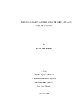
Tải xuống để đọc toàn bộMICROENVIRONMENTAL FORCES REGULATE NOTCH SIGNALING THROUGH INTEGRINS by Michael Allen Detweiler A thesis submitted in partial fulfillment of the requirements for the degree of Master of Science in Biology Boise State University December 2018 © 2018 Michael Allen Detweiler ALL RIGHTS RESERVED BOISE STATE UNIVERSITY GRADUATE COLLEGE DEFENSE COMMITTEE AND FINAL READING APPROVALS of the thesis submitted by Michael Allen Detweiler Thesis Title: Microenvironmental Forces Regulate Notch Signaling Through Integrins Date of Final Oral Examination: 25 October 2018 The following individuals read and discussed the thesis submitted by student Michael Allen Detweiler, and they evaluated his presentation and response to questions during the final oral examination. They found that the student passed the final oral examination. Allan Albig, Ph. Chair, Supervisory Committee Brad E.
Member, Supervisory Committee Troy Rohn, Ph. Member, Supervisory Committee The final reading approval of the thesis was granted by Allan Albig, Ph., Chair of the Supervisory Committee. The thesis was approved by the Graduate College. ACKNOWLEDGEMENTS First and foremost, I would like to thank my advisor, Dr.
Allan Albig, for being an incredible mentor and friend. With his guidance and moral support, I have been able to fully understand and integrate the ethics and investigative nature of science. I would also like to thank Dr. Brad Morrison and Dr.
Troy Rohn for being on my thesis committee and for their very helpful suggestions. Through my time at Boise State I have had the pleasure of working with and learning from my fellow colleagues, and for this I would like to thank the Albig lab members, Jacob Crow, Bryce Lafoya, Jordan Munroe, Shelby Havel, and Dr. I would also like to give thanks to the National Institute of Health for providing the funding to support this thesis. iv ABSTRACT The extracellular microenvironment contributes significantly to a cell’s function and behavior.
For instance, cell-cell interactions, cell-substrate interactions, and physical forces are all factors of the extracellular environment that can alter cellular behavior. Cells can receive these signals and forces through various membrane channels and receptors that transmit the signals from the extracellular to the intracellular space. Canonical Notch signaling is induced by ligand interactions with neighboring cells, but recent evidence has revealed that Notch signaling can occur through a variety of extracellular stimuli including hyperglycemia, hypoxia, multiple growth factors, fluid shear stress, and extracellular matrix (ECM) composition. Although Notch activation through ligand interactions with adjacent cells have been well established, non-canonical Notch signaling through the microenvironment is poorly understood.
Previous evidence suggests a novel activation of Notch signaling through an integrin pathway, proposing Notch as a microenvironmental sensor. Integrins are cell membrane receptors that are mainly recognized for cell-ECM attachment and induction of cellular signaling cascades but have also been shown to respond and transmit signaling through fluid shear stress and ECM stiffness. Since integrins have been shown to regulate Notch signaling and both exhibit a response to fluid shear stress, we hypothesized that Notch signaling responds to fluid shear stress through integrin activation. To test this, we compared Notch activation following exposure to fluid shear stress and Notch activation following shear stress after inhibiting integrin function.
Our data confirms that Notch activation is significantly v upregulated from fluid shear stress compared to a static control and inhibiting integrin function attenuates this response, suggesting integrins are required for Notch’s upregulation from shear stress. Because integrins also respond and transmit signals from varying ECM stiffness and Notch has been shown to be upregulated during conditions of fibrosis, we hypothesized that Notch signaling will be regulated by varying degrees of ECM stiffness through integrin activation. To investigate this hypothesis, we cultured cells on hydrogels with multiple levels of stiffness and measured Notch signaling. Our results indicate that like shear stress, Notch signaling is influenced by ECM stiffness.
Collectively our results indicate that Notch signaling is regulated through microenvironmental forces like fluid shear stress and ECM stiffness and is regulated through integrins. This furthers our understanding of the variations of Notch signaling in response to microenvironmental stimuli and the mechanisms involved. Notch has been implicated in a variety of diseases and our results improve our knowledge of Notch signaling in pathological conditions of the microenvironment including abnormal shear stress (e. atherosclerosis) and tissue stiffness (e.
vi TABLE OF CONTENTS ACKNOWLEDGEMENTS. v LIST OF TABLES .xi LIST OF FIGURES .xii LIST OF ABBREVIATIONS. xvi CHAPTER ONE: INTRODUCTION. 1 The Cellular Microenvironment.
3 Downstream Notch Signaling. 5 Canonical vs Non-Canonical Activation. 6 Notch Signaling and Disease. 8 Integrin Downstream Signaling.
12 Notch and Shear Stress. 18 CHAPTER TWO: INITIAL SHEAR STRESS-INDUCED NOTCH SIGNALING IS REGULATED THROUGH AND INTEGRIN PATHWAY. 24 Notch Signaling is induced by Shear Stress. 24 Notch Activation from Shear Stress Is Regulated by Integrins.
27 Notch Activation from Shear Stress Is Regulated Via Integrin Downstream Pathways Src and Enos. 37 mRNA Expression Analysis:. 38 Western Blot Analysis:. 38 CHAPTER THREE: MATRIX STIFFNESS REGULATES NOTCH ACTIVATION.
42 viii Notch Activation Changes with Varying Degrees of Matrix Stiffness in Endothelial Cells. 63 Notch: A multi-functional integrating system of microenvironmental signals. 64 The Cellular Microenvironment:. 66 Notch as an Integrator of Cellular Microenvironments:.
69 ECM-Notch Interactions:. 70 Direct ECM-Notch Interactions That Control Notch Signaling. 70 Indirect ECM-Notch Interactions that Control Notch Signaling (Transcriptional Mechanisms). 76 Direct ECM Notch Interactions That Control Notch Signaling (Crosstalk Mechanisms).
78 Notch crosstalk with other signaling networks. Integrins, TGF-, WNT, and VEGF:. 80 Crosstalk between Notch and Integrins:. 80 ix Notch and TGF-:.
84 Notch and WNT:. 86 Notch and VEGF:. 88 Other Microenvironment Conditions That Control Notch (Shear stress, hypoxia, and hyperglycemia). 90 Notch and Shear Stress:.
91 Notch and Hypoxia. 93 Notch and Hyperglycemia:. 108 x LIST OF TABLES Table A.1: Basic and proposed mechanisms by which Notch responds to various microenvironmental signals. 98 xi LIST OF FIGURES Figure 1.1: Domains of the Notch receptor.
The extracellular domain (NECD) of Notch contains 36 epidermal growth factor (EGF)-like repeats, followed by a transmembrane portion containing Lin-12-Notch repeats. The intracellular domain (NICD) consists of RAM, Ankyrin repeats, transactivation domain (TAD) and proline, glutamine, serine, and threonine (PEST)-rich domains and is responsible for transcriptional control.2: Blood vessels consist of 3 main layers. The tunica intima is composed of endothelial cells surrounded by a basement membrane and regulates vessel permeability and integrity. The tunica media has smooth muscle cells and matrix proteins and is mainly responsible for contraction/dilation of the vessel, and the tunica adventitia consists of mostly ECM proteins and associates the vessels with the surrounding tissue and provides elasticity.
Illustration showing the proposed method of mechanotransduction. As shear stress forces are implemented to the apical surface of cells, the force applies tension to the cytoskeleton where it will then transmit to the focal adhesions and junctions of the cell. Illustration of experimental hydrogels. A glass coverslip is used to attach polyacrylamide hydrogel.
The hydrogel is then crosslinked to an ECM protein using crosslinkers like L-DOPA or Sulfo- sanpah. The cells are then cultured on the surface attaching to the ECM protein with focal adhesions.1: Notch signaling is induced via shear stress. HMEC1s exposed to ~12dyn/cm^2 of orbital shear stress. A) Notch intracellular domain (N1ICD) and full length Notch1 whole cell lysates detected by Western blot and normalized to Vinculin control.
b) Downstream Notch genes Hey1, Hes1, and Hey2 expression levels detected by qPCR, Klf2 used as a positive shear stress control. Values are mean +/- SEM; * = p < 0. c) Downstream Notch genes Hey1 and Hes1 expression levels detected by qPCR following treatment with Doxycycline in N1ICD transgenic HMEC1s to overexpress Notch signaling.2: Shear Stress-induced Notch is regulated through integrins. HMEC1s exposed to ~12 dyn/cm^2 of shear stress following integrin inhibition.
a) xii Notch downstream genes Hey1 and Hes1 mRNA expression detected by qPCR following treatment with RGD peptides (5 g/ml) and 90 minutes of orbital shear stress. Klf2 used as a positive shear stress control. b) Notch downstream genes Hey1 and Hes1 mRNA expression detected by qPCR following treatment with MBCD (10 mM) and 90 minutes of orbital shear stress. Klf2 used as a positive shear stress control.
Values are mean +/- SEM; * = p < 0. c) N1ICD fragment whole cell lysate detected via western blot and folded to Vinculin following treatment with RGD peptide and MBCD and exposed to orbital shear stress for 45 and 90 minutes.3: Shear stress-induced Notch activation is regulated by integrin downstream pathways, Src & eNOS. HMEC1s exposed to ~12 dyn/cm^2 of shear stress following treatment with AZM and DPI. a) mRNA expression of Hey1 and Hes1 detected by qPCR when AZM (10 m) is folded to vehicle control following shear stress.
b) mRNA expression of Hey1, Hes1, and Hey2 detected by qPCR when DPI (100 M) treatment is folded to static control following shear stress. Values are mean +/- SEM; * = p < 0. c) Western blot analysis of NICD with AZM, DPI, and vehicle control.4: Shear stress induces Notch activation through Integrin-Src-eNOS pathway. Illustration representing the suggested regulation of Notch activation following shear stress via an integrin-Src-eNOS pathway.1: Notch downstream genes are regulated through ECM stiffness.
a) Downstream Notch genes Hey1 and Hes1 were analyzed via qPCR following culturing on polyacrylamide hydrogels with stiffnesses of 10, 20, and 40 kPa. B) Downstream Notch gene Hes5 was analyzed via luciferase assay after being cultured on various stiffness of polyacrylamide hydrogels at 10, 20, & 40 kPa. Values are mean +/- SEM; * = p < .1: Canonical Notch signaling and Notch conservation between human and Monosiga brevicollis. Throughout Figure 1, conservation of Notch proteins or domains between human and M.
brevicollis is indicated by green (positive), yellow (unknown), or red (negative) shading according to references (King, Westbrook et al. 2008) and (Gazave, Lapebie et al. (A) Conservation of mammalian Notch receptor domains in M. Mammalian (human) Notch receptors contain 36 EGF-like repeats and three LNR or NRR (Lin-12 Notch Repeats or Negative Regulatory Region) repeats in the extracellular domain.
The intracellular portion of human Notch contains seven ankyrin domains and a PEST sequence at the C-terminal. For simplicity, the intracellular RAM (RBPj Association Module) domain, two NLS (Nuclear Localization sequence) domains, and TAD (Transactivation Domain) are not shown in this figure. xiii Please refer to references (Kopan and Ilagan 2009, Kopan 2010) for complete details. Three separate proteins (N1, N2, and N3) in M.
brevicollis contain six Ankyrin domains, two LNR domains, and 36 EGF- like repeats respectively (King, Westbrook et al. (B) Model of canonical Notch activation mechanism. Notch receptors are modified in the secretory pathway (ER/golgi) by Furin cleavage (S1 cleavage) and glycosylation of EGF-like domains by O-fucosyltransferase (O-fut), Rumi/Poglut1 (Protein O-Glycosyltransferase 1), and fringe family glycosyltransferases. The Furin cleavage products remain non-covalently associated in the membrane where a pulling force initiated by Notch ligand endocytosis in sending cells enables further cleavage by -secretase (S2 cleavage, NEXT fragment) at the LNR domain, and -secretase (S3 cleavage, NICD fragment) in the membrane of receiving cells.
Several regulatory proteins including Numb, Notchless, and Deltex control Notch availability at the membrane. After -secretase cleavage, the NICD fragment translocates to the nucleus where it displaces the transcriptional co-repressor SMRT from CSL/RBP-jk. NICD participates in a transcriptional complex with CSL, MAML, and p300 to drive transcription of Notch targets such as Hes and Hey genes. NICD steady- state levels are controlled by nuclear export, ubiquitination (Ub) by Sel10, and subsequent degradation in the proteasome.2: Summary of ECM control of Notch signaling.
Canonical activation of Notch receptors by Notch ligands can be manipulated in three ways by cellular interactions with ECM.) Direct interactions between Notch receptors or ligands and various ECM molecules can either inhibit or promote activation of Notch signaling.) Indirect interactions between ECM and Notch are characterized by ECM mediated increased or decreased expression of Notch ligands on sending cells or Notch receptors on receiving cells.
Nội dung được bảo vệ bản quyền — Tải xuống đầy đủ
Trích dẫn luận án này
Michael Allen Detweiler (2018). Microenvironmental forces regulate notch signaling through i [Luận án tiến sĩ, Boise State University]. LuanAn.net. https://luanan.net/sinh-hoc/sinh-hoc-phan-tu-te-bao/microenvironmental-forces-regulate-notch-signaling-through-integr
Câu hỏi thường gặp
Luận án "Microenvironmental forces regulate notch signaling through i" nghiên cứu về vấn đề gì?
Lực vi môi trường điều hòa tín hiệu Notch qua sự tương tác protein. Khám phá cơ chế phân tử quan trọng trong sinh học tế bào.
Luận án "Microenvironmental forces regulate notch signaling through i" được bảo vệ tại trường nào?
Luận án này được bảo vệ tại Boise State University. Năm bảo vệ: 2018.
Luận án "Microenvironmental forces regulate notch signaling through i" thuộc chuyên ngành gì?
Luận án "Microenvironmental forces regulate notch signaling through i" thuộc chuyên ngành Biology. Danh mục: Sinh Học Phân Tử Tế Bào.
Luận án "Microenvironmental forces regulate notch signaling through i" có bao nhiêu trang?
Luận án "Microenvironmental forces regulate notch signaling through i" có 150 trang. Bạn có thể xem trước một phần tài liệu ngay trên trang web trước khi tải về.
Cách tải luận án "Microenvironmental forces regulate notch signaling through i" về máy như thế nào?
Để tải luận án về máy, bạn nhấn nút "Tải xuống ngay" trên trang này, sau đó hoàn tất thanh toán phí lưu trữ. File sẽ được tải xuống ngay sau khi thanh toán thành công. Hỗ trợ qua Zalo: 0559 297 239.