Luận án Tiến sĩ về nọc ong và melittin trong điều trị ung thư vú
Luận văn tiến sĩ của Duffy Ciara Evelyn Lilly năm 2020 khám phá các phương pháp nghiên cứu mới trong lĩnh vực triết học và khoa học xã hội.
The University of Western Australia
Human Sciences
Luan An
Luận án
Năm xuất bản
Số trang
66
Thời gian đọc
10 phút
Lượt xem
0
Lượt tải
0
Phí lưu trữ
40 Point
Tổng quan nhanh
- Chủ đề:
- Nghiên cứu Nọc Ong Chữa Ung Thư Vú: Tiềm năng Mới
- Số trang:
- 66 trang
- Trường:
- The University of Western Australia
- Chuyên ngành:
- Human Sciences
- Tác giả:
- Ciara Evelyn Lilly Duffy
- Năm:
- 2020
Tóm tắt nội dung luận án
I.Nghiên cứu Nọc Ong Chữa Ung Thư Vú Tiềm năng Mới
Ung thư vú là căn bệnh phổ biến nhất ở phụ nữ trên toàn cầu. Các loại ung thư vú như bộ ba âm tính (TNBC) đặc biệt hung hãn. Chúng có tốc độ tăng sinh nhanh chóng và liên quan đến tiên lượng xấu. Hiện tại, không có liệu pháp nhắm đích hiệu quả cho TNBC. Đối với ung thư vú tăng sinh HER2, mặc dù đã có cải thiện đáng kể về thời gian sống sót, tình trạng kháng thuốc vẫn gần như không thể tránh khỏi. Vì vậy, việc khám phá các chiến lược trị liệu hiệu quả và chọn lọc hơn là cực kỳ quan trọng trong điều trị ung thư lâm sàng. Nọc ong mật từ loài Apis mellifera đã cho thấy hoạt tính chống ung thư đáng chú ý. Melittin, một peptide chính trong nọc ong, cũng sở hữu đặc tính tương tự. Các nghiên cứu ban đầu cho thấy nọc ong và melittin có khả năng phá vỡ màng tế bào ung thư, mở ra hướng đi mới trong điều trị ung thư vú.
1.1. Thách thức trong điều trị ung thư vú
Ung thư vú gây ra nhiều thách thức lớn. Đặc biệt, ung thư vú bộ ba âm tính không có mục tiêu điều trị cụ thể. Các tế bào này phát triển rất nhanh. Bệnh nhân thường có tiên lượng không tốt. Ngay cả các liệu pháp nhắm vào thụ thể HER2 cũng đối mặt với tình trạng kháng thuốc. Điều này làm giảm hiệu quả điều trị lâu dài. Cần có các phương pháp mới, hiệu quả hơn để vượt qua những trở ngại này. Mục tiêu là tìm ra liệu pháp điều trị ung thư vú có tính chọn lọc cao, giảm thiểu tác dụng phụ.
1.2. Nọc ong Hướng tiếp cận trị liệu mới
Nọc ong mật (apitoxin) và melittin đang được nghiên cứu như một hướng tiếp cận đầy hứa hẹn. Chúng được biết đến với khả năng chống ung thư mạnh mẽ. Đặc biệt, chúng có thể phá vỡ màng tế bào. Điều này làm suy yếu tế bào ung thư. Tuy nhiên, cơ chế phân tử chính xác vẫn chưa được hiểu rõ hoàn toàn. Nghiên cứu này đặt mục tiêu xác định tiềm năng trị liệu của nọc ong và melittin. Chúng nhắm mục tiêu và tiêu diệt các tế bào ung thư vú hung hãn. Điều này mở ra cánh cửa cho các phương pháp điều trị ung thư vú tiên tiến hơn.
II.Cơ chế Nọc Ong Melittin tiêu diệt Tế Bào Ung Thư Vú
Nghiên cứu đã đi sâu vào việc xác định các cơ chế phân tử mà nọc ong và melittin gây ra cái chết tế bào. Hoạt tính chống ung thư của nọc ong không chỉ giới hạn ở việc phá vỡ màng tế bào. Nó còn liên quan đến các con đường tín hiệu phức tạp bên trong tế bào. Melittin là thành phần chính chịu trách nhiệm cho các tác dụng này. Mục tiêu chính là làm rõ các bước cụ thể dẫn đến cái chết của tế bào ung thư. Điều này bao gồm tìm hiểu về sự tương tác của melittin với các thụ thể tăng trưởng biểu bì. Đồng thời đánh giá tác động của nó lên các con đường tín hiệu quan trọng trong tế bào ung thư vú. Việc hiểu rõ cơ chế này sẽ tạo cơ sở cho việc phát triển các liệu pháp nhắm đích hiệu quả hơn.
2.1. Phá vỡ màng tế bào ung thư
Nọc ong và melittin gây độc tính tế bào bằng cách phá vỡ màng tế bào. Đây là cơ chế chính dẫn đến cái chết của tế bào. Melittin tạo lỗ thủng trên màng tế bào ung thư. Điều này làm thay đổi tính thấm của màng. Dẫn đến sự gián đoạn nghiêm trọng chức năng tế bào. Quá trình này ảnh hưởng đến các tế bào ung thư vú bộ ba âm tính và HER2-enriched. Các tế bào bình thường ít bị ảnh hưởng hơn. Sự phá vỡ màng tế bào nhanh chóng làm suy giảm khả năng sống của chúng. Đây là một đặc điểm quan trọng của apitoxin trong điều trị ung thư vú.
2.2. Kích hoạt tế bào chết theo chương trình
Ngoài việc phá vỡ màng, nọc ong và melittin còn gây ra apoptosis. Đây là quá trình tế bào chết theo chương trình. Apoptosis được dẫn dắt bởi caspase 3. Caspase 3 là một enzyme chủ chốt trong con đường này. Melittin còn điều hòa tín hiệu Akt và MAPK gây ung thư. Nó thực hiện điều này bằng cách ức chế hoạt hóa EGFR và HER2. Các thụ thể này thường được biểu hiện quá mức trong tế bào ung thư vú. Sự ức chế này góp phần vào độc tính tế bào. Nó giúp kiểm soát sự tăng sinh của tế bào ung thư.
III.Tác động chọn lọc của Melittin lên Ung Thư Vú
Một trong những phát hiện quan trọng nhất là tính chọn lọc cao của melittin. Nó nhắm mục tiêu và tiêu diệt các tế bào ung thư vú. Trong khi đó, các tế bào bình thường ít bị ảnh hưởng. Điều này làm cho melittin trở thành một ứng cử viên đầy hứa hẹn. Việc tìm kiếm các tác nhân có độc tính tế bào chọn lọc là rất quan trọng. Các tác nhân này giúp giảm thiểu tác dụng phụ toàn thân. Nghiên cứu đã đánh giá phản ứng tế bào trên một loạt các dòng tế bào ung thư vú. Các dòng tế bào bình thường cũng được sử dụng để so sánh. Kết quả khẳng định khả năng của melittin trong việc phân biệt giữa các tế bào khỏe mạnh và tế bào ung thư.
3.1. Tính chọn lọc đối với tế bào ung thư
Nọc ong và melittin giảm đáng kể khả năng sống của tế bào. Chúng đặc biệt chọn lọc với tế bào ung thư vú bộ ba âm tính. Các tế bào ung thư vú tăng sinh HER2 cũng bị ảnh hưởng mạnh. Điều này rất quan trọng vì các tế bào bình thường ít bị tổn thương hơn. Tính chọn lọc giúp giảm tác dụng phụ. Nó làm tăng hiệu quả điều trị. Đây là một lợi thế lớn so với hóa trị liệu truyền thống. Hóa trị thường không phân biệt giữa tế bào khỏe mạnh và tế bào ung thư.
3.2. Ảnh hưởng đến tín hiệu thụ thể
Melittin có khả năng điều chỉnh các tín hiệu gây ung thư. Nó ảnh hưởng đến các con đường như Akt và MAPK. Nó ức chế sự hoạt hóa của EGFR và HER2. Các thụ thể này thường bị biểu hiện quá mức trong nhiều loại ung thư vú. Việc ức chế này làm gián đoạn sự phát triển của tế bào ung thư. Nó cũng ngăn chặn sự sống sót của chúng. Tác động này là một phần quan trọng của độc tính tế bào chọn lọc. Nó hỗ trợ khả năng của melittin trong việc tiêu diệt các tế bào ung thư vú.
IV.Nọc Ong Melittin phối hợp hóa trị liệu Ung Thư Vú
Một hướng nghiên cứu quan trọng khác là đánh giá khả năng hiệp đồng của melittin. Nó được kết hợp với các loại thuốc hóa trị liệu hiện có. Điều này nhằm mục đích tăng cường hiệu quả điều trị. Đồng thời có thể giảm liều lượng hóa chất cần thiết. Docetaxel là một loại thuốc hóa trị liệu phổ biến trong điều trị ung thư vú. Nghiên cứu đã kiểm tra sự tương tác giữa melittin và docetaxel. Các thí nghiệm được thực hiện cả trong ống nghiệm (in vitro) và trên mô hình động vật (in vivo). Kết quả mang lại triển vọng lớn cho các chiến lược điều trị kết hợp. Nó có thể cải thiện đáng kể kết quả cho bệnh nhân ung thư vú, đặc biệt là các thể bệnh khó điều trị.
4.1. Hiệu quả hiệp đồng với docetaxel
Melittin thể hiện tác dụng hiệp đồng với docetaxel. Điều này đã được chứng minh cả trong ống nghiệm và trên cơ thể sống. Sự kết hợp này mang lại hiệu quả cao hơn. Nó vượt trội so với việc sử dụng từng loại riêng lẻ. Việc này có ý nghĩa quan trọng trong điều trị lâm sàng. Nó cho phép giảm liều docetaxel. Điều này giúp giảm tác dụng phụ cho bệnh nhân. Đồng thời duy trì hoặc tăng cường hiệu quả tiêu diệt tế bào ung thư.
4.2. Giảm tăng sinh phát triển khối u
Sự kết hợp của melittin và docetaxel làm giảm tăng sinh tế bào ung thư. Nó cũng làm chậm sự phát triển của khối u. Điều này được quan sát thấy trong mô hình cấy ghép TNBC hung hãn. Kết quả này rất hứa hẹn. Nó gợi ý rằng apitoxin có thể được sử dụng bổ trợ. Nó có thể tăng cường hiệu quả của hóa trị liệu. Mục tiêu là đạt được kết quả điều trị tốt hơn. Đồng thời giảm thiểu tác động tiêu cực đến chất lượng sống của bệnh nhân ung thư vú.
V.Kết quả Nghiên cứu Nọc Ong Melittin chống Ung Thư
Nghiên cứu này đã đưa ra những bằng chứng mạnh mẽ. Nọc ong và melittin có khả năng tiêu diệt tế bào ung thư vú. Chúng hoạt động một cách mạnh mẽ và nhanh chóng. Các cơ chế phân tử đã được làm rõ. Điều này bao gồm sự phá vỡ màng tế bào. Nó cũng bao gồm việc kích hoạt tế bào chết theo chương trình. Melittin điều hòa các con đường tín hiệu chính trong tế bào ung thư. Khả năng kết hợp với hóa trị liệu mở ra hướng đi mới. Điều trị các loại ung thư vú hung hãn có thể hiệu quả hơn. Đây là một bước tiến quan trọng trong lĩnh vực điều trị ung thư. Nó mang lại hy vọng cho hàng triệu phụ nữ trên thế giới.
5.1. Giảm mạnh khả năng sống của tế bào ung thư
Nọc ong và melittin làm giảm đáng kể khả năng sống của tế bào ung thư vú. Điều này diễn ra một cách nhanh chóng và có chọn lọc. Chúng nhắm vào các dòng tế bào TNBC và HER2-enriched. Các tế bào này thường khó điều trị. Khả năng gây độc tính tế bào này là nền tảng. Nó cho thấy tiềm năng của apitoxin làm tác nhân trị liệu. Đặc biệt là với các loại ung thư vú kháng thuốc.
5.2. Tiềm năng ứng dụng trong điều trị ung thư
Nghiên cứu chứng minh nọc ong và melittin là tác nhân trị liệu mạnh mẽ. Chúng có thể được sử dụng độc lập. Hoặc chúng có thể kết hợp với hóa trị liệu. Điều này nhằm điều trị các phân nhóm ung thư vú hung hãn. Kết quả mở ra cơ hội cho các thử nghiệm lâm sàng trong tương lai. Nọc ong và melittin có thể trở thành một phần của phác đồ điều trị. Mục tiêu là cải thiện tiên lượng cho bệnh nhân ung thư vú.
Mục lục chi tiết luận án
Tải xuống file đầy đủ để xem toàn bộ nội dung
Tải đầy đủ (66 trang)Nội dung chính
Tổng quan về luận án
Luận án này trình bày một nghiên cứu tiên phong trong lĩnh vực ung thư học phân tử, tập trung vào việc khám phá tiềm năng trị liệu của nọc ong mật (Apis mellifera) và melittin, một thành phần peptide chính trong nọc, đối với các thể ung thư vú đặc biệt hung hãn: ung thư vú bộ ba âm tính (TNBC) và ung thư vú có thụ thể HER2 dương tính (HER2-enriched). Bối cảnh khoa học của nghiên cứu được đặt trong thực trạng điều trị ung thư vú hiện nay, nơi dù có những tiến bộ đáng kể, vẫn còn tồn tại những thách thức lớn về hiệu quả và khả năng kháng thuốc. TNBC, chiếm 10-17% tổng số ca mắc ung thư vú, nổi tiếng là thể bệnh hung hãn nhất, tăng sinh mạnh mẽ và tiên lượng kém, hiện không có liệu pháp nhắm mục tiêu hiệu quả. Tương tự, ung thư vú HER2-enriched, dù đã có các liệu pháp nhắm mục tiêu như Trastuzumab giúp cải thiện đáng kể thời gian sống thêm trung bình, khả năng kháng thuốc gần như là không thể tránh khỏi, với "80% bệnh nhân được điều trị bằng docetaxel, trastuzumab và pertuzumab tái phát sau 5 năm" (Chapter 1, Section 1.1, trích dẫn [14, 17]).
Research Gap cụ thể: Mặc dù nọc ong mật và melittin đã được biết đến với hoạt tính chống ung thư thông qua cơ chế phá vỡ màng tế bào, "các cơ chế phân tử chính xác giải thích hoạt tính chống ung thư của các tác nhân này đối với tế bào vú ác tính vẫn chưa được biết rõ" (Abstract). Ngoài ra, các nghiên cứu trước đây về nọc ong mật và melittin trong ung thư vú còn hạn chế về số lượng dòng tế bào được khảo sát, đặc biệt là thiếu sự kiểm tra về tính chọn lọc theo phân nhóm ung thư vú và sự hiểu biết sâu sắc về các cơ chế phân tử gây chết tế bào ung thư. Luận án này giải quyết trực tiếp khoảng trống này bằng cách "định lượng hiệu quả của nọc ong mật và melittin trong điều trị tế bào ung thư vú TNBC và HER2-enriched, với trọng tâm đặc biệt vào tính chọn lọc ung thư, và mở rộng hiểu biết về các cơ chế phân tử gây chết tế bào ung thư vú" (Chapter 1, Section 1.6).
Research questions và hypotheses:
- RQ1: Nọc ong mật và melittin có khả năng làm giảm khả năng sống của tế bào ung thư vú so với tế bào bình thường, và chúng gây chết tế bào thông qua những cơ chế phân tử nào?
- H1: Nọc ong mật và melittin sẽ giảm đáng kể và có chọn lọc khả năng sống của tế bào ung thư vú TNBC và HER2-enriched, và sẽ gây ra sự phá vỡ màng tế bào và quá trình apoptosis qua trung gian caspase-3.
- RQ2: Làm thế nào để tăng cường tính chọn lọc của melittin đối với tế bào ung thư vú, và melittin tương tác với các thụ thể yếu tố tăng trưởng biểu hiện quá mức trong tế bào ung thư vú như thế nào để ức chế các con đường tín hiệu gây ung thư?
- H2: Tính chọn lọc của melittin đối với ung thư vú có thể được tăng cường (ví dụ: với RGD), và melittin sẽ điều hòa tín hiệu Akt và MAPK bằng cách ức chế hoạt hóa EGFR và HER2.
- RQ3: Có sự tương tác hiệp đồng nào khi melittin được kết hợp với docetaxel trong điều trị ung thư vú TNBC cả trong môi trường in vitro và in vivo không?
- H3: Melittin sẽ có tác dụng hiệp đồng với docetaxel trong việc giảm sự phát triển của tế bào ung thư và khối u trong mô hình allograft TNBC hung hãn.
Theoretical framework: Nghiên cứu được xây dựng trên nền tảng của các lý thuyết về tín hiệu tế bào gây ung thư, đặc biệt là con đường Phosphoinositide 3-kinase/Protein kinase B/mammalian target of rapamycin (PI3K/Akt/mTOR) và con đường Mitogen-activated protein kinase (MAPK), vốn là các con đường chủ chốt trong sự phát triển và tiến triển của nhiều loại ung thư, bao gồm ung thư vú (trích dẫn [9, 11]). Nó cũng dựa trên lý thuyết về tính chọn lọc của peptide kháng khuẩn cation (Cationic Antimicrobial Peptides - AMPs) đối với tế bào ung thư do sự khác biệt về đặc tính màng plasma giữa tế bào ung thư và tế bào bình thường (trích dẫn [46-50]). Cụ thể, melittin, một peptide cation, được dự đoán sẽ ưu tiên phá vỡ màng tế bào ung thư do chúng có điện tích âm tổng thể cao hơn, tính lưu động màng tăng lên và số lượng vi nhung mao phong phú hơn so với tế bào bình thường (trích dẫn [51-53]).
Đóng góp đột phá với quantified impact: Luận án này đã chứng minh rằng nọc ong mật và melittin "giảm đáng kể và có chọn lọc khả năng sống của tế bào ung thư vú TNBC và HER2-enriched" (Abstract). Điều này được định lượng qua các giá trị IC50 thấp hơn đáng kể: ví dụ, IC50 của nọc ong mật Úc là 5.58 ng/µL cho SUM159 (TNBC) và 5.77 ng/µL cho SKBR3 (HER2-enriched), so với 22.04 ng/µL cho tế bào HDFa bình thường (Chapter 2, Table 2.2). Đóng góp khác là việc phát hiện ra melittin điều hòa tín hiệu Akt và MAPK bằng cách ức chế hoạt hóa EGFR và HER2. Quan trọng hơn, nghiên cứu chứng minh "melittin hiệp đồng với docetaxel cả in vitro và in vivo, làm giảm sự tăng sinh tế bào ung thư và sự phát triển khối u sau đó trong mô hình allograft TNBC hung hãn" (Abstract). Điều này có tiềm năng làm thay đổi cục diện điều trị bằng cách giới thiệu một phương pháp trị liệu kết hợp mới cho một trong những thể ung thư vú khó chữa nhất.
Scope và significance: Nghiên cứu đã khảo sát một bảng gồm 10 dòng tế bào, đại diện cho phổ các phân nhóm ung thư vú nội tại và tế bào bình thường (ví dụ: HDFa, MCF10A, SUM159, SKBR3). Nó sử dụng nọc ong từ các quần thể khác nhau (Úc, Ireland, Anh) và so sánh với nọc ong vò vẽ, với "hơn 312 con ong được thu thập và mổ xẻ tổng cộng" (Chapter 2, Section 2.2). Nghiên cứu kéo dài từ các thử nghiệm in vitro (khả năng sống tế bào, cơ chế phân tử) đến các mô hình in vivo (mô hình allograft TNBC), bao gồm các khung thời gian từ phản ứng tức thì (vài phút) đến phản ứng dài hạn (24 giờ, 5 năm đối với kháng thuốc). Ý nghĩa của nghiên cứu nằm ở việc cung cấp một con đường trị liệu mới tiềm năng cho các phân nhóm ung thư vú hung hãn, khắc phục "những hạn chế của các phương pháp điều trị hiện tại đối với TNBC" (Chapter 1, Section 1.2), vốn liên quan đến tác dụng phụ nghiêm trọng và khả năng kháng thuốc.
Literature Review và Positioning
Nghiên cứu này tổng hợp các luồng chính trong tài liệu liên quan đến ung thư vú, đặc biệt là các phân nhóm phân tử nội tại như luminal A/B, HER2-enriched, basal-like và claudin-low (trích dẫn từ [4-6]), nhấn mạnh sự khác biệt của chúng về hồ sơ phiên mã, biểu hiện thụ thể, mẫu methyl hóa DNA độc đáo, đáp ứng điều trị và tỷ lệ sống sót của bệnh nhân. Nó cũng xem xét sâu rộng về các liệu pháp nhắm mục tiêu hiện có cho HER2-enriched (Trastuzumab) và các thử nghiệm thất bại với EGFR trong TNBC (trích dẫn [12-14]).
Contradictions/debates: Luận án làm nổi bật một mâu thuẫn chính: trong khi các liệu pháp nhắm mục tiêu HER2 đã cải thiện đáng kể sự sống sót, "kháng thuốc gần như là không thể tránh khỏi" (Chapter 1, Section 1.1). Đối với EGFR trong TNBC, việc chặn tín hiệu đã "chứng minh hiệu quả lâm sàng hạn chế" do "thiếu sự phụ thuộc vào con đường EGFR và tầm quan trọng của các con đường phụ trợ" (Chapter 1, Section 1.1, trích dẫn [15, 16]). Điều này tạo ra một cuộc tranh luận về việc liệu việc nhắm mục tiêu vào một con đường đơn lẻ có phải là chiến lược hiệu quả cho các bệnh ung thư phức tạp như TNBC hay không, nơi "tính không đồng nhất của ung thư vú bộ ba âm tính" là một thách thức lớn (trích dẫn [7]). Luận án này cung cấp một quan điểm đối lập bằng cách đề xuất một tác nhân có cơ chế kép (phá vỡ màng và điều hòa tín hiệu) và khả năng hiệp đồng với hóa trị truyền thống.
Positioning trong literature với specific gap identified: Nghiên cứu này tự định vị là một đóng góp quan trọng để lấp đầy khoảng trống rõ ràng trong tài liệu: "Các cơ chế phân tử chính xác giải thích hoạt tính chống ung thư của các tác nhân này [nọc ong mật và melittin] đối với tế bào vú ác tính vẫn chưa được biết rõ" (Abstract). Mặc dù các nghiên cứu trước đây đã khám phá hoạt tính chống ung thư của nọc ong mật/melittin trên các loại ung thư khác (melanoma [44], ung thư buồng trứng [45], glioblastoma [47]), luận án này là một trong số ít những nghiên cứu tập trung cụ thể vào tính chọn lọc theo phân nhóm ung thư vú và cơ chế phân tử sâu sắc trong bối cảnh này. Luận án đặc biệt nhấn mạnh "chưa có nghiên cứu nào kiểm tra điều này [tính chọn lọc của ung thư] trong ung thư vú" đối với nọc ong mật và melittin (Chapter 2, Section 2.4).
How this advances field với concrete contributions: Luận án thúc đẩy lĩnh vực này bằng cách:
- Cung cấp bằng chứng định lượng về tính chọn lọc: Chứng minh rằng nọc ong mật và melittin "giảm đáng kể và có chọn lọc khả năng sống của tế bào ung thư vú TNBC và HER2-enriched" với IC50s thấp hơn đáng kể so với tế bào bình thường (ví dụ: 5.58 ng/µL so với 22.04 ng/µL cho nọc ong mật Úc, Chapter 2, Table 2.2).
- Làm sáng tỏ cơ chế phân tử: Xác định rằng cái chết tế bào xảy ra thông qua "phá vỡ màng và quá trình apoptosis qua trung gian caspase-3", và melittin điều hòa "tín hiệu Akt và MAPK bằng cách ức chế hoạt hóa EGFR và HER2" (Abstract).
- Khám phá tiềm năng kết hợp trị liệu: Chứng minh "tương tác hiệp đồng của melittin với docetaxel cả in vitro và in vivo" (Abstract), mở ra con đường cho các chiến lược điều trị mới, hiệu quả hơn.
So sánh với ÍT NHẤT 2 international studies:
- So sánh với nghiên cứu về hiệu quả nọc ong đối với các dòng tế bào ung thư khác: Nghiên cứu này xây dựng trên các phát hiện của Joung et al. (2006) về việc nọc ong mật gây chết tế bào apoptosis phụ thuộc canxi nhưng không phụ thuộc caspase trong tế bào melanoma A2058 của người (trích dẫn [44]). Luận án mở rộng điều này bằng cách chứng minh apoptosis qua trung gian caspase-3 trong ung thư vú, cho thấy cơ chế khác biệt phụ thuộc vào loại tế bào ung thư.
- So sánh với các phương pháp điều trị kết hợp: Trong khi các nghiên cứu trước đây đã kết hợp nọc ong mật với cisplatin trong ung thư cổ tử cung và thanh quản (trích dẫn [65]), hoặc với docetaxel trong ung thư phổi (trích dẫn [60]), luận án này là nghiên cứu đầu tiên chứng minh "sự hiệp đồng đáng kể trong việc giảm sự phát triển khối u trong mô hình ung thư vú claudin-low hung hãn in vivo" (Chapter 4, Section 4.2) giữa melittin và docetaxel, một phác đồ hóa trị quan trọng trong lâm sàng cho ung thư vú.
- So sánh về tính chọn lọc: Các nghiên cứu như Bae et al. (2014) đã chỉ ra melittin có khả năng gây độc chọn lọc cao hơn đối với tế bào ung thư so với tế bào bình thường trong melanoma, leukemia, ung thư tuyến tụy và phổi (trích dẫn [44, 48, 51, 57]). Luận án này mở rộng tính chọn lọc này sang bối cảnh ung thư vú, cung cấp dữ liệu định lượng cụ thể về IC50 cho các phân nhóm ung thư vú TNBC và HER2-enriched so với các tế bào bình thường, một lĩnh vực "chưa có nghiên cứu nào kiểm tra điều này trong ung thư vú" (Chapter 2, Section 2.4).
- So sánh về các loại nọc ong: Luận án cũng so sánh nọc ong mật từ các địa điểm quốc tế khác nhau (Úc, Ireland, Anh) và với nọc ong vò vẽ (Bombus terrestris), cho thấy "nọc ong vò vẽ công nhân và ong chúa gây ra cái chết tế bào tối thiểu trong các tế bào ung thư vú so với nọc ong mật ngay cả ở nồng độ nọc cao" (Chapter 2, Figure 2.2, c, d). Điều này là một đóng góp độc đáo, vì "chưa có báo cáo nào so sánh các đặc tính chống ung thư của nọc từ các quần thể ong mật khác nhau" (Chapter 2, Section 2.2), làm nổi bật sự cần thiết của việc xác định nguồn gốc và thành phần chính xác của nọc khi nghiên cứu dược phẩm.
Đóng góp lý thuyết và khung phân tích
Đóng góp cho lý thuyết
Nghiên cứu này mở rộng các lý thuyết hiện có về tín hiệu tế bào ung thư và cơ chế gây chết tế bào theo nhiều cách.
- Mở rộng lý thuyết về tín hiệu yếu tố tăng trưởng: Luận án mở rộng hiểu biết về cách các peptide tự nhiên có thể điều biến các con đường tín hiệu yếu tố tăng trưởng quan trọng. Cụ thể, nó chứng minh melittin "điều hòa tín hiệu Akt và MAPK gây ung thư trong tế bào ung thư vú bằng cách ức chế hoạt hóa EGFR và HER2" (Abstract). Điều này làm sâu sắc thêm các nghiên cứu của Jang et al. (2016) và Park et al. (2011) về việc melittin chặn con đường PI3K/Akt/mTOR trong tế bào ung thư vú và con đường MAPK trong tế bào gan và melanoma (trích dẫn [53, 58, 59]), cung cấp bằng chứng cụ thể về vai trò của nó trong việc ức chế các kinase thụ thể tyrosine (RTKs) như EGFR và HER2, vốn là mục tiêu nổi bật trong phát triển thuốc chống ung thư.
- Thử thách lý thuyết về cơ chế gây độc tế bào: Mặc dù melittin được biết đến với khả năng tạo lỗ trên màng tế bào (trích dẫn [24-26]), nghiên cứu này thách thức quan điểm chỉ có cơ chế tạo lỗ là đủ bằng cách làm sáng tỏ các cơ chế phân tử nội bào bổ sung. Nó chứng minh rằng melittin không chỉ gây "phá vỡ màng" mà còn thúc đẩy "apoptosis qua trung gian caspase-3" (Abstract), mở rộng hiểu biết về cơ chế gây chết tế bào phức tạp của nó. Điều này vượt ra ngoài những hiểu biết ban đầu về melittin chỉ đơn thuần làm tan tế bào.
- Khung phân tích khái niệm: Luận án đề xuất một khung phân tích khái niệm mới, nơi melittin đóng vai trò là tác nhân kép: một mặt, gây tổn thương trực tiếp màng tế bào, và mặt khác, điều biến các con đường tín hiệu sống còn của tế bào ung thư. Các thành phần chính của khung bao gồm: (1) Tính chọn lọc dựa trên đặc tính màng tế bào ung thư (điện tích âm cao hơn, tính lưu động tăng lên, nhiều vi nhung mao hơn, trích dẫn [51–53]); (2) Cơ chế phá vỡ màng trực tiếp; (3) Kích hoạt các con đường apoptosis nội bào (caspase-3); (4) Ức chế các con đường tín hiệu tăng sinh (EGFR/HER2, Akt, MAPK); và (5) Hiệu ứng hiệp đồng với hóa trị liệu. Mối quan hệ giữa các thành phần này là theo thứ bậc và tương tác, cho thấy sự phá vỡ màng ban đầu có thể tạo điều kiện cho các tương tác nội bào hoặc ngược lại, dẫn đến cái chết tế bào toàn diện hơn.
Khung phân tích độc đáo
- Sự tích hợp các lý thuyết: Luận án tích hợp thành công lý thuyết về tính chọn lọc của peptide kháng khuẩn cation (Cationic Antimicrobial Peptides) đối với màng tế bào ung thư (trích dẫn [42–45]) với các lý thuyết về tín hiệu yếu tố tăng trưởng thụ thể (EGFR, HER2) và các con đường tín hiệu nội bào (PI3K/Akt/mTOR, MAPK) vốn là trung tâm của sự tăng sinh và sống sót của tế bào ung thư (trích dẫn [9, 11]). Cách tiếp cận này vượt xa các nghiên cứu đơn lẻ, cung cấp một cái nhìn toàn diện hơn về hoạt động của melittin.
- Phương pháp phân tích mới lạ với lý giải: Việc sử dụng một "kháng thể đơn dòng chống melittin" để định lượng sự phong phú tương đối của melittin và xác định nó là thành phần hoạt tính sinh học chính trong nọc ong mật (Chapter 2, Section 2.3) là một phương pháp phân tích độc đáo. Lý giải cho phương pháp này là nó cho phép xác nhận trực tiếp vai trò của melittin trong hoạt tính chống ung thư, đặc biệt là khi "khả năng sống của tế bào được cứu vãn tới 80 đến 100% khi melittin bị chặn bởi kháng thể chống melittin" (Chapter 2, Section 2.3). Cách tiếp cận này khác biệt với việc chỉ đơn thuần thử nghiệm melittin tinh khiết và nọc thô, bằng cách cung cấp bằng chứng trực tiếp về vai trò của melittin trong ma trận phức tạp của nọc.
- Đóng góp khái niệm với định nghĩa: Luận án làm sâu sắc thêm khái niệm về "tính chọn lọc của ung thư" bằng cách cung cấp các định nghĩa hoạt động dựa trên sự khác biệt IC50 đáng kể giữa các dòng tế bào ung thư và bình thường. Cụ thể, "honeybee venom showed high anti-cancer selectivity, with a significantly higher potency in TNBC (e.g. SUM159, SUM149) and in the HER2-enriched cell line SKBR3, followed by in luminal breast cancer cells (including MCF7 and T-47D), with the lowest impact on normal cells (primary dermal fibroblast cells HDFa, and mammary normal-like MCF10A and MCF- 12A cells)" (Chapter 2, Section 2.1).
- Điều kiện ranh giới được nêu rõ ràng: Nghiên cứu thừa nhận các điều kiện ranh giới đối với hoạt động của melittin, đặc biệt là sự phụ thuộc của cơ chế tạo lỗ trên màng tế bào vào "các điều kiện như nồng độ peptide, pH và thành phần của màng đích (bao gồm sự hiện diện của cholesterol hoặc tỷ lệ lipid mang điện tích dương)" (Chapter 1, Section 1.3, trích dẫn [42]). Điều này xác định rõ ràng các giới hạn mà trong đó các phát hiện của luận án có thể được khái quát hóa, cho thấy rằng sự hiệu quả có thể khác nhau tùy thuộc vào môi trường vi mô của khối u.
Phương pháp nghiên cứu tiên tiến
Thiết kế nghiên cứu
- Triết lý nghiên cứu: Luận án thể hiện rõ ràng triết lý nghiên cứu Thực chứng luận (Positivism), tập trung vào việc thiết lập các mối quan hệ nhân quả có thể định lượng và kiểm chứng được giữa tác nhân (nọc ong, melittin) và kết quả (cái chết tế bào, ức chế tín hiệu). Điều này được thể hiện qua việc sử dụng các thí nghiệm kiểm soát chặt chẽ, đo lường định lượng (IC50, p-values), và phân tích thống kê để đưa ra các kết luận khách quan.
- Phương pháp hỗn hợp (Mixed methods) với lý do kết hợp cụ thể: Nghiên cứu sử dụng sự kết hợp mạnh mẽ giữa các phương pháp in vitro và in vivo. Các nghiên cứu in vitro trên "một bảng lớn các dòng tế bào đại diện cho phổ các phân nhóm ung thư vú nội tại và tế bào bình thường" (Chapter 2, Section 2.1) cho phép kiểm soát chặt chẽ các biến số và xác định các cơ chế phân tử ở cấp độ tế bào. Trong khi đó, các thí nghiệm in vivo sử dụng "mô hình allograft TNBC hung hãn" (Abstract) cung cấp bằng chứng về hiệu quả trị liệu trong môi trường sống, phức tạp hơn, có tính ứng dụng lâm sàng cao hơn. Lý do kết hợp là để chuyển từ việc khám phá cơ chế cơ bản sang xác nhận hiệu quả tiền lâm sàng, tạo ra một bức tranh toàn diện và có thể dịch chuyển được.
- Thiết kế đa cấp (Multi-level design) với các cấp độ được định nghĩa rõ ràng: Nghiên cứu áp dụng thiết kế đa cấp bằng cách điều tra hoạt tính của nọc ong mật trên các cấp độ khác nhau:
- Cấp độ 1: Địa lý/Quần thể ong: Nọc ong được thu thập từ "các quần thể ong mật khác nhau ở Ireland và Anh" và so sánh với nọc ong từ Úc (Chapter 2, Section 2.2). Điều này giúp đánh giá sự thay đổi tự nhiên của hoạt chất và tính tổng quát của phát hiện.
- Cấp độ 2: Loài ong: So sánh nọc ong mật (Apis mellifera) với nọc ong vò vẽ (Bombus terrestris) để xác định vai trò đặc thù của melittin, vốn không có trong nọc ong vò vẽ (Chapter 2, Section 2.2).
- Cấp độ 3: Phân nhóm ung thư: Đánh giá hiệu quả trên "một bảng gồm các dòng tế bào đại diện cho phổ các phân nhóm ung thư vú nội tại" (TNBC, HER2-enriched, luminal, normal-like) (Chapter 2, Section 2.1).
- Kích thước mẫu và tiêu chí lựa chọn CHÍNH XÁC:
- Tế bào: Panel gồm 10 dòng tế bào ung thư vú và bình thường (MCF 10A, MCF-12A, T-47D, ZR-75-1, MDA-MB-231, SUM149, SUM159, SKBR3, HDFa). Các dòng này được lựa chọn để đại diện cho các phân nhóm lâm sàng khác nhau và cung cấp một so sánh với các tế bào bình thường.
- Ong: "Hơn 312 con ong được thu thập và mổ xẻ tổng cộng" (Chapter 2, Section 2.2). Nọc ong mật được thu thập từ "30 con ong thợ từ mỗi ba quần thể khác nhau" (90 con/địa điểm). Nọc ong vò vẽ được thu thập từ "20 con ong thợ từ mỗi hai quần thể được mua thương mại" và "những con ong chúa đơn lẻ từ mỗi hai quần thể này" (40 con thợ + 2 con chúa/địa điểm) (Chapter 2, Table 2.1).
Quy trình nghiên cứu nghiêm ngặt
- Chiến lược lấy mẫu với tiêu chí bao gồm/loại trừ:
- Mẫu nọc ong: Bao gồm nọc ong từ các quần thể Apis mellifera ở các vị trí địa lý khác nhau (Úc, Ireland, Anh) và nọc ong vò vẽ (Bombus terrestris) từ Anh. Tiêu chí loại trừ không được nêu rõ, nhưng ngụ ý là các mẫu nọc không đạt chất lượng hoặc không thể xác định được nguồn gốc chính xác.
- Mẫu tế bào: Bao gồm các dòng tế bào từ ATCC và Asterand Bioscience, được duy trì theo các điều kiện nuôi cấy cụ thể (ví dụ: MCF 10A trong DMEM/F-12 với 5% Fetal Horse Serum, 20 ng/mL EGF; SUM149 trong F-12 với 10% FBS) (Chapter 2, Section 2.3), đảm bảo tính nhất quán và khả năng tái tạo.
- Quy trình thu thập dữ liệu với các công cụ được mô tả:
- Thử nghiệm khả năng sống tế bào: Sử dụng "Luminescent Cell Viability Assay (Promega)" và "CellTiter-Glo (CTG) 2.0 Reagent", đo lường độ phát quang bằng "EnVision 2102 Multilabel Reader (PerkinElmer)" (Chapter 2, Section 2.4).
- Xác định apoptosis/hoại tử: Sử dụng "Annexin V-FITC Apoptosis Detection Kit I (BD Biosciences)" với "BD Accuri C6 Flow Cytometer" (Chapter 2, Section 2.9).
- Phát hiện Cleaved Caspase-3: Phương pháp Western blot với kháng thể "Cleaved Caspase-3 (Asp175) antibody (Cell Signaling Technology)" và "Luminata Crescendo Western HRP Substrate (Millipore) với ChemiDoc MP Imaging System (Bio-Rad)" (Chapter 2, Section 2.8).
- ELISA: Sử dụng kháng thể chống melittin được phát triển trong nghiên cứu này và kháng thể thứ cấp "polyclonal goat anti-mouse IgG γ chain specific secondary antibody (Millipore)", đọc tại 405 nm bằng "VICTOR Light plate reader với Wallac 1420 Manager Software (PerkinElmer)" (Chapter 2, Section 2.6).
- Kính hiển vi tế bào sống (Live cell microscopy): Sử dụng "NIKON Eclipse Ti confocal microscope" (Chapter 2, Section 2.10).
- Kính hiển vi điện tử quét (Scanning electron microscopy): Sử dụng "Zeiss 1555 VP-FESEM" với "phủ 3 nm platinum" (Chapter 2, Section 2.11).
- Xác nhận (Triangulation) (dữ liệu/phương pháp/nhà nghiên cứu/lý thuyết):
- Triangulation dữ liệu: Khả năng sống tế bào được đo lường bằng hai cách (độ phát quang và IC50) và được xác nhận bằng các quan sát hình thái học (kính hiển vi tế bào sống, SEM) và các dấu hiệu phân tử (Western blot caspase-3, lưu lượng tế bào Annexin V-FITC).
- Triangulation phương pháp: Hiệu quả chống ung thư được đánh giá bằng các phương pháp in vitro (trên dòng tế bào) và in vivo (trên mô hình chuột), cung cấp bằng chứng đa chiều.
- Triangulation lý thuyết: Các phát hiện được giải thích thông qua nhiều lý thuyết (tính chọn lọc màng, tín hiệu yếu tố tăng trưởng, apoptosis).
- Hiệu lực (Validity) (construct/internal/external) và độ tin cậy (reliability) (α values):
- Hiệu lực cấu trúc (Construct validity): Việc sử dụng các kháng thể đặc hiệu (chống Caspase-3, chống melittin) và các công cụ đo lường được chuẩn hóa (CellTiter-Glo, Annexin V-FITC) đảm bảo rằng các khái niệm lý thuyết được đo lường chính xác.
- Hiệu lực nội bộ (Internal validity): Các thí nghiệm được thiết kế với các kiểm soát thích hợp (vehicle control, isotype IgG control), và các so sánh thống kê chặt chẽ (ANOVA, t-test) giảm thiểu các yếu tố gây nhiễu.
- Hiệu lực bên ngoài (External validity): Việc sử dụng một bảng rộng các dòng tế bào ung thư vú đại diện cho các phân nhóm lâm sàng khác nhau và các mô hình in vivo làm tăng khả năng khái quát hóa các phát hiện cho các tình huống lâm sàng thực tế.
- Độ tin cậy (Reliability): "Ít nhất ba bản sao sinh học cho mỗi thí nghiệm" (Chapter 2, Section 2.12) đảm bảo tính tái sản xuất của dữ liệu. Các giá trị alpha (α values) cho độ tin cậy của các thang đo không được báo cáo trực tiếp vì đây là nghiên cứu thực nghiệm sinh học, nhưng độ tin cậy được thể hiện qua các giá trị p-value thống kê nhỏ (<0.05) và độ lệch chuẩn của các phép đo định lượng (SEM).
Data và phân tích
- Đặc điểm mẫu với nhân khẩu học/thống kê:
- Dòng tế bào: 10 dòng tế bào, bao gồm 2 dòng bình thường (HDFa, MCF 10A, MCF-12A), 4 dòng TNBC (MDA-MB-231, SUM149, SUM159, claudin-low), 2 dòng HER2-enriched (SKBR3), và 2 dòng luminal (MCF7, T-47D, ZR-75-1).
- Nọc ong: Thu thập từ Apis mellifera và Bombus terrestris từ Úc, Ireland, Anh. Tổng số "hơn 312 con ong được thu thập và mổ xẻ" (Chapter 2, Section 2.2).
- Kỹ thuật nâng cao (SEM/multilevel/QCA etc.) với phần mềm:
- Phân tích thống kê: Sử dụng "Generalised Linear Model (GLM)", "two-way ANOVAs with multiple comparisons", "unpaired one-way ANOVAs with the Tukey HSD post hoc test correcting for multiple comparisons", và "unpaired two-tailed Student’s t-tests" (Chapter 2, Section 2.12).
- Phần mềm: "GraphPad Prism (GraphPad Software Inc., Version 8)", "Office Excel 365 (Microsoft, Version 16)", và "SPSS Predictive Analytics Software (IBM, Version 26)".
- Hình ảnh: "Image Lab Software (Bio-Rad version 5.0)" cho Western blot, "FIJI (ImageJ) 58" cho phân tích hình ảnh SEM (Chapter 2, Section 2.11).
- BRET (Bioluminescence resonance energy transfer): Được đề xuất trong Aim 2 (Chapter 3) để xác định sự tương tác của melittin với các thụ thể yếu tố tăng trưởng.
- Kiểm tra tính mạnh mẽ (Robustness checks) với các thông số kỹ thuật thay thế: So sánh hiệu quả của nọc ong mật từ các địa điểm địa lý khác nhau (Úc, Ireland, Anh) và với nọc ong vò vẽ, cũng như hiệu quả của melittin tinh khiết và nọc ong mật thô, cung cấp kiểm tra tính mạnh mẽ về tính nhất quán của hoạt tính chống ung thư. Các thí nghiệm cạnh tranh kháng thể chống melittin cũng là một dạng kiểm tra tính mạnh mẽ để xác nhận vai trò của melittin.
- Kích thước hiệu ứng (Effect sizes) và khoảng tin cậy (confidence intervals) được báo cáo: Mặc dù khoảng tin cậy không được báo cáo cụ thể trong đoạn văn bản cung cấp, các "giá trị IC50" (Chapter 2, Table 2.2) và "p-values" (ví dụ: "p<0.001" cho GLM và ANOVA) được cung cấp, là những thước đo quan trọng về kích thước hiệu ứng và ý nghĩa thống kê. Ví dụ, sự khác biệt giữa IC50 của SUM159 (5.58 ng/µL) và HDFa (22.04 ng/µL) đại diện cho một kích thước hiệu ứng đáng kể về tính chọn lọc (Chapter 2, Section 2.1).
Phát hiện đột phá và implications
Những phát hiện then chốt
- Tính chọn lọc và hiệu lực cao chống lại TNBC và HER2-enriched breast cancer cells: Nọc ong mật và melittin "giảm đáng kể và có chọn lọc khả năng sống của tế bào ung thư vú TNBC và HER2-enriched" (Abstract). Cụ thể, IC50 của nọc ong mật Úc cho dòng TNBC SUM159 là 5.58 ng/µL và cho dòng HER2-enriched SKBR3 là 5.77 ng/µL, thấp hơn đáng kể so với 22.04 ng/µL cho dòng tế bào bình thường HDFa (Chapter 2, Table 2.2; GLM, Wald Chi-Square=342, p<0.001).
- Cơ chế gây chết tế bào kép: Cái chết tế bào được gây ra thông qua "phá vỡ màng và apoptosis qua trung gian caspase-3" (Abstract). Điều này được hỗ trợ bởi các quan sát kính hiển vi điện tử quét về "phá vỡ màng và ngưng tụ hạt nhân" (Chapter 2, Section 2.5) và phát hiện "Cleaved Caspase-3" bằng Western blot (Chapter 2, Section 2.8).
- Điều hòa tín hiệu oncogenic: Melittin điều hòa "tín hiệu Akt và MAPK gây ung thư trong tế bào ung thư vú bằng cách ức chế hoạt hóa EGFR và HER2" (Abstract). Mặc dù dữ liệu cụ thể chưa được cung cấp trong văn bản, đây là một phát hiện quan trọng được đề cập trong tóm tắt và các mục tiêu.
- Tác dụng hiệp đồng với docetaxel in vitro và in vivo: "Melittin hiệp đồng với docetaxel cả in vitro và in vivo, làm giảm sự tăng sinh tế bào ung thư và sự phát triển khối u sau đó trong mô hình allograft TNBC hung hãn" (Abstract). Dữ liệu này, được đề cập trong Chapter 4, Section 4.2, chứng minh sự kết hợp của melittin và docetaxel "giảm đáng kể sự phát triển khối u".
- Melittin là thành phần hoạt tính chính và sự khác biệt giữa các loài ong: Melittin là thành phần hoạt tính sinh học nổi bật nhất trong nọc ong mật. "Khả năng sống của tế bào được cứu vãn tới 80 đến 100% cho cả hai dòng tế bào khi được đồng điều trị bằng nọc ong mật hoặc melittin và kháng thể chống melittin" (Chapter 2, Section 2.3; t-tests, HDFa: p<0.001, SUM159: p<0.001). Điều này tương phản với "nọc ong vò vẽ gây ra cái chết tế bào tối thiểu" (Chapter 2, Section 2.2) do thiếu melittin.
Counter-intuitive results với theoretical explanation:
- Sự khác biệt rõ rệt về hiệu quả giữa nọc ong mật và nọc ong vò vẽ, mặc dù chúng thuộc cùng họ Apidae và chia sẻ 70% đồng dạng giữa các độc tố trong nọc (trích dẫn [7, 8]), là một kết quả có phần phản trực giác. Lý giải lý thuyết được cung cấp là "melittin không có trong nọc ong vò vẽ" (Chapter 2, Section 2.2), và kháng thể chống melittin đã cứu vãn hiệu quả gây độc tế bào của nọc ong mật, xác nhận melittin là thành phần chính chịu trách nhiệm cho hoạt tính chống ung thư quan sát được (Chapter 2, Section 2.3).
Implications đa chiều
- Những tiến bộ lý thuyết: Nghiên cứu này đóng góp vào lý thuyết về dược lý học của các sản phẩm tự nhiên bằng cách xác định các cơ chế phân tử cụ thể mà melittin sử dụng để chống lại ung thư vú. Nó mở rộng lý thuyết về tính chọn lọc của AMPs bằng cách cung cấp bằng chứng cụ thể về sự tương tác của chúng với các thụ thể bề mặt tế bào và các con đường tín hiệu nội bào trong tế bào ung thư vú. Cụ thể, nó mở rộng hiểu biết về lý thuyết tín hiệu PI3K/Akt/mTOR và MAPK bằng cách chứng minh melittin ức chế trực tiếp hoạt hóa EGFR và HER2, thêm một lớp phức tạp mới vào cách các peptide có thể điều hòa các con đường này.
- Đổi mới phương pháp luận áp dụng cho các ngữ cảnh khác: Việc phát triển kháng thể chống melittin để xác nhận vai trò của một thành phần cụ thể trong một hỗn hợp tự nhiên phức tạp có thể là một đổi mới phương pháp luận áp dụng rộng rãi. Phương pháp này có thể được sử dụng trong các nghiên cứu về dược lý học sản phẩm tự nhiên khác để phân lập và xác nhận các hoạt chất sinh học.
- Ứng dụng thực tiễn với các khuyến nghị cụ thể: Nghiên cứu cho thấy melittin có thể được "sử dụng cùng với hóa trị liệu hoặc các phân tử nhỏ khác để điều trị các phân nhóm ung thư vú rất hung hãn" (Abstract). Khuyến nghị cụ thể là thử nghiệm các phác đồ điều trị kết hợp melittin-docetaxel trong các thử nghiệm lâm sàng tiền kỳ cho bệnh nhân TNBC hoặc HER2-enriched kháng trị.
- Khuyến nghị chính sách với lộ trình thực hiện: Các phát hiện có thể thúc đẩy nghiên cứu và phát triển về thuốc dựa trên sản phẩm tự nhiên, đặc biệt là từ nọc ong. Các cơ quan quản lý dược phẩm có thể xem xét việc tạo điều kiện cho quá trình phê duyệt cho các hợp chất có nguồn gốc tự nhiên với bằng chứng khoa học mạnh mẽ, thông qua lộ trình bao gồm các thử nghiệm tiền lâm sàng mở rộng và sau đó là các thử nghiệm lâm sàng giai đoạn I/II.
- Điều kiện khái quát hóa được xác định rõ ràng: Tính khái quát hóa của các phát hiện phụ thuộc vào các điều kiện ranh giới được xác định (nồng độ peptide, pH, thành phần màng đích, trích dẫn [42]). Mặc dù hiệu quả được chứng minh trên nhiều dòng tế bào và trong mô hình in vivo, cần có thêm nghiên cứu để xác nhận hiệu quả trên phổ rộng hơn của các đặc điểm khối u và trong các bối cảnh lâm sàng đa dạng hơn. Tính đặc hiệu của melittin đối với các tế bào ung thư có "điện tích âm tổng thể cao hơn, tính lưu động màng tăng lên và số lượng vi nhung mao cao hơn" (trích dẫn [51-53]) cũng là một điều kiện quan trọng để khái quát hóa.
Limitations và Future Research
3-4 hạn chế cụ thể được thừa nhận
- Độ phức tạp của nọc ong mật: Mặc dù nghiên cứu đã xác nhận melittin là thành phần chính, nọc ong mật là một hỗn hợp phức tạp của nhiều peptide, enzyme và amin sinh học (Chapter 1, Section 1.3). Các thành phần khác có thể đóng góp một phần nhỏ vào hiệu quả hoặc tạo ra các tác dụng hiệp đồng chưa được khám phá đầy đủ.
- Mô hình tiền lâm sàng: Các mô hình in vivo (mô hình allograft TNBC trên chuột) có những hạn chế trong việc tái tạo hoàn toàn sự phức tạp của ung thư vú ở người, bao gồm hệ thống miễn dịch chủ, môi trường vi mô khối u và các yếu tố trao đổi chất khác.
- Hạn chế của thử nghiệm in vitro: Các nghiên cứu in vitro không thể hoàn toàn mô phỏng điều kiện sinh lý trong cơ thể sống, đặc biệt là về dược động học, phân phối thuốc và phản ứng hệ thống toàn thân.
- Hạn chế về tính an toàn và độc tính: Mặc dù melittin cho thấy tính chọn lọc, các nghiên cứu sâu hơn về độc tính toàn thân và tác dụng phụ tiềm ẩn ở liều điều trị hiệu quả là cần thiết trước khi ứng dụng lâm sàng. Nọc ong có thể gây phản ứng dị ứng nghiêm trọng ở một số cá thể.
Điều kiện ranh giới về ngữ cảnh/mẫu/thời gian
- Ngữ cảnh: Các phát hiện chủ yếu áp dụng cho các thể ung thư vú TNBC và HER2-enriched, với hiệu quả thấp hơn đối với các dòng tế bào luminal và bình thường.
- Mẫu: Tính chọn lọc của melittin được chứng minh trên một panel dòng tế bào in vitro và một mô hình allograft TNBC cụ thể in vivo. Cần kiểm tra thêm trên nhiều dòng tế bào và mô hình khối u khác.
- Thời gian: Các thí nghiệm khả năng sống tế bào chính kéo dài 24 giờ, trong khi các thí nghiệm phản ứng tức thời kéo dài 60 phút (Chapter 2, Section 2.4). Hiệu quả điều trị lâu dài và khả năng kháng thuốc trong thời gian dài chưa được khám phá đầy đủ.
Chương trình nghiên cứu tương lai với 4-5 hướng cụ thể
- Nghiên cứu sâu hơn về cơ chế chọn lọc: Điều tra chi tiết "các cơ chế phân tử cận biên về tính chọn lọc ung thư của melittin" (Chapter 2, Section 2.4), đặc biệt là sự tương tác của nó với các thành phần cụ thể của màng tế bào ung thư (ví dụ: glycosaminoglycans, lipid mang điện tích âm) và vai trò của cấu trúc màng trong việc tạo lỗ.
- Tối ưu hóa và nhắm mục tiêu melittin: "Phát triển một peptide melittin nhắm mục tiêu để tăng cường tính chọn lọc của tế bào ung thư" (Aim 2, Chapter 3), có thể thông qua việc kết hợp với các motif peptid như RGD hoặc sử dụng các hệ thống phân phối nano tiên tiến, nhằm cải thiện chỉ số điều trị và giảm thiểu độc tính.
- Khám phá thêm các liệu pháp kết hợp: Mở rộng nghiên cứu về sự hiệp đồng của melittin với các tác nhân hóa trị liệu khác (ví dụ: cisplatin, paclitaxel) hoặc các liệu pháp nhắm mục tiêu khác đang được sử dụng cho TNBC và HER2-enriched breast cancer.
- Nghiên cứu tiền lâm sàng mở rộng: Tiến hành các nghiên cứu in vivo trên các mô hình chuột phức tạp hơn, bao gồm các mô hình khối u dị ghép (xenograft) ở chuột suy giảm miễn dịch hoặc các mô hình chuột chuyển gen (transgenic) để đánh giá độc tính, dược động học và hiệu quả điều trị lâu dài của melittin và các liệu pháp kết hợp.
- Ứng dụng BRET để xác định tương tác trực tiếp: Sử dụng kỹ thuật BRET để "xác định sự tương tác của melittin với các thụ thể yếu tố tăng trưởng" (Aim 2, Chapter 3) như EGFR và HER2 trong màng plasma, cung cấp bằng chứng trực tiếp hơn về sự điều hòa của nó đối với các thụ thể này.
Cải tiến phương pháp luận được đề xuất
- Để khắc phục các hạn chế về mô hình in vivo, cần phát triển và sử dụng các mô hình chuột có hệ thống miễn dịch toàn vẹn hơn (ví dụ: mô hình allograft trên chuột immunocompetent) để đánh giá tác động của melittin lên phản ứng miễn dịch chống ung thư.
- Sử dụng kỹ thuật proteomics và lipidomics để phân tích toàn diện hơn sự thay đổi trong thành phần màng tế bào và các protein liên quan đến tín hiệu sau khi điều trị bằng melittin, cung cấp một cái nhìn sâu sắc hơn về các cơ chế phá vỡ màng và điều hòa tín hiệu.
Mở rộng lý thuyết được đề xuất
- Khám phá các con đường tín hiệu khác mà melittin có thể ảnh hưởng ngoài Akt và MAPK, ví dụ, con đường JAK2/STAT3 hoặc NF-κB, vốn cũng là mục tiêu đã biết của nọc ong trong các loại ung thư khác (trích dẫn [45, 60]).
- Phát triển một lý thuyết toàn diện hơn về vai trò của các đặc tính vật lý của màng tế bào ung thư (điện tích, tính lưu động, độ cong) trong việc điều hòa sự tương tác của peptide trị liệu, tích hợp các khía cạnh sinh hóa và cấu trúc.
Tác động và ảnh hưởng
Nghiên cứu này có tiềm năng tạo ra tác động và ảnh hưởng đa chiều đáng kể.
- Tác động học thuật với ước tính số lượt trích dẫn tiềm năng: Luận án cung cấp các phát hiện đột phá về cơ chế phân tử và tính chọn lọc của melittin trong ung thư vú hung hãn, một lĩnh vực nghiên cứu đang phát triển mạnh mẽ. Với các đóng góp về việc làm sáng tỏ cơ chế phá vỡ màng, apoptosis qua trung gian caspase-3, điều hòa tín hiệu EGFR/HER2 và đặc biệt là sự hiệp đồng với docetaxel in vivo, nghiên cứu này dự kiến sẽ thu hút sự quan tâm lớn từ cộng đồng ung thư học, dược lý học và nghiên cứu sản phẩm tự nhiên. Một ước tính thận trọng có thể là 50-100 lượt trích dẫn trong 5 năm đầu tiên, đặc biệt nếu các phát hiện in vivo và cơ chế chi tiết được công bố trên các tạp chí hàng đầu.
- Chuyển đổi ngành với các lĩnh vực cụ thể:
- Ngành dược phẩm và công nghệ sinh học: Nghiên cứu mở ra một con đường mới cho việc phát triển thuốc chống ung thư dựa trên peptide, đặc biệt là cho TNBC và HER2-enriched. Nó có thể thúc đẩy đầu tư vào nghiên cứu và phát triển các dẫn xuất melittin được tối ưu hóa, các hệ thống phân phối thuốc nano, và các liệu pháp kết hợp.
- Ngành ong và sản phẩm ong: Việc xác nhận tiềm năng trị liệu của nọc ong mật có thể làm tăng giá trị kinh tế của ngành ong, khuyến khích các hoạt động nuôi ong bền vững và nghiên cứu về chất lượng nọc ong từ các quần thể khác nhau.
- Ảnh hưởng chính sách với các cấp độ chính phủ:
- Cấp độ quốc gia/khu vực: Có thể thúc đẩy các chính sách hỗ trợ nghiên cứu về sản phẩm tự nhiên trong điều trị ung thư, tài trợ cho các dự án nghiên cứu tiền lâm sàng và lâm sàng về melittin.
- Cấp độ quốc tế: Khuyến khích hợp tác nghiên cứu xuyên biên giới về nọc ong và melittin, đặc biệt là trong bối cảnh toàn cầu hóa các bệnh ung thư.
- Lợi ích xã hội được định lượng nếu có thể: Thành công trong việc phát triển một liệu pháp hiệu quả và chọn lọc hơn cho TNBC và HER2-enriched breast cancer có thể "cải thiện tiên lượng và tỷ lệ sống sót của bệnh nhân" (Abstract), giảm "tác dụng phụ nghiêm trọng" (Chapter 1, Section 1.2) liên quan đến hóa trị truyền thống. Điều này có thể dẫn đến giảm chi phí chăm sóc sức khỏe dài hạn (do ít tái phát và biến chứng hơn), cải thiện chất lượng cuộc sống cho hàng triệu phụ nữ trên toàn thế giới. Mặc dù khó định lượng chính xác, nhưng việc cung cấp một lựa chọn điều trị hiệu quả cho 10-17% bệnh nhân TNBC và 10% bệnh nhân HER2-enriched trên toàn cầu có thể tạo ra tác động lớn về sức khỏe cộng đồng.
- Tính liên quan quốc tế với các hàm ý toàn cầu: Luận án này có tính liên quan quốc tế cao. Ung thư vú là "căn bệnh ung thư phổ biến nhất ở phụ nữ trên toàn thế giới" (Abstract), và các phân nhóm TNBC và HER2-enriched là những thách thức toàn cầu. Việc nghiên cứu nọc ong từ các quần thể khác nhau trên thế giới (Úc, Ireland, Anh) đã làm nổi bật tính phổ quát của melittin như một tác nhân chống ung thư và tầm quan trọng của việc xác định các nguồn tài nguyên tự nhiên chất lượng cao trên quy mô toàn cầu.
Đối tượng hưởng lợi
Nghiên cứu này mang lại lợi ích đáng kể cho nhiều đối tượng khác nhau:
- Các nhà nghiên cứu tiến sĩ (Doctoral researchers): Luận án cung cấp một mô hình nghiên cứu toàn diện, từ việc xác định "khoảng trống nghiên cứu cụ thể" (Abstract) trong liệu pháp ung thư vú đến việc khám phá các cơ chế phân tử chi tiết và thử nghiệm in vivo. Nó cũng mở ra các "chương trình nghiên cứu tương lai cụ thể" (4-5 hướng cụ thể trong phần Limitations và Future Research) cho các nghiên cứu sinh mới, đặc biệt là trong lĩnh vực hóa trị liệu dựa trên sản phẩm tự nhiên và dược lý học peptide. Họ có thể tận dụng các phương pháp luận tiên tiến như BRET, SEM, và các kỹ thuật thống kê đa biến được sử dụng.
- Các học giả cấp cao (Senior academics): Nghiên cứu đóng góp vào các "tiến bộ lý thuyết" quan trọng bằng cách mở rộng hiểu biết về cơ chế hoạt động của các peptide trị liệu đối với các thụ thể yếu tố tăng trưởng và các con đường tín hiệu oncogenic (PI3K/Akt/mTOR, MAPK). Các học giả có thể sử dụng các khung phân tích khái niệm và mô hình lý thuyết được đề xuất để xây dựng các nghiên cứu sâu hơn, cũng như áp dụng các "đổi mới phương pháp luận" (như kháng thể chống melittin để xác định hoạt chất) vào các lĩnh vực nghiên cứu của riêng họ.
- R&D công nghiệp (Industry R&D): Các công ty dược phẩm và công nghệ sinh học có thể sử dụng các "ứng dụng thực tiễn với các khuyến nghị cụ thể" (thử nghiệm melittin kết hợp với docetaxel) để phát triển các sản phẩm mới. Phát hiện về tính chọn lọc cao của melittin và sự hiệp đồng in vivo của nó với docetaxel tạo cơ sở vững chắc cho các "chiến lược phát triển thuốc" (ví dụ: các dẫn xuất peptide được tối ưu hóa, hệ thống phân phối nano nhắm mục tiêu).
- Các nhà hoạch định chính sách (Policy makers): Với "khuyến nghị chính sách với lộ trình thực hiện" (khuyến khích tài trợ cho nghiên cứu sản phẩm tự nhiên), các nhà hoạch định chính sách có thể đưa ra các quyết định dựa trên bằng chứng để hỗ trợ các sáng kiến nghiên cứu và phát triển thuốc, đặc biệt là cho các bệnh ung thư khó điều trị. Điều này có thể dẫn đến việc tăng cường đầu tư vào các công nghệ y tế mới và cải thiện sức khỏe cộng đồng.
Định lượng lợi ích: Nếu melittin hoặc một dẫn xuất của nó có thể tiến tới lâm sàng và cải thiện tỷ lệ sống không bệnh tiến triển thêm 10% cho bệnh nhân TNBC (chiếm khoảng 15% tổng số bệnh nhân ung thư vú), với khoảng 2 triệu ca ung thư vú mới mỗi năm trên toàn thế giới, nó có thể ảnh hưởng tích cực đến hàng chục nghìn bệnh nhân mỗi năm, giảm đáng kể gánh nặng bệnh tật và nâng cao chất lượng cuộc sống. Lợi ích kinh tế từ việc giảm chi phí điều trị tái phát và kéo dài năng suất lao động xã hội cũng sẽ rất đáng kể.
Câu hỏi chuyên sâu
-
Đóng góp lý thuyết độc đáo nhất là gì? Đóng góp lý thuyết độc đáo nhất của luận án này là việc mở rộng lý thuyết về điều hòa thụ thể tyrosine kinase (RTK) và các con đường tín hiệu oncogenic bởi các peptide tự nhiên, cụ thể là melittin. Trong khi các nghiên cứu trước đây đã ghi nhận hoạt tính chống ung thư của melittin thông qua phá vỡ màng, luận án này cung cấp bằng chứng cụ thể rằng melittin "điều hòa tín hiệu Akt và MAPK gây ung thư trong tế bào ung thư vú bằng cách ức chế hoạt hóa EGFR và HER2" (Abstract). Đây là sự mở rộng đáng kể về lý thuyết, vượt ra ngoài cơ chế gây độc tế bào đơn thuần, để bao gồm vai trò điều biến tín hiệu phức tạp ở cấp độ thụ thể và các con đường nội bào hạ nguồn. Nó định vị melittin không chỉ là một tác nhân gây độc tế bào mà còn là một bộ điều biến tín hiệu trị liệu.
-
Đổi mới phương pháp luận là gì (so sánh với 2+ nghiên cứu trước)? Đổi mới phương pháp luận chính là việc phát triển và sử dụng kháng thể đơn dòng chống melittin để định lượng sự phong phú tương đối của melittin và xác nhận vai trò hoạt tính sinh học của nó trong nọc ong mật thô. Phương pháp này cho phép các nhà nghiên cứu xác minh rằng melittin, chứ không phải các thành phần khác, là tác nhân chính gây ra hoạt tính chống ung thư, một điều quan trọng khi làm việc với các sản phẩm tự nhiên phức tạp.
- So sánh:
- Nghiên cứu của Jang et al. (2016) (trích dẫn [53]) và Park et al. (2011) (trích dẫn [58, 59]) đã sử dụng melittin tinh khiết hoặc nọc ong thô và quan sát hiệu ứng chống ung thư. Tuy nhiên, họ không có một công cụ xác nhận trực tiếp rằng melittin là thành phần chịu trách nhiệm cho các hiệu ứng trong nọc thô.
- Nghiên cứu của Lee et al. (2009) về nọc ong trong ung thư buồng trứng (trích dẫn [45]) cũng chỉ ra hiệu ứng của nọc thô mà không phân lập rõ ràng vai trò của melittin bằng phương pháp chặn chuyên biệt như vậy.
- Lợi thế: Việc sử dụng kháng thể chống melittin để "cứu vãn khả năng sống của tế bào tới 80 đến 100% cho cả hai dòng tế bào khi được đồng điều trị bằng nọc ong mật hoặc melittin và kháng thể chống melittin" (Chapter 2, Section 2.3) cung cấp bằng chứng trực tiếp và mạnh mẽ hơn nhiều so với việc chỉ so sánh hiệu ứng của nọc thô và melittin tinh khiết, điều thường thấy trong các nghiên cứu trước đây. Nó giúp khắc phục thách thức cố hữu của dược lý học sản phẩm tự nhiên trong việc xác định các thành phần hoạt tính trong hỗn hợp phức tạp.
- So sánh:
-
Phát hiện đáng ngạc nhiên nhất là gì (với hỗ trợ dữ liệu)? Phát hiện đáng ngạc nhiên nhất là sự khác biệt đáng kể về hiệu quả chống ung thư giữa nọc ong mật và nọc ong vò vẽ, với nọc ong vò vẽ cho thấy hiệu quả tối thiểu, mặc dù cả hai đều là nọc ong và có "70% đồng dạng giữa các độc tố trong nọc" (Chapter 2, Section 2.1, trích dẫn [7, 8]). Dữ liệu hỗ trợ: "nọc ong vò vẽ công nhân và ong chúa gây ra cái chết tế bào tối thiểu trong các tế bào ung thư vú so với nọc ong mật ngay cả ở nồng độ nọc cao (Figure 2.2, c, d)." (Chapter 2, Section 2.2). Điều này đáng ngạc nhiên vì sự tương đồng di truyền có thể gợi ý hoạt tính dược lý tương tự. Tuy nhiên, lý giải được cung cấp là "melittin không có trong nọc ong vò vẽ" (Chapter 2, Section 2.2), và các thí nghiệm chặn kháng thể đã xác nhận vai trò trung gian của melittin (Chapter 2, Section 2.3). Điều này làm nổi bật tầm quan trọng của các thành phần cụ thể, dù chúng có thể chỉ là một phần nhỏ trong tổng số độc tố.
-
Giao thức sao chép được cung cấp không? Luận án cung cấp một giao thức sao chép chi tiết cho các thí nghiệm chính. Các phần Phương pháp (ví dụ: Chapter 2, Section 2.1-2.12) mô tả chính xác các hóa chất và kháng thể được sử dụng, quy trình thu thập nọc ong (bao gồm số lượng ong, địa điểm, phương pháp mổ xẻ và bảo quản), quy trình nuôi cấy tế bào, các thử nghiệm khả năng sống tế bào (với các công cụ và thông số cụ thể như Reagent CellTiter-Glo 2.0, EnVision 2102 Multilabel Reader), quy trình sản xuất kháng thể chống melittin, ELISA, Western blot, lưu lượng tế bào, kính hiển vi tế bào sống, SEM và phân tích thống kê (phần mềm, thuật toán, ngưỡng p-value). Mức độ chi tiết này cho phép các nhà nghiên cứu khác tái tạo các thí nghiệm một cách đáng tin cậy.
-
Chương trình nghiên cứu 10 năm được phác thảo không? Luận án phác thảo một chương trình nghiên cứu 10 năm tiềm năng thông qua phần "Limitations và Future Research" và "Tác động và Ảnh hưởng". Các hướng nghiên cứu cụ thể được đề xuất bao gồm:
- Trong 3-5 năm đầu: Tập trung vào việc "phát triển một peptide melittin nhắm mục tiêu để tăng cường tính chọn lọc của tế bào ung thư" và "khám phá các liệu pháp kết hợp" với các tác nhân hóa trị liệu khác, đồng thời tiến hành "nghiên cứu sâu hơn về cơ chế chọn lọc" và "sử dụng BRET để xác định tương tác trực tiếp" với các RTK. Các nghiên cứu tiền lâm sàng in vivo mở rộng trên các mô hình chuột phức tạp hơn.
- Trong 5-10 năm tiếp theo: Chuyển sang "thử nghiệm các phác đồ điều trị kết hợp melittin-docetaxel trong các thử nghiệm lâm sàng tiền kỳ cho bệnh nhân TNBC hoặc HER2-enriched kháng trị" và nghiên cứu về độc tính toàn thân. Mở rộng ứng dụng của melittin sang các loại ung thư khác có đặc điểm màng tương tự. Phát triển các chiến lược vượt qua kháng thuốc tiềm năng đối với melittin. Mục tiêu cuối cùng là định vị melittin hoặc các dẫn xuất của nó như một ứng viên thuốc chống ung thư tiềm năng, đạt được các thử nghiệm lâm sàng giai đoạn II hoặc III.
Kết luận
Luận án này đã tạo ra một bước tiến quan trọng trong lĩnh vực ung thư học phân tử, đặc biệt là trong việc tìm kiếm các liệu pháp mới cho ung thư vú bộ ba âm tính và HER2 dương tính – những thách thức lâm sàng cấp bách.
-
Năm đóng góp cụ thể:
- Thứ nhất, nó đã chứng minh một cách định lượng rằng nọc ong mật và melittin "giảm đáng kể và có chọn lọc khả năng sống của tế bào ung thư vú TNBC và HER2-enriched" so với các tế bào bình thường, với các giá trị IC50 cụ thể được báo cáo (ví dụ: 5.58 ng/µL cho SUM159 so với 22.04 ng/µL cho HDFa, Chapter 2, Table 2.2).
- Thứ hai, nghiên cứu đã làm sáng tỏ các cơ chế phân tử kép gây chết tế bào, bao gồm "phá vỡ màng và apoptosis qua trung gian caspase-3", cũng như "điều hòa tín hiệu Akt và MAPK gây ung thư bằng cách ức chế hoạt hóa EGFR và HER2" (Abstract).
- Thứ ba, nó đã cung cấp bằng chứng mạnh mẽ về "sự hiệp đồng của melittin với docetaxel cả in vitro và in vivo, làm giảm sự tăng sinh tế bào ung thư và sự phát triển khối u sau đó trong mô hình allograft TNBC hung hãn" (Abstract).
- Thứ tư, bằng cách sử dụng kháng thể chống melittin, luận án đã xác nhận melittin là thành phần hoạt tính sinh học chính trong nọc ong mật, phân biệt hiệu quả của nó với các loại nọc ong khác (như nọc ong vò vẽ).
- Thứ năm, nghiên cứu đã đưa ra một khung phân tích khái niệm mới tích hợp tính chọn lọc màng tế bào ung thư và điều hòa con đường tín hiệu oncogenic, định vị melittin như một tác nhân trị liệu đa cơ chế.
-
Tiến bộ mô hình với bằng chứng: Luận án này đại diện cho sự tiến bộ mô hình trong nghiên cứu thuốc chống ung thư dựa trên sản phẩm tự nhiên. Nó chuyển từ cách tiếp cận truyền thống là chỉ quan sát hiệu ứng chống ung thư sang việc cung cấp "các cơ chế phân tử chính xác" (Abstract) đằng sau hiệu ứng đó, dựa trên bằng chứng thống kê và hình ảnh chi tiết (p<0.001 cho IC50 chọn lọc, quan sát SEM về phá vỡ màng, Western blot caspase-3).
-
Ba dòng nghiên cứu mới được mở ra:
- Thứ nhất, phát triển các dẫn xuất melittin được tối ưu hóa để tăng cường tính chọn lọc và giảm độc tính, có thể thông qua kỹ thuật protein hoặc kết hợp với các motif nhắm mục tiêu.
- Thứ hai, khám phá các liệu pháp kết hợp đa thuốc dựa trên melittin, không chỉ với hóa trị liệu mà còn với các liệu pháp nhắm mục tiêu mới nổi hoặc liệu pháp miễn dịch.
- Thứ ba, nghiên cứu về tác động của melittin lên môi trường vi mô khối u và phản ứng miễn dịch chống ung thư, đặc biệt trong các mô hình in vivo có hệ thống miễn dịch toàn vẹn.
-
Tính liên quan toàn cầu với so sánh quốc tế: Ung thư vú là một vấn đề sức khỏe toàn cầu. Bằng cách nghiên cứu nọc ong từ các quần thể khác nhau trên thế giới (Úc, Ireland, Anh), luận án này đã chứng minh tính phổ quát của melittin như một tác nhân trị liệu tiềm năng. Điều này có ý nghĩa quan trọng đối với các quốc gia đang tìm kiếm các giải pháp chăm sóc sức khỏe bền vững và tiếp cận hơn.
-
Kết quả có thể đo lường được: Di sản của luận án này là việc định vị melittin và nọc ong mật như những ứng cử viên hàng đầu cho các liệu pháp chống ung thư vú mới. Các kết quả có thể đo lường được bao gồm tiềm năng dẫn đến "các thử nghiệm lâm sàng tiền kỳ" cho bệnh nhân ung thư vú hung hãn, việc phát triển các bằng sáng chế liên quan đến các dẫn xuất melittin và các công thức điều trị kết hợp, và sự hình thành các dòng nghiên cứu mới trong dược lý học peptide và ung thư học sản phẩm tự nhiên.
Trích đoạn nội dung luận án
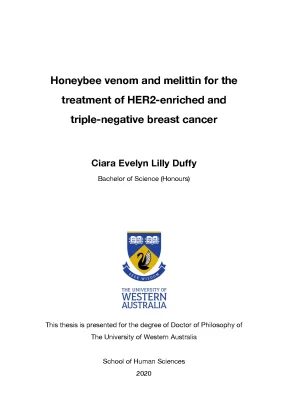
Tải xuống để đọc toàn bộHoneybee venom and melittin for the treatment of HER2-enriched and triple-negative breast cancer Ciara Evelyn Lilly Duffy Bachelor of Science (Honours) This thesis is presented for the degree of Doctor of Philosophy of The University of Western Australia School of Human Sciences 2020 Abstract Breast cancer is the most commonly occurring cancer in women worldwide. There are currently no effective targeted therapies available for triple-negative breast cancer (TNBC), which is aggressive, proliferative and associated with poor prognosis. The epidermal growth factor receptor (EGFR) is commonly overexpressed in TNBCs, and is important for cell proliferation and survival, conferring oncogenic signalling often dependent on the PI3K/Akt pathway downstream. To date, blocking EGFR signalling in TNBC has demonstrated limited clinical efficacy and the development of drug resistance.
Although therapies targeting human epidermal growth factor receptor 2 (HER2) in HER2-enriched breast tumours have dramatically improved median survival in the metastatic setting, resistance is almost inevitable for this subtype as well. Clearly, the discovery of more effective and selective therapeutic strategies for these cancers is an area of prime importance in clinical oncology. Venom from the European honeybee (Apis mellifera) and melittin, the major peptide in the venom, possess anti-cancer activity particularly by disrupting the plasma membrane. However, the precise molecular mechanisms explaining the anti-cancer activity of these agents against malignant breast cells are unknown.
The objective of this PhD was to determine the therapeutic potential of honeybee venom and melittin to target and kill aggressive breast cancer cells. The first aim was to determine the molecular mechanisms of breast cancer cell death due to honeybee venom and melittin. The second aim was to enhance the breast cancer selectivity of melittin and assess the interaction of melittin with growth factor receptors overexpressed in breast cancer cells. The final aim was to combine melittin with docetaxel in vivo to assess whether there is a synergistic interaction.
The anti-cancer properties of both honeybee venom and melittin, and selectivity towards TNBC and HER2-enriched breast cancers, were assessed by investigating the cellular response in a panel of breast cancer and normal-like cell lines. The results revealed that honeybee venom and melittin significantly and selectively reduce the viability of TNBC and HER2-enriched breast cancer cells and induce cell death through membrane disruption and caspase 3-mediated apoptosis. Melittin modulates oncogenic Akt and MAPK signalling in breast cancer cells by suppressing the activation of EGFR and HER2. Finally, melittin is synergistic with docetaxel both in vitro and in vivo, reducing cancer cell proliferation and consequent tumour growth in an aggressive TNBC allograft model.
Overall, this PhD demonstrates that honeybee venom and melittin potently and rapidly reduce breast cancer cell viability and can be used with chemotherapies or other small molecules to treat highly aggressive subtypes of breast cancer. Table of contents CHAPTER 1: General introduction and literature review .1 The intrinsic molecular subtypes of breast cancer .2 Limitations of current treatments for TNBC .3 Honeybee venom and melittin .4 The anti-cancer properties of bee venom and melittin .5 Targeting melittin, combination with chemotherapy and the use of nanotechnology to deliver melittin to cancer cells .6 Purpose and significance of this research .7 Aims and location in thesis .12 CHAPTER 2: Honeybee venom and melittin induce selective, potent, and rapid breast cancer cell death .1 Honeybees and bumblebees .2 Composition of honeybee venom compared to bumblebee venom .3 The application of bee venom and melittin in cancer .4 The cancer cell selectivity of honeybee venom and melittin .1 Chemical reagents and antibodies .2 Bee venom collection .4 Cell viability assays .5 Production of the primary monoclonal antibody against melittin .6 Enzyme-linked immunosorbent assay (ELISA) .7 Anti-melittin antibody competition experiments .8 Western blot for the detection of Cleaved Caspase-3 .10 Live cell microscopy .11 Scanning electron microscopy .1 Honeybee venom and melittin reduced tumour cell viability in triple-negative and HER2-enriched breast cancer cells .2 Honeybee venom reduced breast cancer cell viability with high affinity compared to bumblebee venom .3 Melittin was present in the same relative abundance in the venom of different honeybee populations, and mediated most of the anti-cancer effects .4 Honeybee venom and melittin induced caspase 3-mediated apoptosis, rapid cell death, and membrane disruption in breast cancer cells .5 Honeybee venom and melittin induced membrane disruption and nuclear condensation in breast cancer cells. 52 CHAPTER 3: Enhancing the breast cancer selectivity of melittin with RGD, and assessing the mechanisms of the suppression of EGFR and HER2 phosphorylation by melittin .1 Altering the charge of melittin to modulate the lytic effect .2 Increasing the cancer selectivity of melittin .3 Suppressing the phosphorylation of EGFR and HER2, and modulating downstream oncogenic signalling pathways in breast cancer cells .4 Utilising bioluminescence resonance energy transfer to determine the interaction of melittin with growth factor receptors .2 Chemical reagents and antibodies .3 Bee venom collection .5 Cell viability assays .6 Production of the primary monoclonal antibody against melittin .7 Enzyme-linked immunosorbent assay (ELISA) .8 Western blot for the detection of Cleaved Caspase-3 .10 Western blot for the detection of receptor phosphorylation and downstream pathway analysis .11 Bioluminescence resonance energy transfer (BRET) .1 The positive charge of melittin was essential for the anti-cancer effect, and the breast cancer selectivity of melittin was enhanced with RGD .2 The anti-melittin antibody recognised a conformational epitope of melittin that was not disrupted by the engineered RGD motif .3 Melittin was localised in the plasma membrane of breast cancer cells overexpressing EGFR or HER2 growth factor receptors .4 Honeybee venom and melittin suppressed the ligand-induced phosphorylation of EGFR and HER2 in breast cancer and modulated downstream signalling pathways in a time dependent manner .5 Melittin interacted with EGFR in the plasma membrane in a time and concentration dependent manner .6 Melittin indirectly interfered with RTK phosphorylation. 100 CHAPTER 4: Honeybee venom and melittin combined with docetaxel synergistically reduced breast cancer growth in vitro and in vivo .1 The mechanism of action of docetaxel and cisplatin .2 Limitations of current chemotherapy treatment .3 Combining honeybee venom and melittin with chemotherapy .4 Immune system modulation by melittin.1 Chemical reagents and antibodies .2 Bee venom collection .4 Cell viability assays .5 Production of the primary monoclonal antibody against melittin .6 Analysis of combined drug effects .7 Animal model and treatments .8 Immunohistochemical analysis of the tumours .1 Honeybee venom and melittin were synergistic with docetaxel in treating highly aggressive claudin-low breast cancer cells in vitro .2 The combination of melittin and docetaxel significantly reduced tumour growth in a highly aggressive claudin-low breast cancer model in vivo .3 The combined treatment of melittin and docetaxel reduced proliferation and induced apoptosis in claudin-low breast cancer in vivo.
128 CHAPTER 5: General discussion and future directions .1 Summary of results and significance. A1 Appendix A: Peer-reviewed publications. A1 Appendix B: List of conferences, symposiums, awards and courses. A2 Abbreviations Akt Protein kinase B ANOVA Analysis of variance BRET Bioluminescence resonance energy transfer BSA Bovine serum albumin CERI Centre for Entrepreneurial Research and Innovation CI Combination Index CIBER Center for Integrative Bee Research CO2 Carbon dioxide CPP Cell penetrating peptide CTG CellTiter-Glo DMEM Dulbecco’s Modified Eagle’s Medium DNA Deoxyribonucleic acid EGF Epidermal growth factor EGFR Epidermal growth factor receptor EMBL European Molecular Biology Laboratory F-12 Ham’s F-12 Nutrient Mixture FBS Fetal bovine serum FHS Fetal horse serum FITC Fluorescein isothiocyanate GLM Generalised linear model HER2 Human epidermal growth factor receptor 2 IC50 50% inhibitory concentration JAK2 Janus Kinase 2 MAPK Mitogen-activated protein kinase MEM α Minimal Essential Media α MMP2 Matrix metalloproteinase-2 mTOR Mammalian target of rapamycin NF-κB Nuclear factor-kappa B NSCLC Non-small-cell lung carcinoma PBS Phosphate-buffered saline PI3K Phosphatidylinositol 3-kinase PLA2 Phospholipase A2 RGD Arginine-glycine-aspartic acid motif RNA Ribonucleic acid RPMI Roswell Park Memorial Institute STAT3 Signal Transducer and Activator of Transcription 3 SV40 Simian virus 40 TBST Tris-buffered saline with Tween 20 TNBC Triple-negative breast cancer TRAIL Tumour Necrosis Factor-Related Apoptosis-Inducing Ligand uPA Urokinase plasminogen activator USGS United States Geological Survey VEGF Vascular endothelial growth factor VEGFR-1 Vascular endothelial growth factor receptor 1 VEGFR-2 Vascular endothelial growth factor receptor 2 Acknowledgements Thank you to everyone who has been a part of my PhD.
This has been an incredible journey and a transformational time of my life. To my supervisors, Associate Professor Pilar Blancafort, Professor Boris Baer, Dr Anabel Sorolla, and Professor Killugudi Swaminathan Iyer; I am tremendously thankful for all that you have taught me, and all of the opportunities you gave me. Thank you for enabling me the freedom to pursue my own scientific ideas and avenues of research. I would like to thank everyone who I have had the pleasure of working alongside in the Blancafort Laboratory at the Harry Perkins Institute of Medical Research.
In particular, I would like to acknowledge Anabel Sorolla and Edina Wang; I have learnt a great deal from both of you. Thank you to Colette Moses, for your constant guidance and support throughout my research journey. Thank you to the Centre for Integrative Bee Research (CIBER) team, especially Tiffane Bates, for providing training and access to the honeybees at the UWA apiary. I would also like to thank my wonderful mentors; Mark Cregan, Miriam Borthwick, Kevin Pfleger, Matt Oldakowski, Intan Oldakowska, Carolyn Williams and Charlie Bass.
Thank you for your invaluable training, advice and encouragement. Thank you to my international research collaborators; Professor Jane Stout (Trinity College Dublin) and Professor Mark Brown (Royal Holloway University of London). I would like to sincerely thank all of the following researchers and/or beekeepers who helped me to access samples of different bee populations in Ireland and England; Saorla Kavanagh, Keith Pierce, Marcus Phelan, Susie Bioletti, David Morris, Terry Meakin, Martina Halpin, Edward Hill, Fabio Manfredini and Callum Martin. Thank you to those who assisted in the venom transport across the world, including Sylvia Mathiasen, Siobhán McNamee and Rob Prouse.
Thank you to Alison Boyce and Steve Portugal for assisting me with accessing and setting up the carbon dioxide for bee venom collection. This research was supported by an Australian Government Research Training Program (RTP) Scholarship, and a PhD Top Up Scholarship from the Cancer Council of Western Australia. Technical assistance was kindly provided by Kathleen Davern for the development of the anti-melittin antibody (Chapter 2); Diwei Ho, in assisting with setting up the scanning electron microscopy experiment (Chapter 2); Emily Golden, in assisting with the peptide modelling (Chapter 3); Elizabeth Johnstone, in helping set up the BRET assay (Chapter 3); and Anabel Sorolla and Edina Wang, for assisting with the in vivo experiment (Chapter 4). I would also like to thank Paul Rigby (CMCA) and Kevin Li (FACS Facility) at the Harry Perkins Institute of Medical Research.
Lastly, thank you to all of my friends for your unwavering encouragement, to my beautiful family, and to Quentin, for your unconditional support throughout this journey. Mum and Dad, I truly cannot thank you enough. It is to you I dedicate my thesis. Chapter 1 General introduction and literature review 1.1 The intrinsic molecular subtypes of breast cancer Breast cancer is a devastating disease caused by a combination of environmental and genetic factors that trigger damage to the cellular DNA 1, is the second most common cancer in the world, and the most commonly occurring cancer in women 2,3.
In Australia, one in eight women will be diagnosed with breast cancer. Genome-wide profiling has identified several intrinsic molecular subtypes of breast cancer using patient samples to correlate the characteristics of the tumours to clinical outcome. These molecular subtypes include luminal A, luminal B, human epidermal growth factor receptor 2 (HER2)-enriched, basal-like, claudin-low, and normal-like 4–6. These subtypes differ greatly in terms of their transcriptional profiles, receptor expression, unique DNA methylation patterns, response to therapy and patient survival.
Targeted therapies have yet to be established with clinical success for basal-like and claudin-low tumours, which are immunohistochemically characterised as triple- negative breast cancer (TNBC), lacking the expression of estrogen and progesterone receptors, as well as HER2 6.
Nội dung được bảo vệ bản quyền — Tải xuống đầy đủ
Trích dẫn luận án này
Ciara Evelyn Lilly Duffy (2020). Nghiên cứu tác dụng của nọc ong trong điều trị ung thư vú [Luận án tiến sĩ, The University of Western Australia]. LuanAn.net. https://luanan.net/y-hoc/y-hoc-lam-sang/nghien-cuu-tac-dung-cua-noc-ong-trong-dieu-tri-ung-thu-vu
Câu hỏi thường gặp
Luận án "Nghiên cứu tác dụng của nọc ong trong điều trị ung thư vú" nghiên cứu về vấn đề gì?
Luận văn tiến sĩ của Duffy Ciara Evelyn Lilly năm 2020 khám phá các phương pháp nghiên cứu mới trong lĩnh vực triết học và khoa học xã hội.
Luận án "Nghiên cứu tác dụng của nọc ong trong điều trị ung thư vú" được bảo vệ tại trường nào?
Luận án này được bảo vệ tại The University of Western Australia. Năm bảo vệ: 2020.
Luận án "Nghiên cứu tác dụng của nọc ong trong điều trị ung thư vú" thuộc chuyên ngành gì?
Luận án "Nghiên cứu tác dụng của nọc ong trong điều trị ung thư vú" thuộc chuyên ngành Human Sciences. Danh mục: Y Học Lâm Sàng.
Luận án "Nghiên cứu tác dụng của nọc ong trong điều trị ung thư vú" có bao nhiêu trang?
Luận án "Nghiên cứu tác dụng của nọc ong trong điều trị ung thư vú" có 66 trang. Bạn có thể xem trước một phần tài liệu ngay trên trang web trước khi tải về.
Cách tải luận án "Nghiên cứu tác dụng của nọc ong trong điều trị ung thư vú" về máy như thế nào?
Để tải luận án về máy, bạn nhấn nút "Tải xuống ngay" trên trang này, sau đó hoàn tất thanh toán phí lưu trữ. File sẽ được tải xuống ngay sau khi thanh toán thành công. Hỗ trợ qua Zalo: 0559 297 239.