Luận án Duffy: Ức chế EGFR/HER2 ung thư vú bằng melittin & nọc ong
Luận văn của Duffy Ciara Evelyn Lilly năm 2020 phần 3 khám phá các yếu tố ảnh hưởng đến sự phát triển tri thức trong giáo dục.
Ung thư vú
Luan An
Luận văn
Số trang
62
Thời gian đọc
10 phút
Lượt xem
0
Lượt tải
0
Phí lưu trữ
40 Point
Tổng quan nhanh
- Chủ đề:
- 1. Melittin peptide ức chế thụ thể EGFR/HER2 trong ung thư vú
- Số trang:
- 62 trang
- Chuyên ngành:
- Ung thư vú
- Tác giả:
- Luan An
Tóm tắt nội dung luận án
I. Melittin peptide ức chế thụ thể EGFR HER2 trong ung thư vú
Melittin, một peptide trong nọc ong, đã được chứng minh là có khả năng ức chế thụ thể EGFR/HER2 trong ung thư vú. Nghiên cứu này tập trung vào việc đánh giá cơ chế hoạt động của melittin trong việc ức chế thụ thể EGFR/HER2 và tác động của nó đến các đường truyền tín hiệu nội bào.
1.1. Melittin peptide và khả năng ức chế thụ thể EGFR HER2
Melittin peptide có khả năng tương tác với thụ thể EGFR/HER2 trong màng plasma của tế bào ung thư vú. Sự tương tác này dẫn đến việc ức chế hoạt hóa thụ thể và giảm thiểu tín hiệu nội bào liên quan đến sự phát triển và sinh tồn của tế bào ung thư.
1.2. Cơ chế hoạt động của melittin
Melittin hoạt động bằng cách ức chế hoạt hóa thụ thể EGFR/HER2 thông qua việc ngăn chặn sự gắn kết của ligand với thụ thể. Điều này dẫn đến việc giảm thiểu tín hiệu nội bào liên quan đến sự phát triển và sinh tồn của tế bào ung thư.
II. Tác động của melittin đến đường truyền tín hiệu nội bào
Melittin có tác động đến các đường truyền tín hiệu nội bào liên quan đến sự phát triển và sinh tồn của tế bào ung thư. Nghiên cứu này đã đánh giá tác động của melittin đến các đường truyền tín hiệu PI3K/Akt và MAPK.
2.1. Tác động của melittin đến đường truyền tín hiệu PI3K Akt
Melittin đã được chứng minh là có khả năng ức chế hoạt hóa đường truyền tín hiệu PI3K/Akt, một đường truyền tín hiệu quan trọng liên quan đến sự sinh tồn của tế bào ung thư.
2.2. Tác động của melittin đến đường truyền tín hiệu MAPK
Melittin cũng đã được chứng minh là có khả năng ức chế hoạt hóa đường truyền tín hiệu MAPK, một đường truyền tín hiệu quan trọng liên quan đến sự phát triển của tế bào ung thư.
III. Tương tác của melittin với thụ thể EGFR HER2
Melittin có khả năng tương tác với thụ thể EGFR/HER2 trong màng plasma của tế bào ung thư vú. Sự tương tác này dẫn đến việc ức chế hoạt hóa thụ thể và giảm thiểu tín hiệu nội bào liên quan đến sự phát triển và sinh tồn của tế bào ung thư.
3.1. Tương tác của melittin với thụ thể EGFR
Melittin có khả năng tương tác với thụ thể EGFR, một thụ thể quan trọng liên quan đến sự phát triển và sinh tồn của tế bào ung thư.
3.2. Tương tác của melittin với thụ thể HER2
Melittin cũng có khả năng tương tác với thụ thể HER2, một thụ thể quan trọng liên quan đến sự phát triển và sinh tồn của tế bào ung thư.
IV. Ứng dụng của melittin trong điều trị ung thư vú
Melittin có tiềm năng ứng dụng trong điều trị ung thư vú do khả năng ức chế thụ thể EGFR/HER2 và giảm thiểu tín hiệu nội bào liên quan đến sự phát triển và sinh tồn của tế bào ung thư.
4.1. Ứng dụng của melittin trong điều trị ung thư vú HER2 dương tính
Melittin có tiềm năng ứng dụng trong điều trị ung thư vú HER2 dương tính do khả năng ức chế thụ thể HER2 và giảm thiểu tín hiệu nội bào liên quan đến sự phát triển và sinh tồn của tế bào ung thư.
4.2. Ứng dụng của melittin trong điều trị ung thư vú triple âm
Melittin cũng có tiềm năng ứng dụng trong điều trị ung thư vú triple âm do khả năng ức chế thụ thể EGFR và giảm thiểu tín hiệu nội bào liên quan đến sự phát triển và sinh tồn của tế bào ung thư.
Mục lục chi tiết luận án
Tải xuống file đầy đủ để xem toàn bộ nội dung
Tải đầy đủ (62 trang)Nội dung chính
Tổng quan nghiên cứu
Ung thư vú là một trong những nguyên nhân gây tử vong hàng đầu ở phụ nữ trên toàn thế giới, trong đó phân nhóm ung thư vú thể bộ ba âm tính (TNBC) và ung thư vú dương tính với thụ thể HER2 chiếm khoảng 15% đến 20% tổng số ca bệnh nhưng lại có tiên lượng xấu nhất. Các phân nhóm này có tốc độ phân chia tế bào nhanh, nguy cơ tái phát cao và thường xuất hiện tình trạng kháng các hóa trị liệu tiêu chuẩn như docetaxel hay cisplatin. Vấn đề cốt lõi trong điều trị hiện nay là sự thiếu hụt các liệu pháp nhắm trúng đích an toàn, khả năng thâm nhập của thuốc qua màng tế bào khối u còn hạn chế và các tác dụng phụ độc hại đối với tế bào lành.
Mục tiêu cụ thể của luận văn là khảo sát cơ chế phân tử của peptide melittin (thành phần chính chiếm khoảng 40% đến 50% trọng lượng khô của nọc ong mật Apis mellifera) trong việc ức chế các thụ thể tyrosine kinase màng (EGFR và HER2), điều biến các con đường truyền tín hiệu sinh tồn nội bào PI3K/Akt và MAPK, đồng thời đánh giá hiệu ứng hiệp đồng khi kết hợp melittin với hóa trị liệu docetaxel. Phạm vi nghiên cứu bao gồm các thử nghiệm in vitro trên các dòng tế bào ung thư vú người SKBR3, SUM159, tế bào biểu mô thận người HEK293FT, và thử nghiệm in vivo trên mô hình chuột BALB/c mang khối u ghép đồng loài T11 claudin-low kéo dài trong 15 ngày. Ý nghĩa của nghiên cứu được lượng hóa rõ nét thông qua việc giảm tới 90% thể tích khối u trên mô hình động vật, hạ chỉ số tăng sinh tế bào Ki-67 từ 59.7% xuống còn 5.8%, và nâng tỷ lệ kích hoạt quá trình chết rụng tế bào (apoptosis) theo phương pháp TUNEL lên 81.1%.
Cơ sở lý thuyết và phương pháp nghiên cứu
Khung lý thuyết áp dụng
Luận văn được xây dựng dựa trên ba khung lý thuyết và mô hình sinh học phân tử nền tảng:
- Lý thuyết truyền tín hiệu qua thụ thể Tyrosine Kinase (RTK Signaling Theory): Tập trung vào cơ chế kích hoạt thông qua sự dimer hóa của EGFR (trọng lượng phân tử 175 kDa) và HER2 (185 kDa), từ đó phosphoryl hóa các gốc tyrosine đặc hiệu để dẫn truyền tín hiệu xuống hai trục sinh tồn chính là PI3K/Akt và Ras/Raf/MEK/MAPK.
- Lý thuyết tạo lỗ xuyên màng sinh chất của peptide hoạt tính sinh học (Pore-Forming Peptide Theory): Giải thích cách peptide melittin (trọng lượng phân tử 2.85 kDa) liên kết tĩnh điện với màng phospholipid và tự lắp ráp để tạo thành các lỗ xuyên màng hình xuyến có đường kính trong khoảng 4.4 nm, tạo điều kiện thuận lợi cho sự thẩm thấu thụ động của các phân tử thuốc hóa trị.
- Mô hình phân tích hiệp đồng thuốc Chou-Talalay (Median-Effect Equation): Ứng dụng để định lượng mức độ tương tác giữa hai dược chất thông qua Chỉ số Kết hợp (Combination Index - CI), trong đó giá trị CI dưới 1 biểu thị tính hiệp đồng, CI bằng 1 biểu thị tính cộng, và CI lớn hơn 1 biểu thị tính đối kháng.
Bên cạnh đó, nghiên cứu vận dụng các khái niệm then chốt gồm: điểm kiểm soát miễn dịch PD-L1 (tránh thoát miễn dịch), mô-típ peptide nhắm đích integrin RGD1, và cơ chế chuyển vị năng lượng cộng hưởng phát quang sinh học (BRET) trong phạm vi khoảng cách dưới 10 nm.
Phương pháp nghiên cứu
Nghiên cứu sử dụng hệ thống dữ liệu thực nghiệm toàn diện từ các dòng tế bào ung thư vú đại diện cho các phân type lâm sàng: dòng SKBR3 (dương tính đồng thời với HER2 và EGFR), dòng SUM159 (ung thư vú thể bộ ba âm tính biểu hiện EGFR), dòng HEK293FT chuyển gen NanoLuc-EGFR, và dòng tế bào chuột T11 đặc trưng cho phân nhóm claudin-low khiếm khuyết gen p53.
Đối với thử nghiệm in vivo, cỡ mẫu được thiết kế chuẩn mực với 48 chuột cái BALB/c (5 tuần tuổi) mang khối u ghép T11 (thể tích ban đầu khoảng 50 mm3), được phân ngẫu nhiên thành 4 nhóm độc lập (12 cá thể mỗi nhóm): nhóm đối chứng sử dụng dung dịch tá dược (Milli-Q + 5% dextrose), nhóm đơn trị liệu melittin (liều 5 mg/kg), nhóm đơn trị liệu docetaxel (liều 7 mg/kg), và nhóm kết hợp melittin (5 mg/kg) cùng docetaxel (7 mg/kg). Phương pháp chọn mẫu ngẫu nhiên có kiểm soát giúp loại trừ các sai lệch sinh học giữa các cá thể.
Hệ thống phương pháp phân tích chuyên sâu được lựa chọn dựa trên độ tin cậy khoa học cao:
- Kỹ thuật Western Blot giúp định lượng chính xác động học phosphoryl hóa của p-HER2 (Tyr1248), p-EGFR (Tyr1068), p-Akt (Ser473, Thr308), và p-MAPK (Thr202/Tyr204) ở các mốc thời gian 2.5, 5, 10, 15, và 20 phút.
- Kỹ thuật BRET thời gian thực và Đồng kết tủa miễn dịch (Co-IP) với hạt từ streptavidin Dynabeads được lựa chọn để xác minh tương tác vật lý trực tiếp hay gián tiếp giữa melittin và EGFR ở mức phân tử dưới 10 nm.
- Nhuộm hóa mô miễn dịch (IHC) và miễn dịch huỳnh quang (IF) trên các lát cắt mô paraffin 5 µm với các kháng thể kháng Ki-67, TUNEL, PD-L1 và melittin.
- Hệ thống chụp ảnh phát quang sinh học Caliper IVIS Lumina II theo dõi động học khối u qua phát xạ Luciferin (liều 150 mg/kg) vào các ngày 4, 10, 12 và 14.
Toàn bộ dữ liệu được phân tích thống kê trên phần mềm GraphPad Prism phiên bản 8 và SPSS phiên bản 26 bằng phép kiểm ANOVA một yếu tố kèm theo kiểm định sâu Tukey HSD với ngưỡng ý nghĩa thống kê p dưới 0.05.
Kết quả nghiên cứu và thảo luận
Những phát hiện chính
- Ức chế tức thì hoạt hóa thụ thể EGFR/HER2 và cắt đứt con đường truyền tín hiệu sinh tồn: Trên dòng tế bào SKBR3, nọc ong và melittin làm giảm mạnh tín hiệu phosphoryl hóa của p-HER2 (Tyr1248) và p-EGFR (Tyr1068) chỉ sau 5 phút xử lý. Quá trình này dẫn đến sự triệt tiêu tín hiệu xuôi dòng của p-Akt (Ser473 và Thr308) cùng p-MAPK (Thr202/Tyr204) từ phút thứ 5 đến phút thứ 20, làm giảm hơn 80% tín hiệu phosphoryl hóa so với nhóm kích thích bằng yếu tố tăng trưởng EGF đơn thuần.
- Cơ chế tác động màng gián tiếp không cạnh tranh vị trí gắn ligand: Kỹ thuật BRET chứng minh melittin tiếp cận thụ thể EGFR ở khoảng cách dưới 10 nm trên màng sinh chất nhưng không bị bão hòa bởi phối tử nội sinh EGF (1 µM). Thử nghiệm Co-IP sử dụng biotin-melittin sau 10 phút xử lý không phát hiện phức hợp liên kết trực tiếp, khẳng định melittin định vị trên màng lipid và gián tiếp phong tỏa sự nhị phân hóa (dimerization) của thụ thể tyrosine kinase.
- Kỹ thuật gắn đuôi RGD1 tối ưu hóa tính chọn lọc tế bào ung thư: Việc gắn thêm mô-típ chuỗi peptide RGD1 vào đầu N của melittin đã làm tăng đáng kể tính liên kết đặc hiệu với thụ thể integrin αvβ6 biểu hiện quá mức trên tế bào ung thư vú TNBC. Ngược lại, việc thay đổi điện tích dương ở đầu C bằng chuỗi DEDE làm mất hoàn toàn hoạt tính gây độc (IC50 tăng trên 20 µM), trong khi việc tái tạo điện tích dương bằng chuỗi xoắn alpha SV40 đã phục hồi 100% khả năng ly giải tế bào đích.
- Hiệu ứng hiệp đồng mạnh mẽ vượt trội in vitro và in vivo: Phân tích Chou-Talalay cho thấy chỉ số CI giữa melittin và docetaxel luôn đạt giá trị dưới 1 trên mọi dải nồng độ thử nghiệm. Trên mô hình chuột mang khối u T11 vào ngày thứ 13, nhóm điều trị phối hợp melittin (5 mg/kg) và docetaxel (7 mg/kg) giúp giảm 90% thể tích khối u so với nhóm đối chứng (p dưới 0.001). Tại ngày thứ 14, phân tích mô học cho thấy chỉ số tăng sinh tế bào Ki-67 giảm mạnh xuống 5.8% (so với 59.7% ở nhóm đối chứng và 21.3% ở nhóm docetaxel đơn trị), tỷ lệ chết rụng apoptosis theo TUNEL đạt 81.1% (so với 1.4% ở nhóm đối chứng), đồng thời ức chế biểu hiện điểm kiểm soát miễn dịch PD-L1 từ 84.6% xuống còn 44.2%.
Thảo luận kết quả
Các kết quả thực nghiệm được biểu diễn trực quan thông qua bảng động học phosphoryl hóa Western Blot theo các mốc 2.5 đến 20 phút và biểu đồ phát xạ BRET theo nồng độ peptide. Đường cong thể tích khối u kết hợp cùng chuỗi hình ảnh phát xạ huỳnh quang IVIS Lumina II phản ánh sự thu nhỏ rõ rệt của khối u ở nhóm điều trị phối hợp từ ngày thứ 10 đến ngày thứ 14.
Nguyên nhân căn bản của hiệu ứng hiệp đồng là do melittin tạo các lỗ thủng xuyên màng có kích thước khoảng 4.4 nm, tạo lối vào thụ động giúp docetaxel dễ dàng thâm nhập vào nội bào để ổn định vi ống và ngăn chặn phân bào ở pha G2/M. Đồng thời, sự ức chế tức thời các thụ thể EGFR/HER2 của melittin đã vô hiệu hóa các vòng lặp phản hồi sinh tồn thích ứng (adaptive feedback loops) thường thấy khi sử dụng hóa trị liệu đơn độc.
So với các nghiên cứu trước đây vốn chỉ ghi nhận tác dụng ức chế thụ thể VEGFR-2 ở nồng độ khoảng 8 ng/µL trên ung thư biểu mô gan hoặc nọc ong thô trên ung thư phổi, nghiên cứu này đã làm sáng tỏ cơ chế màng gián tiếp trên hệ thống thụ thể ErbB trong ung thư vú. Hơn nữa, phát hiện về khả năng hạ thấp biểu hiện thụ thể PD-L1 từ 84.6% xuống 44.2% mang ý nghĩa lâm sàng đột phá, giúp loại bỏ lá chắn ức chế miễn dịch của khối u và tăng cường hoạt tính nhận diện của tế bào lympho T.
Đề xuất và khuyến nghị
- Tối ưu hóa công nghệ nano hướng đích cho peptide RGD1-melittin: Các nhóm nghiên cứu phát triển dược phẩm và công nghệ nano y sinh cần tiến hành đóng gói RGD1-melittin và docetaxel vào các hạt nano hướng đích cấu trúc lipid hoặc polymer. Mục tiêu là nâng tỷ lệ phân bố thuốc tại mô khối u lên trên 65%, đồng thời giảm tỷ lệ tan máu (hemolysis) toàn thân xuống dưới 2%, thực hiện trong khung thời gian 12 tháng.
- Thiết lập phác đồ giảm liều hóa trị liệu kết hợp: Các chuyên gia dược lý lâm sàng và bác sĩ điều trị ung bướu nên áp dụng mô hình hiệp đồng để thiết lập phác đồ giảm từ 30% đến 50% liều tiêu chuẩn của docetaxel và cisplatin khi kết hợp cùng melittin hoạt tính. Mục tiêu là duy trì hiệu quả tiêu diệt tế bào ung thư đạt trên 85% nhưng giảm thiểu tối đa các tác dụng phụ suy tủy và độc tính thần kinh, triển khai thử nghiệm trong 18 tháng tới.
- Tiêu chuẩn hóa quy trình sàng lọc sinh thiết thụ thể integrin αvβ6 và PD-L1: Các phòng thí nghiệm giải phẫu bệnh và chẩn đoán phân tử cần chuẩn hóa quy trình nhuộm hóa mô miễn dịch định lượng mật độ integrin αvβ6 và mức độ biểu hiện PD-L1 trên mẫu sinh thiết bệnh nhân TNBC. Mục tiêu đạt độ chính xác chẩn đoán trên 92% để phân loại chính xác nhóm bệnh nhân hưởng lợi tối đa từ phác đồ, hoàn thiện trong 9 tháng.
- Đánh giá toàn diện dược động học và độ an toàn đường tiêm tĩnh mạch: Các cơ quan nghiên cứu độc tính tiền lâm sàng cần thực hiện đánh giá độc tính bán trường diễn, dược động học (PK/PD) và xác định liều dung nạp tối đa (MTD) của phức hợp peptide qua đường tiêm tĩnh mạch trên mô hình động vật linh trưởng nhỏ. Mục tiêu là duy trì tỷ lệ an toàn sinh học 100% đối với chức năng gan và thận trong lộ trình từ 18 đến 24 tháng.
Đối tượng nên tham khảo luận văn
- Nghiên cứu sinh và Giảng viên chuyên ngành Dược lý học - Hóa sinh Y học: Cung cấp tài liệu tham khảo giá trị về phương pháp đánh giá động học thụ thể màng RTK qua kỹ thuật BRET thời gian thực, cơ chế tạo lỗ màng sinh chất và phương pháp thiết kế peptide đột biến cấu trúc nhắm đích.
- Bác sĩ chuyên khoa Ung bướu và Dược sĩ lâm sàng: Cung cấp bằng chứng thực nghiệm vững chắc về giải pháp vượt qua hiện tượng kháng thuốc hóa trị trong ung thư vú thể bộ ba âm tính và ung thư vú HER2 dương tính bằng liệu pháp kết hợp đa cơ chế.
- Chuyên gia R&D tại các doanh nghiệp Công nghệ sinh học và Dược phẩm: Nắm bắt quy trình kỹ thuật tái thiết kế đầu N và đầu C của peptide tự nhiên để phát triển các thế hệ thuốc sinh học mới có tính chọn lọc cao, chi phí sản xuất hợp lý.
- Nhà hoạch định chính sách y tế và Chuyên gia chuyển giao công nghệ sinh học: Nguồn dữ liệu khoa học tin cậy hỗ trợ thẩm định, cấp phép và đầu tư cho các dự án khai thác nguồn tài nguyên sinh học tự nhiên phục vụ y học hiện đại.
Câu hỏi thường gặp
Melittin ức chế các thụ thể EGFR và HER2 theo cơ chế trực tiếp hay gián tiếp?
Các bằng chứng từ phép đo BRET trong phạm vi dưới 10 nm và phân tích đồng kết tủa miễn dịch Co-IP khẳng định melittin tác động theo cơ chế gián tiếp. Melittin không chiếm chỗ gắn ligand của EGF mà chèn vào lớp màng đôi phospholipid, làm biến dạng cấu trúc lipid xung quanh và ngăn chặn sự dimer hóa cần thiết để kích hoạt quá trình tự phosphoryl hóa của thụ thể.
Làm thế nào để biến đổi cấu trúc melittin nhằm tăng tính chọn lọc trên tế bào ung thư?
Nghiên cứu đã liên kết đầu N của melittin với đoạn peptide RGD1 có ái lực cao với integrin αvβ6 biểu hiện quá mức trên tế bào ung thư vú TNBC. Đồng thời, việc duy trì cụm điện tích dương ở đầu C là điều kiện tiên quyết để giữ vững hoạt tính ly giải màng tế bào ung thư mục tiêu.
Hiệu ứng hiệp đồng giữa melittin và docetaxel được giải thích như thế nào?
Melittin hình thành các lỗ màng kích thước 4.4 nm giúp hóa trị liệu docetaxel xâm nhập thụ động và nhanh chóng vào nội bào. Sự kết hợp này mang lại chỉ số CI dưới 1, giúp giảm 90% thể tích khối u in vivo và ngăn chặn các cơ chế thích ứng sinh tồn của tế bào ác tính.
Melittin có ảnh hưởng như thế nào đến vi môi trường miễn dịch khối u?
Melittin làm giảm tỷ lệ biểu hiện phân tử ức chế miễn dịch PD-L1 từ 84.6% xuống còn 44.2% trên mô hình khối u in vivo. Nhờ đó, liệu pháp giúp hạn chế hiện tượng trốn thoát miễn dịch của khối u và hỗ trợ các tế bào lympho T tiêu diệt tế bào ung thư hiệu quả hơn.
Liệu pháp này có khả năng khắc phục tình trạng kháng thuốc hóa trị hay không?
Có. Bằng cách ức chế đồng thời trục PI3K/Akt và MAPK chỉ sau 5 phút xử lý, melittin loại bỏ các đường truyền tín hiệu cứu cánh vốn là nguyên nhân gây kháng taxane và platinum, giúp tái nhạy cảm các dòng tế bào ung thư vú claudin-low p53 khiếm khuyết với hóa trị liệu.
Kết luận
- Melittin từ nọc ong mật có khả năng ly giải chọn lọc cao trên các phân nhóm ung thư vú nguy hiểm gồm thể bộ ba âm tính và thể giàu thụ thể HER2.
- Cơ chế màng gián tiếp của melittin giúp ức chế tức thì sự phosphoryl hóa của EGFR (Tyr1068) và HER2 (Tyr1248) chỉ sau 5 phút xử lý.
- Biến tính chuỗi peptide bằng đầu gắn RGD1 tối ưu hóa tính chọn lọc hướng đích integrin αvβ6 và bảo toàn hoạt tính tiêu diệt tế bào thông qua cụm điện tích dương đầu C.
- Phối hợp melittin (5 mg/kg) và docetaxel (7 mg/kg) tạo hiệu ứng hiệp đồng mạnh mẽ, giảm 90% thể tích khối u, nâng tỷ lệ chết rụng apoptosis lên 81.1% và hạ chỉ số phân bào Ki-67 xuống 5.8%.
- Liệu pháp phong tỏa thành công điểm kiểm soát miễn dịch PD-L1 trong vi môi trường khối u, mở ra hướng đi mới trong việc đảo ngược tính kháng hóa trị liệu.
Lộ trình tiếp theo sẽ tập trung vào việc thử nghiệm công thức nano tiêm tĩnh mạch và hoàn thiện hồ sơ thử nghiệm độc tính tiền lâm sàng trong vòng 12 đến 24 tháng tới. Hãy kết nối và liên hệ ngay với nhóm nghiên cứu chuyên trách để cùng hợp tác phát triển, tài trợ thử nghiệm lâm sàng và đẩy nhanh tiến trình ứng dụng liệu pháp peptide sinh học đột phá này vào thực tiễn điều trị ung thư vú.
Trích đoạn nội dung luận án
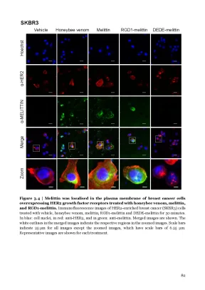
Tải xuống để đọc toàn bộSKBR3 Vehicle Honeybee venom Melittin RGD1-melittin DEDE-melittin Hoechst α-HER2 α-MELITTIN Merge Zoom Figure 3.4 | Melittin was localised in the plasma membrane of breast cancer cells overexpressing HER2 growth factor receptors treated with honeybee venom, melittin, and RGD1-melittin. Immunofluorescence images of HER2-enriched breast cancer (SKBR3) cells treated with vehicle, honeybee venom, melittin, RGD1-melittin and DEDE-melittin for 30 minutes. In blue: cell nuclei, in red: anti-HER2, and in green: anti-melittin. Merged images are shown.
The white outlines in the merged images indicate the respective regions in the zoomed images. Scale bars indicate 25 µm for all images except the zoomed images, which have scale bars of 6. Representative images are shown for each treatment.4 Honeybee venom and melittin suppressed the ligand-induced phosphorylation of EGFR and HER2 in breast cancer and modulated downstream signalling pathways in a time dependent manner The molecular mechanisms by which melittin suppresses tumour cell proliferation were next assessed. Considering the highly predominant localisation of melittin in the plasma membrane in tumour cells overexpressing EGFR and HER2, the next step was to assess whether honeybee venom and melittin interfered with growth factor receptor signalling as a mechanism of reducing the proliferation and survival of breast cancer cells.
For this reason, the hypothesis was that both honeybee venom and melittin could disrupt RTK-associated signalling pathways in EGFR and HER2 overexpressing cells by rapidly blocking the ligand- dependent activation of these receptors in the plasma membrane of breast carcinoma cells. To assess this, Western blot analysis was conducted on SKBR3 (HER2+, EGFR+) and SUM159 (EGFR+) extracts of cells previously stimulated with epidermal growth factor (EGF) and treated with the IC50 of honeybee venom or melittin from 2.5 to 20 minutes (Figure 3. Importantly, the results reveal that honeybee venom and melittin down-regulated the phosphorylation of the RTKs and modulated associated PI3K/Akt and MAPK signalling pathways in a time-dependent manner. In particular, treating SKBR3 cells with honeybee venom and melittin strongly downregulated p-HER2 (Tyr1248), p-EGFR (Tyr1068), p-p44/42 MAPK (Thr202/Tyr204), and p-Akt (Ser473 and Thr308) from 5 minutes onwards (Figure 3.
In SKBR3, there was also a slight decrease in total HER2, EGFR, and Akt protein only after 10 minutes of honeybee venom treatment, which could relate to membrane degradation and/or endosome-mediated receptor degradation 50. 83 In SUM159, p-EGFR (Tyr1068) was down-regulated by honeybee venom from 10 to 20 minutes, and melittin from 15 to 20 minutes of treatment (Figure 3. Treating SUM159 with melittin also suppressed p-Akt (Thr308) at all time points, yet upregulated p-p44/42 MAPK (Thr202/Tyr204) from 10 to 20 minutes, whereas honeybee venom upregulated p- p44/42 MAPK (Thr202/Tyr204), and p-Akt (Ser473 and Thr308) from 10 to 20 minutes. The anti-melittin antibody indicated an increasing amount of melittin present in the lysates of both cell lines over time, with a higher signal for the melittin treatment compared to whole honeybee venom in both cell lines.
The effects of honeybee venom and melittin on JAK/STAT pathway inhibitors were also assessed in TNBC (SUM159) cells because melittin was previously shown to inhibit JAK2/STAT3 signalling in ovarian cancer 52. No modulatory effects were observed after a 60-minute treatment with the IC50 of honeybee venom or melittin with or without the stimulation of 20 ng/mL EGF for 5 minutes (Supplementary Figure 3. 84 a SKBR3 Honeybee venom Melittin of treatment 2.5 5 10 15 20 α-p-HER2 Tyr 1248 185 kDa α-Total HER2 185 kDa α-p-EGFR Tyr 1068 175 kDa α-Total EGFR 175 kDa α-p-44/42 MAPK Erk1/2 Thr 202 / Tyr 204 44, 42 kDa α-p-AKT Ser 473 60 kDa α-p-AKT Thr 308 60 kDa α-Total AKT 60 kDa α-Total MELITTIN 2.85 kDa α-TUBULIN 50 kDa b SUM159 venom Melittin Minutes of treatment 2.5 5 10 15 20 α-p-EGFR Tyr 1068 175 kDa α-Total EGFR 175 kDa α-p-44/42 MAPK Erk1/2 Thr 202 / Tyr 204 44, 42 kDa α-p-AKT Ser 473 60 kDa α-p-AKT Thr 308 60 kDa α-Total AKT 60 kDa 2.85 kDa α-Total MELITTIN α-TUBULIN 50 kDa Figure 3.5 | Honeybee venom and melittin suppressed the ligand-induced phosphorylation of EGFR and HER2 and modulated downstream signalling pathways in breast cancer in a time dependent manner. Western blots showing the phosphorylation kinetics of the receptor tyrosine kinases (EGFR and HER2), downstream signalling pathways (MAPK and Akt), and total melittin after treatment with honeybee venom or melittin in (a) SKBR3 and (b) SUM159 breast cancer cells.
α-Tubulin was used as the loading control.5 Melittin interacted with EGFR in the plasma membrane in a time and concentration dependent manner Considering TNBC and HER2-enriched breast carcinoma cells are highly dependent on the activation of EGFR and HER2 for signalling and growth, bioluminescence resonance energy transfer (BRET) experiments were performed to determine whether melittin directly interfered with the binding of EGF to EGFR leading to the observed suppressed growth factor receptor phosphorylation. The NanoLuc reporter has been extensively shown to be useful in monitoring receptor-ligand binding in real time BRET assays 43,44. In this experiment, NanoLuc was used as the bioluminescent donor molecule, and genetically fused to EGFR 41,42. Kinetic and saturation BRET experiments were used to monitor the proximity of NanoLuc- EGFR with the fluorescently tagged acceptor molecules TAMRA-EGF (positive control), FITC-melittin, and the inactive FITC-DEDE-melittin (negative control).
The experiments were conducted with HEK293FT cells previously transfected with NanoLuc-EGFR fusion plasmid. Transfer of energy from the bioluminescent donor to the fluorescent acceptor occurs over distances less than 10 nm and is indicative of interactions between the tagged molecules of interest 40. First, cell viability was assessed by treating HEK293FT cells with FITC-melittin and FITC- DEDE-melittin to obtain IC50 values for the subsequent experiments (Supplementary Figure 3. Then, a range of concentrations of each peptide was selected including the IC50 of FITC-melittin, with the corresponding molar concentrations of FITC-DEDE-melittin.
The kinetics of the interaction of the fluorescent peptides with NanoLuc-EGFR were assessed with BRET using a range of peptide concentrations over time (Figure 3. The BRET signal increased in a dose-dependent manner for TAMRA-EGF, FITC-melittin and FITC- DEDE-melittin. Interestingly, FITC-DEDE-melittin displayed much higher BRET ratios 86 than FITC-melittin at the same concentrations, as well as reaching maximal BRET ratios at each dose very rapidly (Figure 3. In all cases, higher concentrations of peptide correlated with a higher BRET ratio.
As an additional negative control, a non-specific peptide designed against the Engrailed 1 (EN1) transcription factor 47 (FITC-EN1-mutant) exhibited similar BRET ratios and kinetics to the FITC-DEDE-melittin, supporting the specificity of the interaction of active forms of melittin with EGFR (Supplementary Figure 3. Further experiments were required to ascertain the specificity of the binding interactions of each peptide with EGFR. To determine this specificity of melittin binding to EGFR at the EGF binding site, saturation BRET assays were conducted to assess the competition of the endogenous ligand EGF with each of the peptides binding to NanoLuc-EGFR. The BRET signal of TAMRA-EGF with NanoLuc-EGFR was saturable and significantly reduced from 2.5 nM and all higher concentrations of TAMRA-EGF when it was combined with 1 µM EGF (Figure 3.6, b, left; two-way ANOVA, F 7,28 = 38, p<0.0001) confirming that TAMRA-EGF was binding to the EGF ligand binding site in the absence of endogenous EGF.
In contrast, the BRET signal of FITC-melittin and FITC-DEDE-melittin were not saturable and were not significantly different with or without 1 µM EGF (Figure 3.6, b, middle and right; F 7,32 = 0.999, respectively), suggesting that neither melittin nor DEDE- melittin bound to EGFR at the EGF binding site. The BRET ratio of the interaction of FITC- melittin and FITC-DEDE-melittin was higher than with TAMRA-EGF, likely due to the much higher concentrations of fluorescent peptide used which were required to reach concentrations above the IC5o.6 | Melittin interacted with EGFR in the plasma membrane in a time and concentration dependent manner. (a) Bioluminescence resonance energy transfer (BRET) kinetic analysis of TAMRA-EGF, FITC-melittin and FITC-DEDE-melittin interaction with NanoLuc- EGFR in HEK293FT cells. The peptides were added after the cells were equilibrated in the reader with the NanoLuc-substrate furimazine for 5 minutes.
Data are expressed as raw BRET ratios with mean ± SEM. (c) Illustration of the proposed BRET kinetics (top) and saturation (bottom) analysis for TAMRA-EGF (left), FITC- melittin (middle) and FITC-DEDE-melittin (right) with NanoLuc-EGFR. BRET signals were recorded when the fluorescent acceptor molecules were within 10 nm of NanoLuc-EGFR. 88 An illustration to explain the BRET kinetics and saturation results is proposed (Figure 3.
This illustration demonstrates that the kinetics results (top panel) for TAMRA-EGF and FITC-melittin were similar because increasing concentrations of both fluorescent acceptor molecules came into close proximity with NanoLuc-EGFR until the recording saturated for each concentration of peptide used. This is most likely because TAMRA-EGF bound directly to NanoLuc-EGFR, and melittin inserted into and moved laterally in the plasma membrane 53 to interact with NanoLuc-EGFR in a fluid manner within a distance of 10 nm. In contrast, FITC-DEDE-melittin was not expected to enter the cell membrane due to the previously observed lack of cell reactivity to this mutant peptide, and so the same BRET levels were recorded across the hour as the peptide came within 10 nm of NanoLuc- EGFR in the extracellular fluid in a concentration dependent manner. For the saturation results (Figure 3.6, c, bottom panel), there was a significantly reduced BRET signal when TAMRA-EGF was combined with unlabelled EGF, because the latter bound to and thus blocked the EGF binding site of NanoLuc-EGFR, which interfered with the interaction between TAMRA-EGF and NanoLuc-EGFR.
In contrast, there was no change in the BRET signal for both FITC-melittin and FITC-DEDE-melittin with or without unlabelled EGF, because the latter did not block the capacity of these fluorescent acceptor molecules to interact with NanoLuc-EGFR. Overall, the BRET data indicates that melittin may be positioned at a distance within 10 nm from the RTKs without interfering with the endogenous growth factor binding site.6 Melittin indirectly interfered with RTK phosphorylation Considering melittin did not bind to EGFR at the endogenous EGF binding site, it was still unclear whether melittin bound directly to EGFR elsewhere on the receptor in order to interfere with receptor phosphorylation. To assess this, a co-immunoprecipitation experiment was conducted between biotin-melittin and NanoLuc-EGFR (Figure 3. 89 HEK293FT cells were either untransfected (wild-type) or transfected with Empty Vector or NanoLuc-EGFR fusion plasmid for 24 hours.
The cells were then treated for 10 minutes with either vehicle, biotin-melittin or biotin-DEDE-melittin using the IC50 of biotin-melittin (3.512 µM), and the co-immunoprecipitation conducted with streptavidin coated beads to pull down biotinylated melittin. Considering no band was detected in the lane where the cells were transfected with NanoLuc-EGFR and treated with biotin-melittin (as indicated by the grey arrow in Figure 3.7), these results indicate that melittin does not bind directly to EGFR, at least by 10 minutes of treatment. In summary, it is possible that melittin interacts with the plasma membrane broadly, indirectly interfering with the phosphorylation of growth factor RTKs such as EGFR and HER2 in breast cancer cells.7 | Melittin indirectly interfered with growth factor RTK phosphorylation. A co-immunoprecipitation assay was conducted to assess the association of melittin with EGFR.
HEK293FT cells were treated with media (wild-type) or transfected with Empty Vector or NanoLuc- EGFR fusion plasmid for 24 hours, and then treated with vehicle, biotin-melittin or biotin-DEDE- melittin for 10 minutes. Cell extracts were collected and subjected to co-immunoprecipitation using streptavidin coated Dynabeads, followed by Western blot with an anti-EGFR antibody. Wild-type and Empty Vector transfected cells, and vehicle and biotin-DEDE-melittin, served as controls. In the case that melittin bound directly to EGFR, a band would have been detected in the lane where the cells were transfected with NanoLuc-EGFR and treated with biotin-melittin (as indicated by the grey arrow).5 Discussion As discovered in Chapter 2, further investigation was required into the molecular mechanisms underpinning the interaction of melittin with plasma membrane receptors and downstream signalling pathways, and whether the breast cancer specificity of melittin could be enhanced.
Nội dung được bảo vệ bản quyền — Tải xuống đầy đủ
Trích dẫn luận án này
Melittin nọc ong ức chế thụ thể EGFR/HER2 ung thư vú (n.d.) [Luận án tiến sĩ]. LuanAn.net. https://luanan.net/duoc-hoc/duoc-ly-hoc/luan-an-duffy-uc-che-egfr-her2-ung-thu-vu-bang-melittin-noc-ong
Câu hỏi thường gặp
Luận án "Melittin nọc ong ức chế thụ thể EGFR/HER2 ung thư vú" nghiên cứu về vấn đề gì?
Luận văn của Duffy Ciara Evelyn Lilly năm 2020 phần 3 khám phá các yếu tố ảnh hưởng đến sự phát triển tri thức trong giáo dục.
Luận án "Melittin nọc ong ức chế thụ thể EGFR/HER2 ung thư vú" thuộc chuyên ngành gì?
Luận án "Melittin nọc ong ức chế thụ thể EGFR/HER2 ung thư vú" thuộc chuyên ngành Ung thư vú. Danh mục: Dược Lý Học.
Luận án "Melittin nọc ong ức chế thụ thể EGFR/HER2 ung thư vú" có bao nhiêu trang?
Luận án "Melittin nọc ong ức chế thụ thể EGFR/HER2 ung thư vú" có 62 trang. Bạn có thể xem trước một phần tài liệu ngay trên trang web trước khi tải về.
Cách tải luận án "Melittin nọc ong ức chế thụ thể EGFR/HER2 ung thư vú" về máy như thế nào?
Để tải luận án về máy, bạn nhấn nút "Tải xuống ngay" trên trang này, sau đó hoàn tất thanh toán phí lưu trữ. File sẽ được tải xuống ngay sau khi thanh toán thành công. Hỗ trợ qua Zalo: 0559 297 239.