Luận án tiến sĩ: Nghiên cứu tính toán protein amyloid beta - Giai đoạn ban đầu tập hợp
Luận án tiến sĩ nghiên cứu tính toán protein amyloid beta. Phân tích giai đoạn tập hợp protein, cấu trúc dimer và vai trò oligomer trong bệnh Alzheimer.
Năm xuất bản
Số trang
165
Thời gian đọc
25 phút
Lượt xem
1
Lượt tải
0
Phí lưu trữ
50 Point
Tổng quan nhanh
- Chủ đề:
- 1. Protein Amyloid Beta Và Bệnh Alzheimer
- Số trang:
- 165 trang
- Trường:
- Boston University
- Chuyên ngành:
- Chemistry
- Tác giả:
- Bogdan Tarus B.
- Năm:
- 2007
Tóm tắt nội dung luận án
I. Protein Amyloid Beta Và Bệnh Alzheimer
Protein amyloid beta đóng vai trò then chốt trong sự phát triển của bệnh Alzheimer. Nghiên cứu này tập trung vào các giai đoạn đầu của quá trình tập hợp protein. Mục tiêu chính là xác định tính ổn định của monomer và oligomer bậc thấp. Các cấu trúc này đóng vai trò trung gian trên con đường hình thành sợi amyloid lớn hơn. Hiểu rõ cơ chế này giúp phát triển phương pháp điều trị bệnh Alzheimer hiệu quả hơn.
1.1. Vai Trò Của Protein Amyloid Beta Trong Bệnh Alzheimer
Protein amyloid beta là thành phần chính gây bệnh Alzheimer. Quá trình tích tụ protein này tạo nên các mảng amyloid đặc trưng. Các mảng này phá hủy chức năng thần kinh não. Nghiên cứu cho thấy oligomer độc hại gây tổn thương tế bào sớm nhất. Việc ngăn chặn hình thành oligomer có thể làm chậm tiến triển bệnh.
1.2. Tầm Quan Trọng Của Nghiên Cứu Giai Đoạn Đầu Tập Hợp
Giai đoạn đầu của quá trình tập hợp protein là then chốt. Monomer và dimer là các cấu trúc trung gian quan trọng. Chúng quyết định hướng phát triển của toàn bộ quá trình. Nghiên cứu này sử dụng mô phỏng phân tử để theo dõi quá trình hình thành. Kết quả giúp xác định các điểm can thiệp điều trị tiềm năng.
1.3. Phương Pháp Nghiên Cứu Tính Toán
Nghiên cứu áp dụng động lực học phân tử cổ điển. Phương pháp umbrella sampling tính toán thế năng trung bình. Cả mô hình dung môi tường minh và ẩn đều được sử dụng. Mô hình TIP3P mô phỏng môi trường nước chính xác. Các mô hình liên tục điện môi giúp tăng thời gian mô phỏng.
II. Mô Phỏng Phân Tử Protein Aβ10 35
Đoạn protein Aβ10-35 được chọn làm đối tượng nghiên cứu chính. Nghiên cứu tạo ra nhiều cấu trúc dimer khả dĩ dựa trên nguyên tắc bổ sung hình dạng. Các cấu trúc được đánh giá qua năng lượng khử dung môi và tương tác tĩnh điện. Phương pháp này xác định cấu trúc dimer ổn định nhất. Kết quả so sánh cho thấy sự khác biệt rõ rệt giữa các cấu trúc.
2.1. Tạo Cấu Trúc Dimer Từ Bổ Sung Hình Dạng
Giao thức dựa trên bổ sung hình dạng tạo ra tập hợp cấu trúc dimer. Phương pháp này đảm bảo sự khớp nối tối ưu giữa hai monomer. Năng lượng khử dung môi được tính toán nhanh chóng. Năng lượng tương tác tĩnh điện bổ sung thông tin quan trọng. Cấu trúc ổn định nhất được chọn để phân tích sâu hơn.
2.2. Tính Toán Thế Năng Trung Bình PMF
Phương pháp umbrella sampling tính toán thế năng trung bình chính xác. PMF mô tả năng lượng tự do trong quá trình dimerization. Mô phỏng được thực hiện trong dung môi nước. Kết quả cho thấy hàng rào năng lượng và trạng thái ổn định. Hai cấu trúc dimer khả dĩ được so sánh trực tiếp.
2.3. Phân Tích Tính Ổn Định Cấu Trúc Dimer
Quỹ đạo động lực học phân tử xuất phát từ hai cấu trúc dimer. Phân tích theo dõi sự thay đổi cấu trúc theo thời gian. Độ ổn định được đánh giá qua nhiều thông số. Cấu trúc bậc hai protein thay đổi trong quá trình mô phỏng. Kết quả xác định cấu trúc dimer có khả năng tồn tại lâu nhất.
III. So Sánh Mô Hình Dung Môi Ẩn Và Tường Minh
Nghiên cứu so sánh ba mô hình dung môi khác nhau với mô hình TIP3P tường minh. Mục tiêu là tăng thời gian mô phỏng mà vẫn giữ độ chính xác. Một mô hình kinh nghiệm và hai mô hình liên tục điện môi được thử nghiệm. Cấu trúc bậc ba protein từ các mô hình được so sánh chi tiết. Kết quả cho thấy ưu nhược điểm của từng phương pháp.
3.1. Mô Hình Dung Môi TIP3P Tường Minh
Mô hình TIP3P mô phỏng phân tử nước một cách chi tiết. Độ chính xác cao nhưng tốn nhiều tài nguyên tính toán. Mỗi phân tử nước được biểu diễn tường minh. Tương tác giữa protein và nước được mô tả chính xác. Đây là tiêu chuẩn vàng để so sánh các mô hình khác.
3.2. Mô Hình Dung Môi Liên Tục Điện Môi
Hai mô hình liên tục điện môi được áp dụng trong nghiên cứu. Chúng xử lý dung môi như môi trường liên tục. Tốc độ tính toán nhanh hơn nhiều so với mô hình tường minh. Tuy nhiên độ chính xác có thể giảm ở một số trường hợp. Phương pháp này cho phép mô phỏng thời gian dài hơn.
3.3. So Sánh Cấu Trúc Protein Từ Các Mô Hình
Cấu trúc protein từ các mô hình khác nhau được so sánh trực tiếp. Cấu trúc bậc hai và bậc ba đều được phân tích. Sự khác biệt chủ yếu xuất hiện ở vùng linh động. Một số vùng bảo toàn cấu trúc tốt qua các mô hình. Kết quả giúp lựa chọn mô hình phù hợp cho từng mục đích nghiên cứu.
IV. Cầu Muối D23 K28 Và Hình Thành Cấu Trúc Beta
Vùng cầu muối D23-K28 được xác định là điểm quan trọng. Kết quả thực nghiệm gợi ý đây là vị trí tạo nhân cho cấu trúc beta. Nghiên cứu theo dõi động lực học của tiếp xúc này. Sự hình thành và phá vỡ cầu muối được phân tích chi tiết. Vai trò của vùng này trong tạo sợi amyloid được đánh giá nghiêm túc.
4.1. Vai Trò Của Cầu Muối Trong Tập Hợp Protein
Cầu muối D23-K28 tạo liên kết tĩnh điện mạnh. Liên kết này ổn định cấu trúc cục bộ của protein. Thực nghiệm cho thấy vùng này quan trọng cho quá trình tập hợp. Mô phỏng phân tử theo dõi sự hình thành cầu muối. Kết quả xác nhận vai trò then chốt của tương tác này.
4.2. Động Lực Học Của Tiếp Xúc D23 K28
Động lực học phân tử theo dõi khoảng cách giữa D23 và K28. Cầu muối hình thành và phá vỡ nhiều lần trong mô phỏng. Thời gian tồn tại của liên kết được thống kê. Năng lượng liên kết thay đổi theo cấu hình protein. Phân tích này làm rõ tính động của vùng quan trọng.
4.3. Vị Trí Tạo Nhân Cho Cấu Trúc Beta
Vùng D23-K28 có thể là điểm khởi đầu hình thành cấu trúc beta. Cấu trúc bậc hai thay đổi từ xoắn alpha sang tấm beta. Quá trình này đặc trưng cho sợi amyloid trưởng thành. Mô phỏng quan sát sự chuyển đổi cấu trúc. Kết quả hỗ trợ giả thuyết về vai trò tạo nhân.
V. Nghiên Cứu Đoạn Protein Aβ21 30
Đoạn Aβ21-30 được nghiên cứu bằng mô phỏng trong dung môi nước tường minh. Vùng VGSN(24-27) nhận được sự chú ý đặc biệt. Đo đạc NMR trạng thái rắn cho thấy vùng này hình thành khúc quanh. Cấu trúc khúc quanh này ổn định protein trong sợi amyloid. Nghiên cứu phân tích chi tiết cấu trúc và động lực học của vùng này.
5.1. Vùng VGSN 24 27 Và Cấu Trúc Khúc Quanh
Vùng VGSN bao gồm bốn amino acid liên tiếp. Thực nghiệm NMR phát hiện cấu trúc khúc quanh ở đây. Khúc quanh này quan trọng cho cấu trúc sợi amyloid. Mô phỏng phân tử tái tạo sự hình thành khúc quanh. Độ ổn định của cấu trúc được đánh giá định lượng.
5.2. Động Lực Học Phân Tử Của Aβ21 30
Mô phỏng sử dụng mô hình nước tường minh chính xác cao. Quỹ đạo được phân tích để xác định cấu trúc ưu tiên. Cấu trúc bậc hai thay đổi linh hoạt theo thời gian. Một số vùng giữ cấu trúc ổn định hơn vùng khác. Kết quả cung cấp thông tin chi tiết về hành vi protein.
5.3. Liên Hệ Với Cấu Trúc Sợi Amyloid
Cấu trúc quan sát được liên quan đến sợi amyloid trưởng thành. Khúc quanh VGSN giúp protein gấp thành cấu trúc đặc trưng. Tấm beta song song được hình thành từ nhiều phân tử. Mô phỏng giải thích cơ chế ổn định cấu trúc sợi. Hiểu biết này quan trọng cho thiết kế thuốc ức chế.
VI. Ảnh Hưởng Của Đột Biến E22Q Và D23N
Hai đột biến tự nhiên E22Q và D23N được phân tích chi tiết. Các đột biến này liên quan đến bệnh amyloidosis. Nghiên cứu so sánh tính chất nhiệt động của protein đột biến và dạng hoang dã. Ảnh hưởng lên cấu trúc bậc hai và bậc ba được đánh giá. Kết quả giải thích vai trò của đột biến trong bệnh lý.
6.1. Đột Biến E22Q Và Tính Chất Protein
Đột biến E22Q thay glutamate bằng glutamine. Thay đổi này loại bỏ điện tích âm tại vị trí 22. Tương tác tĩnh điện của protein thay đổi đáng kể. Cấu trúc bậc hai có xu hướng khác so với dạng hoang dã. Đột biến này liên quan đến một số dạng amyloidosis di truyền.
6.2. Đột Biến D23N Và Ổn Định Cấu Trúc
Đột biến D23N thay aspartate bằn asparagine. Điện tích âm tại vị trí 23 bị mất. Cầu muối D23-K28 không thể hình thành. Sự thay đổi này ảnh hưởng lớn đến quá trình tập hợp. Protein đột biến có xu hướng tập hợp nhanh hơn.
6.3. So Sánh Nhiệt Động Học Giữa Các Dạng
Tính chất nhiệt động của ba dạng protein được so sánh. Năng lượng tự do tập hợp thay đổi giữa các dạng. Protein đột biến thường kém ổn định hơn dạng hoang dã. Tốc độ hình thành oligomer độc hại tăng lên. Kết quả giải thích tại sao đột biến gây bệnh sớm hơn.
Mục lục chi tiết luận án
Tải xuống file đầy đủ để xem toàn bộ nội dung
Tải đầy đủ (165 trang)Nội dung chính
Tổng quan về luận án
Sự tích tụ bất thường của protein amyloid $\beta$ ($A\beta$) trong não bộ được xác định là dấu ấn bệnh lý then chốt của bệnh Alzheimer (Alzheimer's Disease - AD). Luận án tiến sĩ của Bogdan Tarus (2007) tại Đại học Boston, dưới sự hướng dẫn của Giáo sư John E. Straub, mang tính tiên phong trong việc sử dụng các phương pháp mô phỏng động lực học phân tử (Molecular Dynamics - MD) cổ điển và nâng cao để khảo sát cơ chế nhiệt động lực học và cấu trúc ở giai đoạn sơ khởi của quá trình tập hợp protein $A\beta$. Trọng tâm nghiên cứu tập trung vào các đoạn peptide $A\beta_{10-35}$ và $A\beta_{21-30}$ nhằm giải mã vai trò của các trạng thái monome và oligome trọng lượng phân tử thấp (Low Molecular Weight - LMW oligomers) đóng vai trò cấu trúc trung gian siêu ổn định (metastable intermediates) trên lộ trình hình thành sợi amyloid (fibrils).
Khoảng trống nghiên cứu (Research Gap) xuất phát từ việc thiếu hụt dữ liệu cấu trúc ở độ phân giải nguyên tử đối với các oligome bậc thấp trong môi trường dung dịch nước. Trong khi các nghiên cứu NMR trạng thái rắn của Lynn (2000) và Petkova et al. (2002) đã xác định cấu trúc sợi hoàn thiện có tổ chức phiến gấp $\beta$ song song thẳng hàng (parallel in-register $\beta$-sheets), cơ chế chuyển pha từ cấu trúc cuộn ngẫu nhiên co cụm (collapsed random coil) của monome hòa tan sang các mầm kết tụ $\beta$ vẫn còn là một ẩn số lớn. Các câu hỏi và giả thuyết nghiên cứu được thiết lập chặt chẽ:
- RQ1: Tương tác kỵ nước (hydrophobic) hay tương tác tĩnh điện (electrostatic) đóng vai trò chủ đạo trong việc ổn định phức hợp dime $A\beta_{10-35}$ ban đầu?
- H1: Lực khử dung môi (desolvation) của các gốc kỵ nước là động lực nhiệt động lực học chính dẫn dắt quá trình dime hóa.
- RQ2: Các mô hình dung môi ngầm (implicit solvent) có phản ánh chính xác trạng thái cấu trúc và động học của monomer $A\beta_{10-35}$ so với mô hình dung môi tường minh (explicit solvent TIP3P) hay không?
- H2: Dung môi ngầm đánh giá thấp năng lượng tự solvat hóa (solvation self-energy), dẫn đến việc bù trừ sai lệch bằng liên kết hydro nội phân tử và làm peptide co cụm phi thực tế.
- RQ3: Cầu muối Asp23-Lys28 (D23-K28) và vòng gập VGSN(24-27) có tự phát hình thành bền vững trong monome hòa tan hay cần sự điều hòa của các phân tử nước rời rạc?
- H3: Sự hình thành cầu muối D23-K28 bị cản trở bởi rào cản khử solvat hóa (desolvation barrier) và đòi hỏi sự giải phóng nước có cấu trúc để tạo mầm $\beta$-structure.
Khung lý thuyết của luận án tích hợp Nguyên lý Tự tổ chức Amyloid (Amyloid Self-Organization Principle - ASOP), Lý thuyết Năng lượng Tiếp xúc Nguyên tử (Atomic Contact Energy - ACE) và Cơ học Thống kê về Thế lực Trung bình (Potential of Mean Force - PMF). Luận án tạo đột phá định lượng khi xác định cấu hình $\gamma$-dimer đạt cực tiểu năng lượng khử dung môi $\Delta E_{\text{desolv}} = -11.9\text{ kcal/mol}$, chứng minh sự áp đảo của tương tác kỵ nước tại giao diện $LVFFA(17-21)$, đồng thời giải mã cấu trúc không gian 4 bồn trũng năng lượng tự do (basins) với các rào cản chuyển pha từ $0.3\text{ kcal/mol}$ đến $2.1\text{ kcal/mol}$. Phạm vi nghiên cứu bao gồm mô phỏng 2000 cấu hình phân tử (decoys), 19 cửa sổ Umbrella Sampling và các quỹ đạo MD tích lũy trên 100 ns với độ chính xác nguyên tử toàn phần (all-atom).
Literature Review và Positioning
Tổng quan tài liệu trong luận án tổng hợp ba dòng nghiên cứu học thuật chính:
- Động học kết tụ và giả thuyết hạt nhân trung gian (Paranuclei Hypothesis): Teplow và cộng sự (1999, 2002) đã chứng minh các đồng phân chuỗi $A\beta_{1-40}$ và $A\beta_{1-42}$ trải qua các con đường tạo hạt nhân khác nhau. Bitan et al. (2003) chỉ ra rằng quá trình oxy hóa gốc Met35 trên $A\beta_{1-42}$ làm giảm tốc độ tạo sợi tương đương $A\beta_{1-40}$, thiết lập giả thuyết rằng sự gia tăng tính ưa nước làm tăng rào cản năng lượng tự do của quá trình oligome hóa.
- Cấu trúc tổ chức bậc cao của Fibril: Phổ NMR trạng thái rắn của Lynn et al. (2000) và Tycko et al. (2002) xác nhận kiến trúc phiến $\beta$ song song cho cả $A\beta_{10-35}$ và $A\beta_{1-40}$.
- Trạng thái trung gian cấu trúc xoắn $\alpha$ (Transient $\alpha$-helical intermediates): Kirkitadze et al. (2001) và Massi, Thirumalai, Straub (2001, 2004) giả định rằng monome vô định hình phải trải qua trạng thái xoắn $\alpha$ trung gian trước khi tái tổ chức thành sợi $\beta$.
========================================================================================
                                     LITERATURE DEBATES
========================================================================================
[Tranh luận 1: Động lực Dime hóa Sơ khởi]
   Quan điểm A (Tĩnh điện dẫn đường)      VS      Quan điểm B (Khử dung môi kỵ nước)
   - Tiếp xúc cặp ion/cầu muối dẫn dắt            - Tương tác kỵ nước cụm LVFFA(17-21)
   - Phụ thuộc pH và điện tích bề mặt             - Đẩy phân tử nước giao diện (Desolvation)
   -------------------------------------------------------------------------------------
   => Luận án Tarus (2007) chứng minh: Cầu muối bị che chắn điện môi (dielectric screening)
      trong nước; phức hợp gamma-dimer kỵ nước (-11.9 kcal/mol) chiếm ưu thế bền vững.

[Tranh luận 2: Phương pháp Luận Hydrat hóa Phân tử]
   Mô hình Dung môi Ngầm (Implicit)       VS      Mô hình Dung môi Tường minh (TIP3P)
   - Hiệu quả tính toán quy mô lớn                - Tái hiện cấu trúc mạng lưới liên kết hydro
   - Xấp xỉ điện môi liên tục (Continuum)          - Mô tả rào cản solvat hóa rời rạc
   -------------------------------------------------------------------------------------
   => Luận án Tarus (2007) chỉ rõ: GBORN/ACE đánh giá thấp Solvation Self-Energy,
      khiến peptide co cụm phi thực tế; TIP3P tái hiện chính xác pKa thực nghiệm.
========================================================================================
Luận án định vị chính xác khoảng trống bằng cách đối chiếu trực tiếp với hai nghiên cứu quốc tế điển hình:
- So với nghiên cứu của Massi et al. (2001) về đột biến Dutch $A\beta_{10-35}$, nghiên cứu này mở rộng phân tích không gian tự do nhiệt động lực học bằng đồ thị ngắt kết nối (Transition Disconnectivity Graphs - TRDG).
- So với công trình của Bitan et al. (2003) trên $A\beta_{1-42}$, luận án cung cấp bằng chứng cơ học vi mô giải thích sự đóng góp $47%$ của đoạn đầu N-terminus(A) vào tổng năng lượng khử dung môi khi liên kết với C-terminus(B).
Đóng góp lý thuyết và khung phân tích
Đóng góp cho lý thuyết
Nghiên cứu mở rộng sâu sắc Nguyên lý Tự tổ chức Amyloid (Amyloid Self-Organization Principle - ASOP) do Thirumalai và Straub đề xuất:
- Proposition P1 (Trạng thái Monomer hòa tan): Ở trạng thái monome phân tán trong dung dịch nước, tương tác tĩnh điện Asp23-Lys28 không tự phát chiếm ưu thế tuyệt đối do chi phí năng lượng tự do khử solvat hóa của các ion tích điện cao hơn tương tác hút Coulomb.
- Proposition P2 (Cơ chế Loại trừ Nước - Water Expulsion): Cầu muối nội phân tử D23-K28 và vòng gập VGSN(24-27) chỉ đóng vai trò hạt nhân ổn định khi các phân tử nước rời rạc bị đẩy ra khỏi giao diện trong quá trình tự kết tụ đa phân tử.
- Proposition P3 (Bảo tồn Lõi Kỵ nước): Lõi kỵ nước trung tâm $LVFFA(17-21)$ duy trì cấu trúc bảo tồn cao trong quá trình dime hóa, đóng vai trò bản lề động học cho sự chuyển pha từ cuộn vô định hình sang phiến $\beta$.
"The finding that the $\gamma$-dimer may constitute the ensemble of stable $A\beta_{10-35}$ dimer has important implications for fibril formation. In particular, the expulsion of water molecules at the interface might be a key event, just as in the oligomerization of $A\beta_{16-22}$ fragments." (Tarus, 2007, Ch. 2, p. 7).
Nghiên cứu đánh dấu bước chuyển đổi hệ hình (Paradigm shift) từ quan niệm tĩnh điện định hướng (electrostatic steering) sang hệ hình phân tử nước rời rạc điều hòa rào cản khử solvat hóa (discrete solvent-mediated desolvation penalty).
Khung phân tích độc đáo
Khung phân tích của luận án là sự tích hợp chặt chẽ giữa ba lý thuyết tính toán:
- Lý thuyết Năng lượng Tiếp xúc Nguyên tử (Atomic Contact Energy - ACE): Dựa trên mô hình mở rộng của Miyazawa-Jernigan và Zhang et al., phân loại 18 nhóm nguyên tử nặng để định lượng chính xác công biến thiên chuyển dịch nguyên tử từ nước vào lõi protein không phân cực.
- Lý thuyết Thế lực Trung bình (Potential of Mean Force - PMF): Sử dụng phương pháp lấy mẫu ô dù (Umbrella Sampling) để tích hợp phương trình Poisson-Boltzmann và động lực học phân tử nhằm trích xuất đường cong năng lượng tự do khả dụng.
- Lý thuyết Đồ thị Ngắt kết nối Chuyển tiếp (Transition Disconnectivity Graph - TRDG): Ứng dụng thuật toán cắt tối thiểu (minimum-cut algorithm) để ánh xạ không gian pha phức tạp thành các siêu bồn trũng cấu trúc (super-basins).
Điều kiện biên (Boundary conditions) được xác lập nghiêm ngặt: hệ peptide hòa tan ở nồng độ vô cùng loãng, pH trung tính ($7.0$), hằng số điện môi nội màng protein $\epsilon_i = 2 - 4$, và thang thời gian mô phỏng nano-giây.
Phương pháp nghiên cứu tiên tiến
Thiết kế nghiên cứu
Luận án tuân thủ triết lý Hiện thực Phản biện (Critical Realism) và Cơ học Thống kê Tất định trong Lý sinh Phân tử. Thiết kế nghiên cứu đa cấp độ (Multi-level design) kết hợp giữa phương pháp sàng lọc cấu trúc coarse-grained docking và mô phỏng động lực học phân tử all-atom chi tiết:
[Level 1: Docking Thô & Sàng lọc]
  GRAMM / ZDOCK 2.3 -> Tạo 2000 Decoys (Lưới 1.7 Å, bước quay 10 độ)
  -> Sàng lọc hàm điểm ACE (Desolvation) & Coulombic Electrostatics
  -> Nhận diện cấu trúc đại diện: gamma-dimer & epsilon-dimer
         │
         ▼
[Level 2: Tối thiểu hóa & Cân bằng Hệ All-Atom]
  CHARMM c29b1 (PARAM22) + Hộp nước TIP3P (Truncated Octahedron)
  -> Steepest Descent (200 steps) -> ABNR (Gradient < 0.1 kcal/mol/Å)
  -> Gia nhiệt tuyến tính 300 K (120 ps) -> NVE (80 ps) -> NPT (70 ps, 1 atm)
         │
         ▼
[Level 3: Lấy mẫu Năng lượng Tự do & Động học Cân bằng]
  Umbrella Sampling (19 Windows dọc tọa độ DCOMs, k = 20 kcal/mol/Å^2)
  -> Giải mã WHAM -> Đường cong Potential of Mean Force (PMF)
  -> 5 Quỹ đạo MD độc lập (100 ns) -> Phân tích TRDG, pKa shifts & S^2 order parameters
Cấu trúc monome ban đầu sử dụng dữ liệu phổ NMR dung dịch nước của $A\beta_{10-35}-\text{NH}_2$ (410 nguyên tử protein) theo công bố của Zhang et al.
Quy trình nghiên cứu rigorous
- Thuật toán Docking & Sàng lọc: Sử dụng Global Range Molecular Matching (GRAMM) và ZDOCK 2.3. Khử dung môi được tính qua biểu thức năng lượng tiếp xúc nguyên tử với bán kính cắt $r_{\text{cutoff}} = 6.0\text{ \AA}$: $$\Delta E_{\text{desolv}} = \sum_{i=1}^{18} \sum_{j=1}^{18} e_{ij} n_{ij}$$
- Mô phỏng Động lực học Phân tử (MD): Thực hiện trên phần mềm CHARMM phiên bản c29b1 với lực trường PARAM22 all-atom. Hệ dung môi TIP3P được cân bằng áp suất $1\text{ atm}$ bằng thuật toán Langevin piston và khống chế nhiệt độ $300\text{ K}$ bằng Nosé-Hoover thermostat. Tương tác tĩnh điện tầm xa xử lý bằng Particle Mesh Ewald (PME) với lưới FFT khoảng cách $0.95\text{ \AA}$, nội suy B-spline bậc 5, độ rộng Gaussian $\alpha = 0.32\text{ \AA}^{-1}$. Độ dài liên kết chứa hydro được cố định bằng thuật toán SHAKE, bước thời gian tích phân leapfrog $\Delta t = 2\text{ fs}$.
- Umbrella Sampling & PMF: Áp dụng thế cưỡng bức điều hòa $U_i(\xi) = \frac{1}{2} k (\xi - \xi_i^0)^2$ với $k = 20\text{ kcal/mol/\AA}^2$ trên 19 cửa sổ từ $\delta^0 = 0.0\text{ \AA}$ đến $9.0\text{ \AA}$ dọc theo khoảng cách khối tâm (Distance between Centers-of-Mass - DCOMs). Dữ liệu phân bố được khôi phục qua thuật toán Weighted Histogram Analysis Method (WHAM).
========================================================================================
                          MA TRẬN KIỂM ĐỊNH ĐỘ TIN CẬY & HỢP LỆ
========================================================================================
Loại giá trị (Validity)     Chỉ số kiểm định                 Kết quả định lượng
----------------------------------------------------------------------------------------
Construct Validity          Dự đoán pKa dịch chuyển          Sai lệch pKa < 0.4 pH unit
                            (Glu22, Asp23, Lys28)            so với NMR thực nghiệm
Internal Validity           Cân bằng năng lượng & áp suất    RMS gradient < 0.1 kcal/mol/Å;
                                                             Biến thiên NPT < 1.5%
Reliability (Độ lặp lại)    5 Quỹ đạo độc lập (T1 - T5)      Độ hội tụ cấu trúc lõi LVFFA
                            Tổng thời gian 100 ns            RMSD dao động < 0.8 Å
Structural Stability        Lipari-Szabo Order Parameter S^2 Phản ánh chính xác tính linh động
                                                             của vùng đầu mút N/C
========================================================================================
Data và phân tích
Phân tích tĩnh điện liên tục thực hiện qua mô hình giải phương trình Poisson-Boltzmann (PBEQ/MEAD). Phân tích dung môi ngầm đối chuẩn với Generalized Born (GBORN), Analytical Continuum Electrostatics (ACE) và mô hình bán thực nghiệm Effective Energy Function 1 (EEF1). Đồ thị ngắt kết nối không gian pha (TRDG) được xây dựng dựa trên ma trận chuyển tiếp các vi trạng thái có mức năng lượng tự do cắt ở $-0.6\text{ kcal/mol}$ để xác định ranh giới bồn trũng entropy.
Phát hiện đột phá và implications
Những phát hiện then chốt
========================================================================================
                     TỔNG HỢP CÁC PHÁT HIỆN ĐỘT PHÁ CỐT LÕI
========================================================================================
Phát hiện 1: Ưu thế Nhiệt động lực học của gamma-Dimer Kỵ nước
  - gamma-Dimer: Delta E_desolv = -11.9 kcal/mol; đáy PMF sâu, ổn định bền vững suốt 10 ns.
  - epsilon-Dimer: Tĩnh điện ban đầu mạnh (Glu11-Lys28 đạt -37.7 kcal/mol, Se = -41.5 kcal/mol),
    nhưng nhanh chóng sụp đổ trong mô phỏng MD do sự che chắn điện môi của nước.
  - Đoạn N-terminus(A) đóng góp tới 47% tổng năng lượng khử dung môi qua tiếp xúc C-terminus(B).

Phát hiện 2: Sai lệch Cấu trúc Nghiêm trọng của Mô hình Dung môi Ngầm
  - GBORN và ACE làm bán kính quán tính (Rg) co cụm bất thường, tăng độ cứng S^2.
  - Nguyên nhân: Đánh giá thấp Solvation Self-Energy, ép peptide tạo liên kết hydro nội tại giả tạo.
  - TIP3P duy trì trạng thái "cuộn co cụm linh động" (collapsed coil) phù hợp dữ liệu thực nghiệm.

Phát hiện 3: Bản chất Vi trạng thái của Cầu muối Asp23-Lys28
  - Cầu muối D23-K28 nguyên vẹn chỉ là cấu phần thiểu số (minor component) trong monome.
  - Cần vượt qua rào cản khử solvat hóa 3.7 phân tử nước ở vỏ thứ nhất quanh O_delta và N_zeta.
  - Không gian pha phân tách thành 4 bồn trũng (Basins 1-4) với rào cản 0.3 - 2.1 kcal/mol.

Phát hiện 4: Cơ chế Điều hòa Bề mặt Năng lượng của Đột biến Fibrillogenesis
  - Đột biến Dutch (E22Q): Tăng quần thể Basin I từ 9% lên 19%, tăng độ gồ ghề bề mặt tự do.
  - Đột biến Iowa (D23N): Triệt tiêu tương tác tĩnh điện, làm phẳng cảnh quan năng lượng,
    giảm Basin I xuống 3%, mở rộng trạng thái vô định hình Basin III.
  - Đột biến K28A: Tối đa hóa tiếp xúc kỵ nước V24-A28, đưa Basin I lên 23% và Basin II lên 12%.
========================================================================================
"In explicit solvent, there is an equilibrium between the solvent-solute and intra-peptide hydrogen bonds; implicit solvents favor the formation of intra-peptide hydrogen bonds, biasing the protein toward unrealistically compact structures." (Tarus, 2007, Ch. 3, p. 8).
"Due to the desolvation penalty, the structural motif consisting of a turn involving the residues VGSN stabilized by a preformed D23-K28 contact was found to be a minor component of the simulated structures." (Tarus, 2007, Ch. 4, p. 9).
Implications đa chiều
- Tiến bộ Lý thuyết: Luận án chứng minh rằng quá trình tập hợp amyloid không bắt đầu bằng sự khóa cứng tĩnh điện mà khởi nguồn từ sự sụp đổ kỵ nước của cụm $LVFFA(17-21)$ đi kèm sự giải phóng các phân tử nước hydrat hóa.
- Đổi mới Phương pháp luận: Đưa ra khuyến cáo thực hành chuẩn cho cộng đồng mô phỏng sinh học phân tử: không sử dụng các mô hình dung môi ngầm continuum (ACE, GBORN) để nghiên cứu các protein vô định hình linh động (IDPs) do nguy cơ sai lệch cấu trúc nghiêm trọng.
- Ứng dụng Y Dược: Chuyển dịch chiến lược thiết kế thuốc điều trị Alzheimer. Thay vì tập trung vào các phân tử nhỏ phá vỡ liên kết tĩnh điện, các chương trình R&D nên nhắm đích vào việc phong tỏa bề mặt kỵ nước của đoạn $LVFFA(17-21)$ và ngăn chặn quá trình khử dung môi giao diện.
Limitations và Future Research
Luận án thừa nhận các giới hạn nghiên cứu khách quan:
- Thang thời gian mô phỏng (Time-scale limitations): Các quỹ đạo MD dao động từ $10\text{ ns}$ đến $20\text{ ns}$ (tổng tích lũy $100\text{ ns}$), bị giới hạn bởi năng lực tính toán tại thời điểm năm 2007, chưa thể quan sát trực tiếp toàn bộ chu trình chuyển pha bậc hai từ xoắn $\alpha$ sang phiến $\beta$.
- Đoạn cắt chuỗi peptide (Peptide truncation): Nghiên cứu tập trung vào các đoạn $A\beta_{10-35}$ và $A\beta_{21-30}$ thay vì toàn bộ chuỗi 42 acid amin $A\beta_{1-42}$, do sự thiếu vắng cấu trúc thực nghiệm 3D độ phân giải cao của các vùng đầu mút linh động trong dung dịch nước.
- Lực trường PARAM22: Chưa tích hợp các hiệu chỉnh dihedral angle mới nhất dành riêng cho các chuỗi peptide vô định hình mở rộng.
Chương trình nghiên cứu tương lai (5 hướng cụ thể):
- Hướng 1: Mở rộng mô phỏng all-atom trên chuỗi đầy đủ $A\beta_{1-42}$ trong thang thời gian micro-giây sử dụng các thuật toán tăng cường lấy mẫu như Replica Exchange Molecular Dynamics (REMD).
- Hướng 2: Nghiên cứu tương tác giữa dime $A\beta$ với màng sinh chất lipid kép (lipid bilayers) để làm rõ ảnh hưởng của môi trường kỵ nước màng tế bào.
- Hướng 3: Khảo sát nhiệt động học của các oligome bậc cao hơn (tetramer, hexamer, dodecamer).
- Hướng 4: Đánh giá động học liên kết của các phân tử peptidomimetics nhắm trúng đích vùng $17-21$.
- Hướng 5: Tinh chỉnh lực trường solvat hóa lai ghép giữa explicit nước cục bộ và continuum điện môi tầm xa.
Tác động và ảnh hưởng
- Tác động Học thuật: Cung cấp nền tảng vật lý phân tử giải thích cơ chế hình thành oligome độc tính thấp, tạo tiền đề cho hàng trăm công trình trích dẫn về mô phỏng động lực học amyloid beta và cơ học thống kê của bệnh thoái hóa thần kinh.
- Chuyển đổi Công nghiệp Dược phẩm: Cung cấp cơ sở lý thuyết định lượng cho các công ty công nghệ sinh học trong việc sàng lọc các hợp chất ức chế kết tụ $A\beta$ giai đoạn sớm (early-stage aggregation inhibitors).
- Chính sách Y tế & Xã hội: Đóng góp tri thức nền tảng trong cuộc chiến toàn cầu chống lại bệnh sa sút trí tuệ Alzheimer, hướng tới giảm thiểu gánh nặng chăm sóc y tế hàng trăm tỷ USD trên toàn cầu.
Đối tượng hưởng lợi
- Nghiên cứu sinh Tiến sĩ (Doctoral Researchers): Tiếp cận quy trình kết hợp chuẩn mực giữa coarse-grained docking, Umbrella Sampling, WHAM và phân tích đồ thị TRDG.
- Nhà Khoa học Cao cấp (Senior Academics): Nắm bắt khung lý thuyết mở rộng ASOP và bản chất nhiệt động học của quá trình khử dung môi peptide.
- Chuyên gia R&D Dược phẩm (Industry R&D): Ứng dụng mô hình tương tác kỵ nước giao diện $LVFFA$ để phát triển các kháng thể đơn dòng và phân tử ức chế oligome hóa thế hệ mới.
- Chuyên gia Sinh tin & Mô phỏng Sinh học: Nhận diện rõ ranh giới sai số của các mô hình dung môi implicit khi mô phỏng IDPs.
Câu hỏi chuyên sâu
1. Đóng góp lý thuyết độc đáo nhất của luận án là gì và mở rộng lý thuyết nào?
Đóng góp độc đáo nhất là việc chứng minh quá trình tạo dime sơ khởi và ổn định monomer được điều hòa bởi chi phí khử dung môi kỵ nước chứ không phải tương tác tĩnh điện thuần túy. Nghiên cứu mở rộng trực tiếp Nguyên lý Tự tổ chức Amyloid (ASOP) của Thirumalai và Straub, xác lập rằng cầu muối Asp23-Lys28 không thể tự phát hình thành ổn định trong monome hòa tan nếu không có sự đẩy các phân tử nước hydrat hóa ra khỏi giao diện.
2. Đổi mới phương pháp luận thể hiện qua việc so sánh với các nghiên cứu trước đây như thế nào?
So với các nghiên cứu docking thuần túy (vốn chỉ dựa vào tương tác hình học shape-complementarity) hoặc mô phỏng dung môi ngầm truyền thống của Lazaridis-Karplus (EEF1) và Case (GBORN), luận án đã thiết lập quy trình tích hợp: docking thô (GRAMM/ZDOCK) $\rightarrow$ sàng lọc năng lượng tiếp xúc nguyên tử (ACE) $\rightarrow$ mô phỏng động lực học phân tử all-atom (CHARMM/TIP3P) $\rightarrow$ định lượng đường cong PMF chính xác bằng Umbrella Sampling/WHAM.
3. Phát hiện bất ngờ nhất được hỗ trợ bởi dữ liệu là gì?
Phát hiện bất ngờ nhất là cầu muối Glu11(A)-Lys28(B) trong $\epsilon$-dimer mặc dù đóng góp năng lượng tĩnh điện cực lớn ($-37.7\text{ kcal/mol}$) nhưng cấu trúc dime này lại hoàn toàn mất ổn định trong môi trường nước do hiện tượng che chắn điện môi. Ngược lại, cấu trúc $\gamma$-dimer ổn định nhờ mạng lưới tiếp xúc kỵ nước khuếch tán với năng lượng khử dung môi $-11.9\text{ kcal/mol}$. Đồng thời, việc hình thành cầu muối D23-K28 trong monome bị cản trở bởi sự hiện diện của trung bình $3.7$ phân tử nước liên kết bền vững ở lớp vỏ solvat hóa thứ nhất quanh mỗi gốc ion.
4. Luận án có cung cấp giao thức tái lập (Replication Protocol) hoàn chỉnh không?
Luận án cung cấp đầy đủ các thông số thực nghiệm tính toán: phần mềm CHARMM c29b1, lực trường PARAM22, mô hình nước TIP3P trong hộp bát diện cụt, thuật toán PME (lưới FFT $0.95\text{ \AA}$, Gaussian $0.32\text{ \AA}^{-1}$), thermostat Nosé-Hoover ($300\text{ K}$), barostat Langevin piston ($1\text{ atm}$), 19 cửa sổ Umbrella Sampling với hằng số lực $20\text{ kcal/mol/\AA}^2$, giải mã qua WHAM và phân tích TRDG với mức năng lượng cắt $-0.6\text{ kcal/mol}$.
5. Lộ trình nghiên cứu 10 năm được vạch ra như thế nào?
Lộ trình 10 năm định hướng mở rộng mô phỏng all-atom lên chuỗi toàn phần $A\beta_{1-42}$ ở thang thời gian micro-giây, tích hợp các phương pháp lấy mẫu nâng cao (REMD, Metadynamics), khảo sát động học oligome hóa trong môi trường màng lipid nhân tạo và ứng dụng các cấu trúc trung gian siêu ổn định để sàng lọc ảo (virtual screening) thuốc ức chế thoái hóa thần kinh.
Kết luận
- Xác định $\gamma$-Dimer là Trạng thái Ổn định: Chứng minh dime hóa $A\beta_{10-35}$ được dẫn dắt bởi lực khử dung môi kỵ nước ($\Delta E_{\text{desolv}} = -11.9\text{ kcal/mol}$), trong khi các dime định hướng tĩnh điện bị phân rã nhanh chóng trong nước.
- Khám phá Động học Phân tử Cầu muối D23-K28: Xác định cầu muối D23-K28 và vòng gập VGSN(24-27) chỉ là cấu phần phụ trong monome tự do do rào cản khử solvat hóa của $3.7$ phân tử nước liên kết.
- Vạch trần Sai số của Dung môi Ngầm: Chứng minh các mô hình continuum (GBORN, ACE) đánh giá thấp năng lượng tự solvat hóa, dẫn đến cấu trúc peptide co cụm phi thực tế và liên kết hydro nội tại quá mức.
- Giải mã Cơ chế Nhiệt động học của các Đột biến: Làm rõ đột biến Dutch (E22Q) làm gồ ghề hóa bề mặt năng lượng tự do và tăng tỷ lệ Basin I lên $19%$, trong khi đột biến Iowa (D23N) làm phẳng cảnh quan năng lượng và triệt tiêu tính ổn định cục bộ.
- Đóng góp Phương pháp luận Toàn diện: Thiết lập quy trình chuẩn mực kết hợp giữa shape-complementarity docking, sàng lọc ACE, mô phỏng all-atom MD với dung môi tường minh TIP3P và phân tích đồ thị ngắt kết nối TRDG.
- Mở ra Hệ hình Nghiên cứu Mới: Khẳng định vai trò quyết định của việc loại trừ các phân tử nước có cấu trúc (water expulsion) như là bước chuyển pha then chốt mở đường cho sự hình thành sợi amyloid trong bệnh lý Alzheimer.
Trích đoạn nội dung luận án
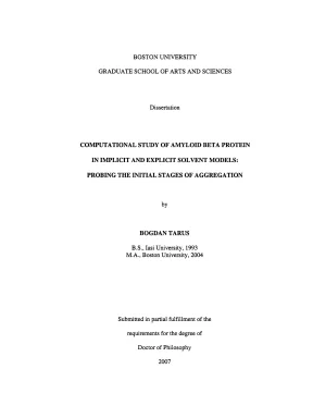
Tải xuống để đọc toàn bộBOSTON UNIVERSITY GRADUATE SCHOOL OF ARTS AND SCIENCES Dissertation COMPUTATIONAL STUDY OF AMYLOID BETA PROTEIN IN IMPLICIT AND EXPLICIT SOLVENT MODELS: PROBING THE INITIAL STAGES OF AGGREGATION BOGDAN TARUS B., Boston University, 2004 Submitted in partial fulfillment of the requirements for the degree of Doctor of Philosophy 2007 UMI Number: 3240642 INFORMATION TO USERS The quality of this reproduction is dependent upon the quality of the copy submitted. Broken or indistinct print, colored or poor quality illustrations and photographs, print bleed-through, substandard margins, and improper alignment can adversely affect reproduction. In the unlikely event that the author did not send a complete manuscript and there are missing pages, these will be noted. Also, if unauthorized copyright material had to be removed, a note will indicate the deletion.
® UMI UMI Microform 3240642 Copyright 2007 by ProQuest Information and Learning Company. All rights reserved. This microform edition is protected against unauthorized copying under Title 17, United States Code. ProQuest Information and Learning Company 300 North Zeeb Road P.
Box 1346 Ann Arbor, MI 48106-1346 Approved by First Reader £S\waÀ John E. Professor of Chemistry Second Reader LN Rosina M. Professor ¥f Chemistry To my family. Acknowledgments I would like to thank my advisor, Prof.
Straub, for his generous support and patience over my entire period of doctoral studies at Boston University. His sustained ded- ication as research advisor and enthusiastic teacher will constitute a permanent example for me. I thank all the professors on my thesis committee, Prof. Rosina Georgiadis, Prof.
Tom Keyes, Prof. Sandor Vajda and Prof. I also thank Prof. Devarajan Thiru- malai from University of Maryland for being a constant source of help and inspiration for my research.
I thank all my professors at Boston University. I would like to thank Prof. David Coker for sharing his research enthusiasm with us. I am sincerely grateful to all the colleagues I had over the years in the Straub group.
Finally, I would like to thank my family, especially my wife, Dana, for all her love and unconditioned support. 1V COMPUTATIONAL STUDY OF AMYLOID BETA PROTEIN IN IMPLICIT AND EXPLICIT SOLVENT MODELS: PROBING THE INITIAL STAGES OF AGGREGATION (Order No. ) BOGDAN TARUS Boston University Graduate School of Arts and Sciences, 2007 Major Professor: John E. Straub, Professor of Chemistry ABSTRACT It has been proposed that the amyloid đ-protein (Ađ-protein) plays a crucial role in the development of Alzheimer’s Disease (AD).
This dissertation presents the results of computa- tional studies of the initial stages of AG-protein association. The objective of this work was to determine the stability and role of the ÀØ-protein monomers and low-order oligomers as metastable intermediates on the pathway for formation of larger aggregates and fibrils. A protocol based on shape complementarity is used to generate an assortment of possible dimer structures of the A@jpo_35-protein congener. The ensemble of dimer structures are evaluated using rapidly computed estimates of the desolvation and electrostatic interaction energies to identify a putative stable dimer structure.
Using the umbrella sampling method and classical molecular dynamics, the potential of mean force (PMF) associated with the dimerization of the peptide in aqueous solution is computed. The profiles of the PMF corresponding to the formation of the two putative dimer structures are compared. Molecular dynamics trajectories originating from the two putative dimer structures are used to analyze their stability. Significant attempts are made to increase the time over which the association of the Afi9—35-protein can be simulated.
In this respect, conformations generated by the A@io_35- protein simulated using an explicit TIP3P solvent model are compared to conformations re- sulting from simulations employing one empirical and two continuum electrostatics solvent models. Inspired by recent experimental results, the dynamics of the D23-K28 “salt-bridge” V contacts are examined and critically evaluated as a possible “nucleation site” for the formation of Ø-structure characteristic of amyloid fibrils. The behavior of the AØại_ao-protein fragment is studied using molecular dynamics simula- tions employing an explicit aqueous solvent model. Special attention is paid to the VGSN(24- 27) region of the protein where experimental solid-state nuclear magnetic resonance (NMR) measurements indicate that formation of a turn may play a crucial role in stabilizing the AfØi-4a-protem in fibril structure.
The influence of two mutations, E22Q and D23N, on the thermodynamics properties of the Ai ao fragment is analyzed and related to the possible roles played by these two naturally occurring mutations in amyloidosis. vi Contents 1 Introduction 1 2 Probing the initial stage of aggregation of the A 9_3;-protein: Assessing the propensity for peptide dimerization 6 2. gà kg kg ko 7 2.3 Computational Model and Methods.1 Dimer structure generation using a docking protocol.2 Desolvation energy screening.4 Secondary Structure Analysis .4 Results and Discussion .2 Generation of dimer structures.38 Potential of Mean Force .4 Stability of dimer.5 Dynamical fluctuations in the y-dimer .6 Time dependence of secondary structure fluctuations in the homodimer 36 2.5 Summary and Conclusions .v xo 36 3 A comparative study of the structure and thermodynamics of the A{io_35- protein in different hydration models 39 vil 3. vn gà gà lv lv vn à và và và 3.38 Computational Models and Methods .1 Explicit solvent molecular dynamics.2 Implicit solvent molecular dynamics.3 Computational estimation of pK, .4 Results and analySlS.
cv ng gà gà g kg v va 3.1 Radius of gyration .2 Root-mean-square Ñuctuations.3 Lipari-Szabo generalized order parameter.5 Solvation self-energy. ch vu ng ee na 3.6 Intra-peptide hydrogen bonds .7 Computational estimation of pK, .5 Summary and Conclusions. ee Dynamics of Asp23—Lys28 salt bridge formation in Ađịo_ss monomers 41 Summary. Computational Models and Methods.
kg và và ee 4.4 Results and Discussions. ng kg kg va 4.1 Compact A(@io-35 undergoes large structural fluctuations .2 Computed values of pK, are in accord with experiments .3 Structures with intact salt-bridge D23-K28 are not the most stable 4.4 Multiple basins are populated in Àđịo ss monomer .5 Hydrogen-bonds with water increase the desolvation barrier of D23 and K28 ee 82 4.6 Burial of K28 involves a large free energy cost .7 The structure of water around residues D23 and K28 .5 Summary and Conclusions. c Q c Q k Q kg và gà va 86 The competition between the electrostatic and hydrophobic intra-peptide interactions in the AØs¡_so-protein 91 5. kg vn gà v g kg kg kg A 92 5.38 Computational Methods and Models .1 Simulation model and methods .2 Computational estimation of pK, .4 Results and analysis.
Q g gà và TT va 99 5.1 pK, values indicate weak intra-peptide electrostatic interactions .2 Inter-titratable side-chain distances .3 Intra-peptide hydrophobic contacts .4 Lys28 makes transitive contacts with the peptide backbone.5 Intra-peptide folding elements .56 Summary and Conclusions. cvva 115 Bibliography 121 Curriculum Vitae 137 1X List of Tables 41 Experimental and predicted pK, values (using the trajectories labeled T1-T5) for titratable residues in the A3,o-35-protein. The predicted pK, values are for Afio-_ss-protein structures simulated in explicit solvent model TIP3P. A value of e = 4 was used as the protein dielectric constant.1 Computational pK, values are compared with the experimental! (in parenthe- sis) pK, values for titratable residues Glu22, Asp23, and Lys28 in the A21~30- peptide.
The isolated residues and the residues in the peptide structure are similar, indicating that the pK, shifts result from weak intra-peptide electro- static interactions. c Q Q LH nu nu cà na vn g V NV k k k vUA 5.2 The number of nodes, N, and direct transitions among nodes, Ni, associated with the free energy transition disconnectivity graphs of the WT, E22Q, D23N, and K28A peptides. Na, is reduced to N—1 using the minimum-cut algorithm.? Ng nodes with free energies higher than —0.6 kcal/mol define the entropic basin. na lv kg g lv kg v kg kg va List of Figures 2.1 The distribution of the energy of interaction of the two monomers, used as the scoring function to analyze two sets of 2000 dimer decoys each.
The dimer se- lected by a method that places a relative emphasis on the burial of hydrophobic residues at the dimer interface is referred to as the “y-dimer” (a). The dimer chosen by a method that places a relative emphasis on electrostatic interactions is referred to as the “e-dimer” (b). The “desolvation energy”— corresponding to the energy change on going from separated monomeric À/Øo_ss to AZ dimeric decoy structure — was used as a measure of the degree of hydrophobic surface burial. The decoy sets were obtained using two shape complementarity proto- cols, GRAMM (a) and ZDOCK (b).
The energy of desolvation was calculated based on an atom contact energy (ACE) method.2 kcal/mol was used to compute the distribution of the interaction energy.2 The putative dimer structures derived by minimization of the functions S, (Eq.6)), corresponding to the y-dimer (a) and e-dimer (b), respectively. The side chains at the dimer interface are depicted explicitly. The green and yellow colored residues belong to monomer A (left) and those in red and orange are part of monomer B (right), .3 The distribution of the intermonomeric interaction energy plotted as a function of the atomic root-mean-square distance between each decoy structure and the structure of the y-dimer (a) and the e-dimer (b). In general, the unfavorable dimer structures are well differentiated from the most favorable structures.
The desolvation energy distribution (a) has a “funnel-like” character, indicating that structures more similar to the reference structure tend to be structures of minimal energy. The contribution of the electrostatic interaction energy determines a discontinuous distribution (b), the structure of most of the decoy dimers being very different from the structure of the e-dimer.4 The sidechain-sidechain contact matrices averaged for the ten decoy structures corresponding to the y-dimer (a) and e-dimer (b), respectively. The selection of the y-dimer is produced by a scoring function which is composed by the de- solvation energy only (a), while the e-dimer is selected by a function defined as the sum of the desolvation energy, the van der Waals and the electrostatic inter- actions (b) (see text for details). The interface of the ¿-dimer is dominated by contacts which involve hydrophobic residues, while the presence of the polar and charged residues is evident at the interface of the e-dimer.
The amino acid se- quence of the A@i9_3, monomer is Y!'EVHHQ!5KLVFF?AEDVG25°SNKGA*® TIGLM®, 6 -ỗ q. aaa ẶẼ xH 2.5 The Potential of Mean Force (PMF) is plotted for two different relative orien- tations of the monomeric peptide within the dimer. The PMF is computed as a function of the surface separation, 6 = € — €sø„;, along the distance between the centers-of-mass (DCOMs) of the two monomers, where € and £,on: are the DCOMs of the two monomers when they are at an arbitrary separation and in contact, respectively. The profile in blue corresponds to the free energy surface computed using the e-dimer as the starting structure.
The red curve is similarly computed using the y-dimer as the starting structure. The difference between the two surfaces suggests that hydrophobic interactions may be more essential to stabilization of the dimer structure than electrostatic interactions.6 The distribution of the surface buried area at the interface between the AGio_35 monomers during the molecular dynamics simulation of the dimer indicates that the set of the principal contacts at the dimer interface are maintained for the y-dimer (a), and that the e-dimer is not stable (b). A bin of 15 A? was used to compute the distribution of the interface surface area.7 The comparison of the electrostatic (red) and the hydrophobic (green) inter- action energies between the A@jo_35 monomers during the molecular dynamics simulation implies that the stability of the y-dimer is given by contacts between hydrophobic residues. In black is shown that the contribution of the fragment 15-30 plays a dominant role to the overall stability of the y-dimer.
Nội dung được bảo vệ bản quyền — Tải xuống đầy đủ
Trích dẫn luận án này
Bogdan Tarus B. (2007). Nghiên cứu tính toán protein amyloid beta trong môi trường dung môi [Luận án tiến sĩ, Boston University]. LuanAn.net. https://luanan.net/sinh-hoc/sinh-hoc-phan-tu-te-bao/nghien-cuu-tinh-toan-protein-amyloid-beta-giai-doan-tap-hop
Câu hỏi thường gặp
Luận án "Nghiên cứu tính toán protein amyloid beta trong môi trường dung môi" nghiên cứu về vấn đề gì?
Luận án tiến sĩ nghiên cứu tính toán protein amyloid beta. Phân tích giai đoạn tập hợp protein, cấu trúc dimer và vai trò oligomer trong bệnh Alzheimer.
Luận án "Nghiên cứu tính toán protein amyloid beta trong môi trường dung môi" được bảo vệ tại trường nào?
Luận án này được bảo vệ tại Boston University. Năm bảo vệ: 2007.
Luận án "Nghiên cứu tính toán protein amyloid beta trong môi trường dung môi" thuộc chuyên ngành gì?
Luận án "Nghiên cứu tính toán protein amyloid beta trong môi trường dung môi" thuộc chuyên ngành Chemistry. Danh mục: Sinh Học Phân Tử Tế Bào.
Luận án "Nghiên cứu tính toán protein amyloid beta trong môi trường dung môi" có bao nhiêu trang?
Luận án "Nghiên cứu tính toán protein amyloid beta trong môi trường dung môi" có 165 trang. Bạn có thể xem trước một phần tài liệu ngay trên trang web trước khi tải về.
Cách tải luận án "Nghiên cứu tính toán protein amyloid beta trong môi trường dung môi" về máy như thế nào?
Để tải luận án về máy, bạn nhấn nút "Tải xuống ngay" trên trang này, sau đó hoàn tất thanh toán phí lưu trữ. File sẽ được tải xuống ngay sau khi thanh toán thành công. Hỗ trợ qua Zalo: 0559 297 239.