Di cư tế bào neural crest sọ trong phôi chim: Nghiên cứu vai trò EPH-A4 và Ephrin-A5
Luận án tiến sĩ nghiên cứu di cư tế bào neural crest sọ trong phôi chim. Phân tích vai trò EPH-A4 và Ephrin-A5 trong điều hướng quỹ đạo tế bào.
Sinh học phát triển
Luan An
Luận án
Năm xuất bản
Số trang
129
Thời gian đọc
20 phút
Lượt xem
2
Lượt tải
0
Phí lưu trữ
40 Point
Tổng quan nhanh
- Chủ đề:
- 1. Di cư tế bào Neural Crest sọ: Tổng quan cơ bản
- Số trang:
- 129 trang
- Trường:
- California Institute of Technology
- Chuyên ngành:
- Sinh học phát triển
- Tác giả:
- Carole Chih-Chen Lu
- Năm:
- 2007
Tóm tắt nội dung luận án
I. Di cư tế bào Neural Crest sọ Tổng quan cơ bản
Tế bào neural crest là quần thể tế bào tạm thời trong phôi thai động vật có xương sống. Các tế bào này di chuyển từ ống thần kinh lưng về phía các vùng khác nhau của phôi. Quá trình di cư diễn ra theo các luồng rời rạc, được kiểm soát chặt chẽ bởi nhiều yếu tố phân tử. Nghiên cứu về cranial neural crest cells mở ra hiểu biết về phát triển sọ mặt và các bệnh lý bẩm sinh. Tế bào neural crest sọ đóng vai trò thiết yếu trong hình thành cấu trúc краниофациальный. Cell migration của chúng tuân theo các tín hiệu dẫn đường phức tạp.
1.1. Đặc điểm tế bào Neural Crest
Neural crest cells xuất hiện từ vùng lưng ống thần kinh. Các tế bào này có khả năng di chuyển xa và biệt hóa thành nhiều loại mô khác nhau. Cranial neural crest di chuyển theo ba luồng chính, liền kề với rhombomere chẵn (r2, r4, r6). Quá trình này đòi hỏi sự phối hợp giữa tín hiệu nội tại và ngoại tại. Tế bào phải nhận biết môi trường xung quanh để chọn lộ trình di chuyển chính xác.
1.2. Vai trò trong phát triển phôi
Di cư tế bào neural crest là bước quan trọng trong phát triển краниофациальный. Các tế bào này tạo nên xương sọ, sụn, và các cấu trúc mặt. Sự rối loạn trong cell migration dẫn đến dị tật bẩm sinh nghiêm trọng. Hiểu rõ cơ chế di chuyển giúp phát triển liệu pháp điều trị. Nghiên cứu trên phôi gà cung cấp mô hình lý tưởng cho quan sát quá trình này.
1.3. Phương pháp nghiên cứu hiện đại
Kính hiển vi confocal time-lapse cho phép theo dõi từng tế bào riêng lẻ. Kỹ thuật cell tracking analysis cung cấp dữ liệu định lượng về quỹ đạo di chuyển. Việc cấy rào cản vật lý vào trung mô giúp kiểm tra phản ứng của tế bào. Các thí nghiệm này tiết lộ khả năng thích ứng đáng kinh ngạc của neural crest cells.
II. EPH A4 Receptor Cơ chế tín hiệu phân tử
EPH receptor thuộc họ receptor tyrosine kinase lớn nhất trong động vật có vú. EPH-A4 đóng vai trò quan trọng trong dẫn đường axon và di cư tế bào. Receptor này tương tác với các phân tử Ephrin để truyền tín hiệu hai chiều. Tương tác EPH-Ephrin điều khiển sự đẩy lùi hoặc hút dẫn giữa các tế bào. Cơ chế này thiết yếu cho việc phân chia các luồng di cư neural crest. Nghiên cứu về EPH-A4 mở ra hiểu biết về kiểm soát không gian trong phát triển phôi.
2.1. Cấu trúc và chức năng EPH A4
EPH-A4 là receptor tyrosine kinase xuyên màng với miền ngoại bào liên kết ligand. Miền nội bào có hoạt tính kinase phosphoryl hóa các protein đích. Receptor này được biểu hiện trong nhiều mô đang phát triển, đặc biệt hệ thần kinh. EPH-A4 tham gia vào nhiều quá trình sinh học từ phát triển phôi đến bệnh lý người lớn. Hoạt động của nó phụ thuộc vào sự hiện diện của Ephrin ligand.
2.2. Tín hiệu hai chiều EPH Ephrin
Tương tác EPH-Ephrin tạo ra tín hiệu cả trong tế bào mang receptor và tế bào mang ligand. Forward signaling xảy ra khi EPH receptor được kích hoạt bởi Ephrin. Reverse signaling diễn ra khi Ephrin ligand truyền tín hiệu ngược lại. Cơ chế hai chiều này tạo ra sự phối hợp phức tạp giữa các tế bào lân cận. Cell migration được điều chỉnh tinh vi qua hệ thống tín hiệu này.
2.3. Vai trò trong dẫn đường tế bào
EPH-A4 tạo ra tín hiệu đẩy lùi khi gặp Ephrin-A5 trên bề mặt tế bào khác. Tín hiệu này ngăn cản neural crest cells xâm nhập vào vùng cấm. Cơ chế dẫn đường axon và di cư tế bào có nhiều điểm tương đồng. EPH-A4 giúp duy trì ranh giới rõ ràng giữa các luồng di cư. Sự mất chức năng của receptor này dẫn đến xáo trộn mô hình di chuyển.
III. Ephrin A5 Ligand Phân tử dẫn đường quan trọng
Ephrin-A5 là thành viên của họ Ephrin ligand gắn màng qua liên kết GPI. Phân tử này hoạt động như tín hiệu đẩy lùi cho các tế bào biểu hiện EPH-A4. Ephrin-A5 được biểu hiện trong các vùng cụ thể của phôi đang phát triển. Mô hình biểu hiện tạo ra các vùng cho phép và vùng cấm cho cell migration. Tương tác với EPH receptor tạo nên bản đồ phân tử dẫn đường cranial neural crest. Nghiên cứu Ephrin-A5 tiết lộ cơ chế kiểm soát không gian trong phát triển краниофациальный.
3.1. Đặc điểm phân tử Ephrin A5
Ephrin-A5 gắn vào màng tế bào thông qua neo GPI thay vì miền xuyên màng. Cấu trúc này cho phép Ephrin-A5 di động cao trên bề mặt tế bào. Ligand này liên kết đặc hiệu với các EPH receptor nhóm A, đặc biệt EPH-A4. Tương tác EPH-Ephrin kích hoạt các con đường tín hiệu điều chỉnh cytoskeleton. Sự linh hoạt của Ephrin-A5 quan trọng cho chức năng dẫn đường.
3.2. Mô hình biểu hiện trong phôi
Ephrin-A5 được biểu hiện trong rhombomere lẻ (r3, r5) của não sau đang phát triển. Mô hình này tạo ra các vùng cấm cho neural crest cells biểu hiện EPH-A4. Cranial neural crest di chuyển qua các luồng liền kề rhombomere chẵn để tránh Ephrin-A5. Sự phân bố không đồng đều tạo nên cấu trúc phân đoạn đặc trưng. Mô hình biểu hiện này được bảo tồn qua nhiều loài động vật có xương sống.
3.3. Cơ chế đẩy lùi tế bào
Khi neural crest cells gặp Ephrin-A5, EPH-A4 trên bề mặt được kích hoạt. Tín hiệu này gây co rút cytoskeleton và thay đổi hướng di chuyển. Tế bào né tránh vùng có nồng độ Ephrin-A5 cao, tạo ra ranh giới rõ ràng. Cơ chế đẩy lùi này hoạt động như rào cản phân tử cho cell migration. Sự phối hợp giữa tín hiệu hút dẫn và đẩy lùi định hình lộ trình di chuyển chính xác.
IV. Thí nghiệm rào cản Phân tích quỹ đạo tế bào
Nghiên cứu sử dụng phương pháp cấy rào cản vật lý vào trung mô phôi gà. Rào cản được đặt xa vị trí xuất phát của neural crest cells để quan sát phản ứng. Kính hiển vi confocal time-lapse ghi lại quá trình di chuyển theo thời gian thực. Cell tracking analysis cho phép định lượng quỹ đạo từng tế bào riêng lẻ. Kết quả cho thấy đa số tế bào có khả năng vượt qua hoặc đi vòng rào cản. Thí nghiệm này tiết lộ sự cân bằng giữa tín hiệu nội tại và ngoại tại trong dẫn đường tế bào.
4.1. Thiết kế thí nghiệm
Rào cản vật lý được cấy vào trung mô, xa điểm xuất phát của cranial neural crest. Vị trí cấy được chọn để không ảnh hưởng trực tiếp đến tế bào khi mới bắt đầu di chuyển. Kỹ thuật này cho phép đánh giá khả năng thích ứng khi tế bào gặp chướng ngại vật. Phôi gà là mô hình lý tưởng nhờ dễ tiếp cận và quan sát. Thí nghiệm được thiết kế để tách biệt tín hiệu nội tại và ngoại tại.
4.2. Kỹ thuật theo dõi và phân tích
Time-lapse confocal microscopy ghi hình liên tục trong nhiều giờ với độ phân giải cao. Các neural crest cells được đánh dấu huỳnh quang để dễ theo dõi. Cell tracking analysis sử dụng phần mềm chuyên dụng để trích xuất quỹ đạo từng tế bào. Dữ liệu định lượng bao gồm tốc độ, hướng di chuyển và thời gian phản ứng. Phương pháp này cung cấp thông tin chi tiết về động lực học cell migration.
4.3. Kết quả và ý nghĩa
Đa số neural crest cells vượt qua rào cản bằng cách điều chỉnh quỹ đạo di chuyển. Một số tế bào dừng lại tạm thời rồi tìm đường đi vòng. Kết quả chứng minh tế bào có khả năng đánh giá môi trường và ra quyết định linh hoạt. Tín hiệu nội tại mạnh mẽ giúp duy trì hướng di chuyển tổng thể. Thí nghiệm này làm sáng tỏ cơ chế thích ứng trong di cư tế bào phát triển.
V. Tương tác EPH Ephrin trong di cư Neural Crest
Tương tác EPH-Ephrin đóng vai trò trung tâm trong việc phân chia các luồng cranial neural crest. EPH-A4 trên neural crest cells nhận biết Ephrin-A5 trong rhombomere lẻ. Tín hiệu đẩy lùi từ tương tác này ngăn tế bào xâm nhập vùng cấm. Cơ chế này tạo ra mô hình phân đoạn đặc trưng của di cư tế bào sọ. Cell migration được điều khiển bởi sự cân bằng giữa nhiều tín hiệu phân tử. Nghiên cứu về receptor tyrosine kinase này mở ra hiểu biết về phát triển краниофациальный bình thường và bệnh lý.
5.1. Cơ chế phân chia luồng di cư
Cranial neural crest di chuyển theo ba luồng rời rạc liền kề r2, r4, và r6. Rhombomere lẻ (r3, r5) biểu hiện Ephrin-A5 tạo vùng cấm. Neural crest cells biểu hiện EPH-A4 nhận tín hiệu đẩy lùi từ các vùng này. Tương tác EPH-Ephrin duy trì ranh giới sắc nét giữa các luồng di chuyển. Cơ chế này đảm bảo tế bào đến đúng vị trí đích trong phát triển phôi.
5.2. Vai trò trong dẫn đường axon
Tương tác EPH-Ephrin không chỉ điều khiển cell migration mà còn dẫn đường axon. Cả hai quá trình đều dựa vào cơ chế đẩy lùi và hút dẫn tương tự. Axon thần kinh sử dụng EPH-A4 để tránh vùng có Ephrin-A5 cao. Sự tương đồng này cho thấy nguyên tắc chung trong dẫn đường tế bào và sợi trục. Nghiên cứu về neural crest cells cung cấp mô hình cho hiểu biết về phát triển hệ thần kinh.
5.3. Ứng dụng trong y học
Hiểu rõ tương tác EPH-Ephrin giúp giải thích các dị tật bẩm sinh краниофациальный. Đột biến gen EPH-A4 hoặc Ephrin-A5 có thể gây rối loạn di cư neural crest. Kiến thức này mở ra khả năng phát triển liệu pháp can thiệp sớm. Nghiên cứu về receptor tyrosine kinase còn liên quan đến ung thư và tái tạo mô. Ứng dụng tiềm năng trải rộng từ phát triển phôi đến y học tái tạo.
VI. Ý nghĩa nghiên cứu và hướng phát triển tương lai
Nghiên cứu về di cư cranial neural crest và vai trò EPH-A4/Ephrin-A5 đóng góp quan trọng vào sinh học phát triển. Kết quả làm sáng tỏ cơ chế phân tử điều khiển cell migration trong phôi thai. Phương pháp kết hợp thí nghiệm rào cản và cell tracking analysis mở ra hướng tiếp cận mới. Hiểu biết về tương tác EPH-Ephrin có ý nghĩa rộng lớn cho nhiều lĩnh vực y sinh học. Nghiên cứu tương lai cần khám phá chi tiết hơn về mạng lưới tín hiệu điều khiển neural crest cells. Kiến thức này là nền tảng cho phát triển liệu pháp điều trị dị tật bẩm sinh và bệnh lý liên quan.
6.1. Đóng góp khoa học chính
Nghiên cứu chứng minh vai trò thiết yếu của tương tác EPH-Ephrin trong phân chia luồng cranial neural crest. Thí nghiệm rào cản tiết lộ khả năng thích ứng đáng kinh ngạc của neural crest cells. Cell tracking analysis cung cấp dữ liệu định lượng về động lực học di cư tế bào. Kết quả làm rõ sự cân bằng giữa tín hiệu nội tại và ngoại tại trong dẫn đường. Nghiên cứu này là nền tảng cho hiểu biết sâu hơn về phát triển краниофациальный.
6.2. Hạn chế và thách thức
Nghiên cứu chủ yếu thực hiện trên phôi gà, cần xác nhận trên các loài khác. Cơ chế phân tử chi tiết của tín hiệu EPH-Ephrin vẫn chưa được làm rõ hoàn toàn. Tương tác giữa nhiều receptor tyrosine kinase và ligand tạo ra độ phức tạp cao. Khó khăn trong việc quan sát in vivo với độ phân giải tế bào đơn trong thời gian dài. Cần công nghệ tiên tiến hơn để theo dõi toàn bộ quá trình phát triển phôi.
6.3. Hướng nghiên cứu tương lai
Nghiên cứu sâu hơn về mạng lưới tín hiệu phân tử điều khiển neural crest cells. Phát triển mô hình 3D và kỹ thuật hình ảnh tiên tiến cho quan sát chi tiết hơn. Khám phá ứng dụng của kiến thức về EPH-Ephrin trong y học tái tạo. Nghiên cứu liên quan đến dị tật bẩm sinh và phát triển liệu pháp can thiệp. Tích hợp dữ liệu từ nhiều mô hình động vật để hiểu nguyên tắc chung của cell migration.
Mục lục chi tiết luận án
Tải xuống file đầy đủ để xem toàn bộ nội dung
Tải đầy đủ (129 trang)Nội dung chính
Tổng quan về luận án
Luận án này tiên phong trong việc khám phá những cơ chế phức tạp chi phối sự di chuyển của tế bào mào thần kinh sọ (cranial neural crest cells - NCCs) trong phôi chim, một quá trình nền tảng cho sự hình thành kiểu mẫu sọ mặt ở động vật có xương sống. Nghiên cứu này đi sâu vào động lực học spatio-temporal của quá trình di chuyển tế bào, sử dụng phương pháp tiếp cận hai nhánh đột phá: can thiệp vật lý và phân tích phân tử, nhằm làm sáng tỏ vai trò tương tác của các tín hiệu nội tại và ngoại sinh. Bối cảnh khoa học của nghiên cứu này nằm ở sự hiểu biết về cách một phôi phức tạp phát triển từ một tế bào duy nhất, đặc biệt là quá trình hình thành kiểu mẫu và phối hợp tế bào thành các cấu trúc. NCCs là một quần thể tế bào đa tiềm năng tạm thời, di chuyển ra khỏi ống thần kinh lưng để tạo ra nhiều loại tế bào, bao gồm tế bào thần kinh đệm, tế bào thần kinh, sụn và xương. Do đó, những khiếm khuyết trong sự phát triển của NCCs có thể dẫn đến các dị tật bẩm sinh ở vùng sọ mặt, làm nổi bật tầm quan trọng của việc hiểu rõ sinh học của chúng.
Mặc dù có những tiến bộ đáng kể trong việc xác định các phân tử hướng dẫn và quỹ đạo tế bào, một khoảng trống nghiên cứu cụ thể vẫn tồn tại trong lĩnh vực này: "However, we still do not understand how individual cells react to changes in the environment in real-time." (Lu, 2007, tr. 36). Các nghiên cứu trước đây đã gợi ý về tính dẻo dai của các con đường di chuyển của NCCs, đặc biệt là khi đối mặt với các nhiễu loạn di truyền hoặc vật lý lớn (Saldivar et al., 1997; Kulesa et al., 2000; Trainor and Krumiauf, 2000; Trainor et al., 2002; Golding et al., 2004), nhưng cơ chế cụ thể mà các tế bào riêng lẻ thay đổi hành vi di chuyển của chúng vẫn chưa được giải thích rõ ràng. Luận án này giải quyết trực tiếp khoảng trống đó bằng cách giới thiệu một rào cản vật lý trơ dọc theo con đường di chuyển của NCCs và theo dõi hành vi của từng tế bào trong thời gian thực, cung cấp một góc nhìn sâu sắc chưa từng có về khả năng điều hòa di chuyển của NCCs.
Nghiên cứu được thúc đẩy bởi các câu hỏi và giả thuyết chính sau:
- RQ1: Mức độ quỹ đạo của tế bào mào thần kinh sọ có được xác định nội tại hay chúng có thể điều chỉnh theo những thay đổi nhỏ trong môi trường?
- H1: Quỹ đạo của tế bào mào thần kinh sọ không được xác định nội tại và chúng có khả năng điều chỉnh theo những thay đổi nhỏ trong môi trường.
- RQ2: Các cơ chế phân tử cụ thể nào (ví dụ: tín hiệu Eph/ephrin) điều hòa sự di chuyển của tế bào mào thần kinh sọ và liệu các thành phần khác nhau của con đường tín hiệu này có đóng vai trò riêng biệt trong việc định hướng và mức độ di chuyển hay không?
- H2: Tín hiệu Eph/ephrin đóng một vai trò đa dạng trong sự di chuyển của tế bào mào thần kinh sọ, với ephrin-A5 tham gia vào việc duy trì di chuyển và EphA4 tham gia vào cả việc định hướng và điều hòa mức độ di chuyển.
- RQ3: Các tế bào dẫn đầu và tế bào theo sau trong các dòng di chuyển của NCCs có vai trò cố định hay vai trò của chúng có thể thay đổi để đáp ứng với các nhiễu loạn môi trường?
- H3: Vai trò của tế bào dẫn đầu và tế bào theo sau không phải là cố hữu và có thể thay đổi trong quá trình di chuyển của tế bào mào thần kinh sọ.
Khung lý thuyết của luận án này được neo giữ bởi lý thuyết phân đoạn (segmentation theory) trong phát triển phôi học (Lumsden & Krumlauf, 1996) và các mô hình về cơ chế di chuyển tế bào nội tại (như cái chết tế bào cục bộ, áp lực quần thể, và ái lực khác biệt) và ngoại sinh (như tín hiệu hướng dẫn hấp dẫn hoặc đẩy lùi) (Le Douarin & Kalcheim, 1999; Newgreen et al., 1979). Nghiên cứu mở rộng lý thuyết bằng cách cung cấp bằng chứng thực nghiệm về tính linh hoạt của NCCs, thách thức quan điểm về một con đường di chuyển hoàn toàn được lập trình sẵn và nhấn mạnh tầm quan trọng của các tương tác tế bào-tế bào và tế bào-môi trường.
Một đóng góp đột phá của luận án này là chứng minh rằng "cranial neural crest cell trajectories are not intrinsically determined. NCCs can respond to minor alterations in the environment to retarget a peripheral destination." (Lu, 2007, tr. v). Phát hiện này đã định lượng được tác động bằng cách chỉ ra rằng phần lớn NCCs có thể vượt qua các rào cản vật lý, cho thấy một mức độ điều hòa và khả năng định hướng lại con đường đáng kinh ngạc. Trong các trường hợp rào cản chỉ chặn 25% con đường di chuyển của r4, các tế bào NCCs gặp rào cản di chuyển nhanh hơn 38% và giảm hướng đi 68% so với phía đối diện (Lu, 2007, tr. 33-34). Một đóng góp quan trọng khác là việc làm sáng tỏ các vai trò khác nhau của Ephrin-A5 và EphA4 trong việc định hướng di chuyển, với Ephrin-A5 điều hòa mức độ di chuyển và EphA4 liên quan đến cả định hướng và mức độ, cung cấp những hiểu biết phân tử cụ thể vào các cơ chế ngoại sinh.
Phạm vi nghiên cứu bao gồm việc sử dụng phôi chim (gà Leghorn trắng) ở các giai đoạn phát triển 7-9 somite, cho phép quan sát trực tiếp và thao tác in ovo. Kích thước mẫu cụ thể được đề cập là n=15 cho một phân tích theo dõi tế bào, với các thí nghiệm khác bao gồm nhiều lần lặp lại (ví dụ, 3/7 trường hợp khi rào cản chặn 100% không thành công). Khung thời gian của các phiên ghi hình thời gian thực thường kéo dài đến 24 giờ sau khi đặt rào cản. Tầm quan trọng của nghiên cứu nằm ở việc nó không chỉ nâng cao hiểu biết cơ bản về sinh học phát triển mà còn có ý nghĩa tiềm năng đối với y học tái tạo, đặc biệt là trong việc điều trị các dị tật bẩm sinh ở vùng sọ mặt, bằng cách cung cấp một mô hình cho tính dẻo dai của tế bào khi đối mặt với các nhiễu loạn.
Literature Review và Positioning
Phần tổng quan tài liệu của luận án đã tổng hợp một cách tinh tế các dòng nghiên cứu chính trong lĩnh vực di chuyển của tế bào mào thần kinh sọ (cranial neural crest cell migration), với sự chú ý đặc biệt đến các cơ chế nội tại và ngoại sinh. Các nghiên cứu ban đầu của Le Douarin và Kalcheim (1999) đã thiết lập NCCs là một quần thể tế bào đa tiềm năng với khả năng di chuyển rộng khắp, trong khi các công trình của Lumsden và Krumlauf (1996) nhấn mạnh tầm quan trọng của sự phân đoạn (segmentation) ở não sau trong việc định hình các con đường di chuyển của NCCs thành ba dòng riêng biệt.
Luận án này đã thảo luận về các mâu thuẫn và tranh luận hiện có, đặc biệt là về việc liệu các dòng di chuyển của NCCs có được hình thành chủ yếu bởi các tín hiệu nội tại hay ngoại sinh. Một quan điểm cho rằng các cơ chế nội tại, như cái chết tế bào cục bộ trong r3 và r5 (Ellies et al., 2000; Graham et al., 1993) hoặc các điểm thoát cụ thể (Birgbauer et al., 1995; Lumsden et al., 1991), thiết lập các con đường di chuyển từ bên trong ống thần kinh. Tuy nhiên, quan điểm đối lập, được hỗ trợ bởi các nghiên cứu ở gà, chuột và cá ngựa vằn (Schilling & Kimmel, 1994; Birgbauer et al., 1995; Kulesa & Fraser, 1998a; Trainor & Krumlauf, 2000b), chỉ ra rằng NCCs từ r3 và r5 thực sự di chuyển theo đường chéo để tham gia vào các dòng từ các rhombomere chẵn. Điều này cho thấy tính dẻo dai đáng kể của NCCs để điều chỉnh con đường của chúng theo các tín hiệu môi trường.
Luận án này định vị nghiên cứu của mình trong bối cảnh tranh luận này bằng cách giải quyết khoảng trống cụ thể về việc "how individual cells react to changes in the environment in real-time" (Lu, 2007, tr. 36). Nghiên cứu không chỉ tổng hợp các mô hình nội tại và ngoại sinh mà còn cung cấp bằng chứng thực nghiệm trực tiếp về cách NCCs điều hòa con đường di chuyển của chúng khi đối mặt với một nhiễu loạn vật lý. Nó tiến bộ lĩnh vực này bằng cách chứng minh rằng "cranial neural crest cell trajectories are not intrinsically determined," và các tế bào này có khả năng "retarget a peripheral destination" ngay cả khi đối mặt với các rào cản vật lý (Lu, 2007, tr. v). Điều này đại diện cho một bước tiến quan trọng so với các nghiên cứu trước đây vốn chủ yếu tập trung vào các can thiệp di truyền hoặc phẫu thuật lớn hơn.
So sánh với các nghiên cứu quốc tế, công trình này có điểm tương đồng và khác biệt đáng kể. Các nghiên cứu về NCCs ở cá ngựa vằn (Danio rerio) của Schilling và Kimmel (1994) đã cung cấp những hiểu biết quan trọng về sự phân đoạn và di chuyển trong phôi cá, cho thấy NCCs có thể di chuyển từ các rhombomere lẻ sang các dòng chẵn. Tương tự, các nghiên cứu trên chuột (Mus musculus) của Trainor và Krumlauf (2000b) đã khám phá tính dẻo dai của NCCs trong việc điều chỉnh biểu hiện gen Hox và con đường di chuyển. Tuy nhiên, luận án hiện tại khác biệt ở chỗ nó sử dụng các rào cản vật lý trơ kết hợp với chụp ảnh thời gian thực độ phân giải cao để theo dõi hành vi của từng tế bào. Trong khi các nghiên cứu của Smith et al. (1997) trên Xenopus đã chỉ ra rằng việc ức chế tín hiệu Eph/ephrin có thể dẫn đến việc NCCs chuyển hướng sang các cung mang khác, luận án này của Lu (2007) đi sâu hơn bằng cách phân biệt vai trò riêng biệt của Ephrin-A5 và EphA4 trong việc định hướng và duy trì di chuyển trong hệ thống phôi gà, cung cấp một mức độ chi tiết phân tử chưa từng có trong ngữ cảnh của các can thiệp động học. Sự chú ý đến các tương tác tế bào-tế bào khi các tế bào vượt qua rào cản cũng cung cấp một khía cạnh cơ học mà các nghiên cứu trước đây chưa thực sự định lượng một cách hiệu quả.
Đóng góp lý thuyết và khung phân tích
Đóng góp cho lý thuyết
Luận án này đã đưa ra những đóng góp đáng kể cho lý thuyết phát triển bằng cách mở rộng và thách thức các lý thuyết hiện có về sự di chuyển của tế bào mào thần kinh sọ. Cụ thể, nó mở rộng lý thuyết về sự phân đoạn ở não sau (Lumsden & Krumlauf, 1996) bằng cách chỉ ra rằng mặc dù có một mô hình di chuyển khuôn mẫu từ các rhombomere chẵn, quỹ đạo của các tế bào riêng lẻ không phải là bất biến. Nghiên cứu thách thức các giả định cho rằng các con đường di chuyển được xác định hoàn toàn nội tại bởi các gen được biểu hiện sớm trong ống thần kinh (Hunt et al., 1991a), cung cấp bằng chứng thực nghiệm mạnh mẽ cho tính dẻo dai của NCCs. Như đã nêu, "cranial neural crest cell trajectories are not intrinsically determined" (Lu, 2007, tr. v), điều này trực tiếp thách thức các mô hình định mệnh hóa di truyền của sự di chuyển tế bào. Thay vào đó, nó ủng hộ một quan điểm tổng hợp hơn, trong đó các tín hiệu ngoại sinh và các tương tác tế bào-tế bào đóng vai trò quyết định trong việc điều hòa các con đường di chuyển.
Khung khái niệm của luận án mô tả sự di chuyển của NCCs là một quá trình linh hoạt, nơi các tế bào phản ứng với các vi môi trường thay đổi bằng cách điều chỉnh tốc độ và hướng đi của chúng. Các thành phần chính bao gồm (1) tín hiệu từ môi trường (ví dụ: rào cản vật lý, vùng đẩy lùi r3), (2) các phản ứng hành vi của tế bào (ví dụ: kéo dài filopodia, thay đổi hướng, thay đổi vai trò dẫn đầu/theo sau), và (3) các yếu tố phân tử (ví dụ: tín hiệu Eph/ephrin) điều hòa các phản ứng này. Mối quan hệ giữa các thành phần này là động, với các yếu tố ngoại sinh có thể kích hoạt các thay đổi trong hành vi của tế bào được điều hòa bởi các con đường tín hiệu phân tử.
Mô hình lý thuyết được đề xuất bao gồm các giả thuyết chính như sau:
- Proposition 1: Các tế bào mào thần kinh sọ thể hiện khả năng định hướng lại con đường đáng kể, cho phép chúng vượt qua các rào cản vật lý đáng kể để đạt được các đích đến ngoại vi.
- Proposition 2: Vai trò của các tế bào dẫn đầu và tế bào theo sau trong các dòng di chuyển của NCCs là linh hoạt và có thể thay đổi để đáp ứng với các nhiễu loạn môi trường.
- Proposition 3: Tín hiệu Eph/ephrin thể hiện các chức năng đa dạng trong NCCs, với ephrin-A5 chủ yếu liên quan đến việc duy trì di chuyển và EphA4 liên quan đến cả định hướng và điều hòa mức độ di chuyển.
- Proposition 4: Tương tác tế bào-tế bào đóng một vai trò quan trọng trong việc cho phép NCCs vượt qua các tín hiệu môi trường bất lợi, chẳng hạn như xâm nhập vào các vùng đẩy lùi.
Những phát hiện của luận án gợi ý một sự thay đổi mô hình nhỏ từ một quan điểm coi sự di chuyển của NCCs là một quá trình chủ yếu được xác định nội tại sang một mô hình nhấn mạnh sự điều hòa liên tục thông qua các tín hiệu ngoại sinh và các tương tác tế bào năng động. Bằng chứng từ các phát hiện cho thấy "the neural crest cells are able to migrate and to overcome environmental cues by migrating on top of each other" (Lu, 2007, tr. 35), điều này chỉ ra rằng các cơ chế hành vi tập thể có thể vượt qua các tín hiệu hóa học đẩy lùi đã biết, một sự thay đổi đáng chú ý.
Khung phân tích độc đáo
Luận án tích hợp một cách khéo léo các lý thuyết từ sinh học phát triển, sinh học tế bào và động lực học quần thể để xây dựng một khung phân tích độc đáo. Nó kết hợp các nguyên tắc từ lý thuyết phân đoạn của Hunt et al. (1991a) và Lumsden và Guthrie (1991) với các khái niệm về áp lực quần thể (Le Douarin & Kalcheim, 1999) và ái lực khác biệt (Fraser et al., 1990) để giải thích sự phức tạp của di chuyển NCC. Khung này vượt ra ngoài việc kiểm tra các tín hiệu đơn lẻ bằng cách xem xét tương tác giữa các yếu tố tế bào (tế bào dẫn đầu/theo sau), vật lý (rào cản) và phân tử (Eph/ephrin).
Phương pháp phân tích mới lạ nằm ở việc kết hợp thực nghiệm việc đặt rào cản vật lý với chụp ảnh confocal thời gian thực độ phân giải cao và phân tích theo dõi tế bào định lượng. Cách tiếp cận này cho phép quan sát trực tiếp các "repeatable series of events by which the cells overcome the barriers" (Lu, 2007, tr. 36), điều này không thể thực hiện được bằng các phương pháp tĩnh hoặc các can thiệp di truyền lớn hơn. Nó cung cấp một cái nhìn chi tiết về các phản ứng hành vi của từng tế bào, chẳng hạn như cách các tế bào dẫn đầu bị chặn tạm thời, trong khi "follower cells find a novel pathway around a barrier and become de novo leaders of a new stream" (Lu, 2007, tr. v).
Các đóng góp khái niệm bao gồm việc định nghĩa lại tính linh hoạt của NCCs không chỉ là khả năng điều chỉnh biểu hiện gen (Trainor et al., 2002) mà còn là khả năng thay đổi hành vi di chuyển ngay lập tức. Khái niệm về "de novo leaders" đại diện cho một sự tiến bộ đáng kể trong việc hiểu vai trò của các tế bào trong các dòng di chuyển tập thể.
Các điều kiện biên được nêu rõ ràng: nghiên cứu tập trung vào sự di chuyển của NCCs trong phôi chim, đặc biệt là dòng r4, và các can thiệp được giới hạn ở việc đặt các rào cản vật lý hoặc điều chỉnh hoạt động của Eph/ephrin trong các NCC di chuyển. Các kết quả có thể tổng quát hóa cho các hệ thống động vật có xương sống khác, vì NCCs được bảo tồn tiến hóa, nhưng các chi tiết cụ thể về điều hòa phân tử có thể thay đổi giữa các loài. Việc nghiên cứu các tương tác tế bào-tế bào khi các tế bào vượt qua rào cản bằng cách di chuyển "on top of each other" cũng giới hạn điều kiện biên cho các tình huống mật độ tế bào cao, nơi các tương tác vật lý trực tiếp trở nên nổi bật.
Phương pháp nghiên cứu tiên tiến
Thiết kế nghiên cứu
Luận án áp dụng triết lý nghiên cứu thực nghiệm-dương tính (positivist/post-positivist philosophy), nhằm thiết lập các mối quan hệ nguyên nhân-kết quả thông qua việc thao tác có kiểm soát các biến số và đo lường khách quan các kết quả. Triết lý này được phản ánh rõ ràng trong việc sử dụng các rào cản vật lý trơ để kiểm tra các giả thuyết cụ thể về hành vi di chuyển của tế bào và việc định lượng các thông số tế bào như vận tốc và hướng đi.
Thiết kế nghiên cứu kết hợp các phương pháp hỗn hợp theo nghĩa nó bao gồm cả can thiệp vật lý (đặt rào cản) và can thiệp phân tử (điều chỉnh tín hiệu Eph/ephrin), được phân tích bằng cách sử dụng các kỹ thuật định lượng nghiêm ngặt. Sự kết hợp cụ thể này cho phép các nhà nghiên cứu khám phá các cơ chế hành vi ở cấp độ quần thể và tế bào riêng lẻ, đồng thời làm sáng tỏ các con đường phân tử cơ bản. Cụ thể, thiết kế "hai nhánh" của luận án, kiểm tra phản ứng với rào cản vật lý và sau đó là nhiễu loạn phân tử, cung cấp một khung toàn diện để hiểu sự di chuyển của NCCs.
Mặc dù không phải là thiết kế đa cấp theo nghĩa thống kê truyền thống, nghiên cứu này tích hợp các quan sát ở các cấp độ khác nhau của tổ chức sinh học: từ hành vi của từng tế bào (phân tích theo dõi tế bào), động lực học của dòng tế bào (sự hình thành dòng mới, vai trò dẫn đầu/theo sau), cho đến phản ứng của phôi toàn bộ (sự xâm nhập của cung mang). Điều này cho phép một cách tiếp cận toàn diện để hiểu sự di chuyển của NCCs, liên kết các sự kiện vi mô với các kết quả phát triển vĩ mô. Kích thước mẫu cụ thể cho việc theo dõi tế bào là "n=15" (Lu, 2007, tr. 34) cho một phân tích cụ thể, trong khi các thí nghiệm chặn 100% con đường di chuyển r4 được báo cáo là "3/7" trường hợp không thành công, cho thấy các nhóm thí nghiệm lớn hơn cho các quan sát tổng quát. Tiêu chí lựa chọn bao gồm phôi chim giai đoạn 7-9 somite (ss) (Hamburger và Hamilton, 1951), đảm bảo một giai đoạn phát triển nhất quán để bắt đầu di chuyển của NCCs.
Quy trình nghiên cứu nghiêm ngặt
Chiến lược lấy mẫu bao gồm việc lựa chọn phôi gà Leghorn trắng khỏe mạnh ở giai đoạn 7-9 somite, đảm bảo tính đồng nhất về mặt phát triển. Tiêu chí đưa vào là các phôi có cấu trúc rõ ràng và không có dị tật rõ ràng, trong khi tiêu chí loại trừ là các phôi bị tổn thương trong quá trình thao tác hoặc không biểu hiện nhãn huỳnh quang thành công.
Các giao thức thu thập dữ liệu được thiết kế tỉ mỉ:
- Gắn nhãn tế bào mào thần kinh tiền di chuyển: Tiêm áp suất 0.5 µg/µl CM-Dil (Molecular Probes C-7000) vào khoang ống thần kinh hoặc tiêm tiêu điểm 5 µg/µl CM-Dil. Điện di các cấu trúc DNA như pca-GFP (5 µg/µl) cũng được sử dụng để gắn nhãn tế bào (Itasaki et al., 1999).
- Đặt rào cản vật lý: Dao mổ sắc được sử dụng để cắt rào cản lá tantalum (dày 7.5 µm) hoặc rào cản thấm từ màng Millicell-CM (kích thước ~100x100 µm hoặc ~400x100 µm). Các kim thủy tinh và kẹp nhỏ được sử dụng để đặt rào cản song song và gần ống thần kinh.
- Chụp ảnh confocal thời gian thực: Phôi được nuôi cấy nguyên vẹn trong hệ thống nuôi cấy được điều khiển nhiệt độ (38°C) và được chụp ảnh bằng kính hiển vi confocal quét laser Zeiss LSM 410 kết nối với kính hiển vi hợp chất đảo ngược Zeiss Axiovert. Hình ảnh được thu thập kỹ thuật số cứ 2 phút một lần trong nhiều giờ, tạo ra các bộ dữ liệu động học phong phú (Kulesa và Fraser, 1998b).
- Nhiễu loạn phân tử: Điện di các cấu trúc DNA mã hóa ephrin-A5 toàn chiều dài, thụ thể EphA4 hoạt động liên tục bị cắt ngắn, và thụ thể EphA4 kinase-dead bị cắt ngắn vào các NCCs di chuyển để điều tra vai trò của tín hiệu Eph/ephrin.
Tam giác hóa được áp dụng thông qua các phương pháp khác nhau để tăng cường tính hợp lệ của phát hiện. Cụ thể, tam giác hóa phương pháp được thể hiện qua việc sử dụng cả rào cản vật lý và nhiễu loạn phân tử để kiểm tra tính linh hoạt của NCCs. Tam giác hóa dữ liệu được hỗ trợ bởi các quan sát hành vi của từng tế bào (từ theo dõi tế bào), động lực học của dòng (từ chụp ảnh thời gian thực), và phân phối tổng thể của tế bào (từ hình ảnh tĩnh).
Tính hợp lệ và độ tin cậy được giải quyết một cách nghiêm ngặt. Tính hợp lệ cấu trúc được đảm bảo bằng cách sử dụng các chỉ dấu tế bào và hình ảnh tiêu chuẩn. Tính hợp lệ nội bộ được duy trì thông qua các nhóm đối chứng (phía đối diện của phôi, các điều kiện di chuyển bình thường) và việc lặp lại các thí nghiệm. Tính hợp lệ bên ngoài được tăng cường bởi bản chất tiến hóa được bảo tồn của NCCs ở động vật có xương sống. Độ tin cậy của các phép đo vận tốc và hướng đi của tế bào được hỗ trợ bởi phần mềm theo dõi tế bào chuyên biệt (TRACKIT) và phân tích thống kê (t-test không ghép cặp). Mặc dù các giá trị α (alpha) cụ thể cho độ tin cậy không được báo cáo trực tiếp trong bản tóm tắt/giới thiệu, việc sử dụng các kỹ thuật định lượng tiêu chuẩn và kiểm tra thống kê ngụ ý cam kết về độ tin cậy.
Dữ liệu và phân tích
Đặc điểm mẫu được mô tả bao gồm phôi chim giai đoạn 7-9 somite, tập trung vào NCCs từ các rhombomere 2, 4, và 6, di chuyển đến các cung mang 1, 2, và 3. Phân tích nhân khẩu học cụ thể (ví dụ: tuổi, giới tính) không áp dụng cho phôi nhưng giai đoạn phát triển là một thống kê quan trọng.
Các kỹ thuật phân tích tiên tiến được sử dụng bao gồm:
- Phân tích theo dõi hạt (Particle-tracking analysis): Sử dụng phần mềm TRACKIT (phiên bản cập nhật của XVTRACK, phát triển bởi S. Solomon) để theo dõi các tế bào riêng lẻ dựa trên độ sáng, hình dạng và vị trí không gian (Lu, 2007, tr. 29). Điều này cho phép định lượng chính xác vận tốc trung bình và hướng đi của tế bào.
- Phân tích thống kê: T-test không ghép cặp (sử dụng InStat v3.0a, GraphPad Software, Inc.) được thực hiện để so sánh các giá trị giữa hai quần thể tế bào (ví dụ: các tế bào gặp rào cản so với các tế bào đối chứng).
- Phân tích hình ảnh: Adobe Photoshop và NIH Image 1.62 được sử dụng để điều chỉnh độ sáng/độ tương phản và xử lý bộ lọc để giảm nhiễu (Lu, 2007, tr. 29).
Kiểm tra độ vững chắc được thực hiện bằng cách sử dụng các rào cản có cấu hình khác nhau (lá tantalum so với rào cản thấm) và các vị trí khác nhau (ví dụ: r3 so với r4) để đảm bảo rằng các kết quả không chỉ là hiện vật của một thiết lập thực nghiệm cụ thể. Các thông số kỹ thuật thay thế (ví dụ: các vị trí rào cản khác nhau) đã cung cấp thêm bằng chứng cho thấy hành vi di chuyển là nhạy cảm với các nhiễu loạn cục bộ.
Kích thước hiệu ứng và khoảng tin cậy được báo cáo cho các phát hiện định lượng chính. Ví dụ, "Our cell tracking analysis of these cells show a 38% increase in average velocity and a 68% decrease in directionality (n=15) when compared to r4 neural crest cells that migrate on the contralateral side of the embryo." (Lu, 2007, tr. 33-34). Mức độ ý nghĩa thống kê (giá trị p) được ngụ ý thông qua việc sử dụng t-test không ghép cặp, với các kết quả được mô tả là "significantly faster but less directly" hoặc "do not show significant changes" (Lu, 2007, tr. v, 34).
Phát hiện đột phá và implications
Những phát hiện then chốt
Luận án này đã đưa ra một số phát hiện then chốt, cung cấp bằng chứng cụ thể từ dữ liệu:
- Tính dẻo dai của NCCs: Phần lớn NCCs có thể vượt qua các rào cản vật lý và đến được các cung mang ngoại vi. "The majority of neural crest cells reaches and populates branchial arch 2 (BA2) comparable to normal (Figure 2.2)." (Lu, 2007, tr. 31). Điều này thách thức quan điểm về các con đường di chuyển được xác định nội tại một cách cứng nhắc.
- Động lực học của vai trò lãnh đạo/theo sau: Các tế bào dẫn đầu và theo sau có vai trò thay thế cho nhau. Khi các tế bào dẫn đầu bị chặn, "follower cells find a novel pathway around a barrier and become de novo leaders of a new stream" (Lu, 2007, tr. v). Điều này chỉ ra rằng vai trò lãnh đạo không phải là cố hữu của một quần thể tế bào cụ thể.
- Phản ứng khác biệt với vị trí rào cản: Các rào cản ở các vị trí khác nhau gây ra các hành vi di chuyển khác nhau. NCCs gặp rào cản ở r3 di chuyển "significantly faster but less directly" so với các tế bào gặp rào cản ở r4, chúng di chuyển bình thường (Lu, 2007, tr. v). Cụ thể, một rào cản ở r3 dẫn đến "a 38% increase in average velocity and a 68% decrease in directionality (n=15)" (Lu, 2007, tr. 33-34).
- Sự xâm nhập vào vùng đẩy lùi: NCCs có thể di chuyển vào các vùng đẩy lùi bình thường (như vùng r3) bằng cách di chuyển "on top of each other" (Lu, 2007, tr. 35). Đây là một hiện tượng mới, cho thấy các tương tác tế bào-tế bào có thể vượt qua các tín hiệu môi trường bất lợi.
- Vai trò khác biệt của Eph/ephrin: Ephrin-A5 điều hòa mức độ di chuyển của NCCs r6 nhưng không ảnh hưởng đến hướng đi, trong khi hoạt động EphA4 lạc chỗ gây ra lỗi định hướng và hành vi di chuyển thất thường (tăng tốc độ, giảm hướng đi). Phát hiện này là bằng chứng cụ thể cho các chức năng điều hòa khác biệt của các thành phần tín hiệu Eph/ephrin.
So sánh với các nghiên cứu trước đây, phát hiện về tính dẻo dai của NCCs nhất quán với các nghiên cứu về việc Ablation/transplantation NCCs ở gà của Kulesa et al. (2000) và Trainor et al. (2002), nhưng luận án này cung cấp chi tiết động học ở cấp độ tế bào riêng lẻ. Phát hiện về vai trò thay thế của tế bào dẫn đầu/theo sau thách thức một số mô hình về áp lực quần thể hoặc tế bào dẫn đầu chuyên biệt được gợi ý bởi Le Douarin (1982). Hành vi di chuyển ngược trực giác (counter-intuitive) của việc di chuyển nhanh hơn nhưng ít trực tiếp hơn khi gặp rào cản ở r3 cho thấy một cơ chế điều hòa phức tạp, có thể là sự tăng cường thăm dò để tìm đường vòng.
Implications đa chiều
- Tiến bộ lý thuyết: Luận án đóng góp cho lý thuyết về sự di chuyển của tế bào bằng cách cung cấp bằng chứng thực nghiệm mạnh mẽ cho lý thuyết về tín hiệu ngoại sinh và tương tác tế bào-tế bào trong việc định hình quỹ đạo tế bào (Lu, 2007, tr. 36-37). Nó mở rộng các lý thuyết về sự phân đoạn và hình thành kiểu mẫu bằng cách chứng minh rằng các cơ chế sinh học phân tử có thể bổ sung cho các cấu trúc giải phẫu vĩ mô. Nghiên cứu này tăng cường hiểu biết của chúng ta về lý thuyết về tín hiệu Eph/ephrin bằng cách làm rõ các vai trò cụ thể của ephrin-A5 và EphA4 trong việc định hướng và mức độ di chuyển (Lu, 2007, tr. 10).
- Đổi mới phương pháp luận: Việc sử dụng phương pháp đặt rào cản vật lý kết hợp với chụp ảnh confocal thời gian thực độ phân giải cao và phân tích theo dõi tế bào nghiêm ngặt có thể áp dụng cho các ngữ cảnh nghiên cứu di chuyển tế bào khác. Phương pháp này cung cấp một công cụ mạnh mẽ để kiểm tra tính dẻo dai của tế bào khi đối mặt với các nhiễu loạn vi môi trường.
- Ứng dụng thực tiễn: Các phát hiện có thể có ý nghĩa đối với y học tái tạo và kỹ thuật mô. Hiểu được tính dẻo dai của NCCs có thể cung cấp thông tin cho các chiến lược để hướng dẫn sự tái tạo mô trong các khiếm khuyết sọ mặt hoặc điều hòa sự di chuyển của các tế bào ung thư bắt chước hành vi di căn của NCCs.
- Khuyến nghị chính sách: Việc làm sáng tỏ các cơ chế điều hòa di chuyển của NCCs có thể cung cấp thông tin cho các chính sách sức khỏe cộng đồng liên quan đến dị tật bẩm sinh. Ví dụ, việc xác định các con đường tín hiệu cụ thể liên quan đến các lỗi định hướng có thể dẫn đến việc phát triển các liệu pháp can thiệp sớm. Việc hiểu được sự tương tác giữa các yếu tố nội tại và ngoại sinh là rất quan trọng để phát triển các phương pháp điều trị dựa trên bằng chứng.
- Điều kiện tổng quát hóa: Các phát hiện về tính dẻo dai của NCCs và động lực học của Eph/ephrin có thể tổng quát hóa cho các hệ thống di chuyển tế bào khác ở động vật có xương sống, đặc biệt là những hệ thống liên quan đến quá trình phát triển phôi và các hiện tượng bệnh lý như di căn ung thư. Tuy nhiên, các điều kiện biên bao gồm bối cảnh phôi chim và các dòng NCCs cụ thể, có thể có những biến thể ở các loài hoặc mô khác.
Limitations và Future Research
Nghiên cứu này, mặc dù đột phá, vẫn có những hạn chế cụ thể cần được thừa nhận một cách trung thực:
- Độ phân giải phân tử của rào cản vật lý: Các rào cản vật lý là trơ và không trực tiếp mô phỏng các tín hiệu phân tử phức tạp mà NCCs gặp phải in vivo. Do đó, phản ứng của tế bào với rào cản có thể không hoàn toàn đại diện cho các con đường sinh lý thông thường.
- Khả năng mở rộng của phân tích theo dõi tế bào: Phân tích theo dõi tế bào được thực hiện trên một số lượng tế bào giới hạn (ví dụ: n=15 cho một phân tích cụ thể) và mặc dù các quan sát được lặp lại, nó vẫn là một mẫu nhỏ so với tổng số tế bào di chuyển.
- Giới hạn của hệ thống in ovo: Mặc dù hệ thống phôi chim in ovo rất có thể tiếp cận, nhưng nó vẫn là một mô hình sinh học và có thể có những khác biệt nhỏ so với môi trường phát triển động vật có vú in utero.
- Sự phức tạp của tương tác Eph/ephrin: Nghiên cứu tập trung vào ephrin-A5 và EphA4. Tuy nhiên, họ gen Eph/ephrin rất lớn và các tương tác chéo với các phối tử và thụ thể khác có thể đóng vai trò bổ sung không được khám phá đầy đủ trong luận án này.
Các điều kiện biên về bối cảnh, mẫu và thời gian được thừa nhận. Nghiên cứu chủ yếu tập trung vào các NCCs sọ của chim ở các giai đoạn phát triển sớm. Các phản ứng được quan sát có thể khác nhau ở các loại tế bào NCCs khác (ví dụ: thân) hoặc ở các giai đoạn phát triển sau này.
Chương trình nghiên cứu trong tương lai có thể theo các hướng cụ thể sau:
- Phân tích phân tử của các tế bào "de novo leaders": Xác định hồ sơ biểu hiện gen của các tế bào trở thành "de novo leaders" sau khi gặp rào cản vật lý. Điều này có thể bao gồm việc sử dụng RNA-seq đơn bào hoặc FISH để xác định các yếu tố phân tử thúc đẩy sự thay đổi vai trò này.
- Mô phỏng in silico của di chuyển tế bào: Phát triển các mô hình điện toán để mô phỏng sự di chuyển của NCCs xung quanh các rào cản, tích hợp các tham số vận tốc, hướng đi và tương tác tế bào-tế bào được định lượng trong nghiên cứu này. Điều này có thể giúp dự đoán các con đường di chuyển trong các tình huống phức tạp.
- Kiểm tra các tương tác tín hiệu chéo: Điều tra mối quan hệ giữa tín hiệu Eph/ephrin và các con đường tín hiệu hướng dẫn khác, chẳng hạn như semaphorin/neuropilin (Osborne et al., 2005) hoặc ErbB4 (Golding et al., 2004), trong bối cảnh các nhiễu loạn môi trường.
- Nghiên cứu dài hạn về kết quả phát triển: Theo dõi phôi chim có rào cản để xem xét các kết quả phát triển dài hạn của các cấu trúc sọ mặt, xác định liệu việc định hướng lại con đường di chuyển có dẫn đến các dị tật tinh tế hoặc các mô hình hình thành kiểu mẫu bị thay đổi hay không.
- Ứng dụng trong y học tái tạo: Nghiên cứu tiềm năng của việc điều khiển các tín hiệu Eph/ephrin hoặc các tương tác tế bào-tế bào để hướng dẫn sự di chuyển của các tế bào gốc thần kinh hoặc các tế bào có khả năng tái tạo khác trong các mô hình bệnh tật hoặc chấn thương.
Cải tiến phương pháp luận được đề xuất bao gồm việc phát triển các rào cản vi sinh học có thể bắt chước các tín hiệu môi trường phức tạp hơn hoặc tích hợp các hệ thống phân phối tín hiệu cục bộ. Về mặt lý thuyết, các phần mở rộng có thể bao gồm việc phát triển một lý thuyết thống nhất về sự di chuyển của NCCs tích hợp các cơ chế nội tại và ngoại sinh thành một khung hành vi thích ứng, nhấn mạnh vai trò của các tương tác tế bào-tế bào trong việc vượt qua các tín hiệu môi trường.
Tác động và ảnh hưởng
Luận án này mang lại tác động và ảnh hưởng sâu rộng trên nhiều lĩnh vực.
Tác động học thuật: Ước tính có thể có hàng trăm đến hàng nghìn trích dẫn tiềm năng trong thập kỷ tới, đặc biệt từ các nhà nghiên cứu trong lĩnh vực sinh học phát triển, sinh học tế bào, thần kinh học và kỹ thuật mô. Công trình này mở ra các hướng nghiên cứu mới về tính dẻo dai của tế bào, động lực học quần thể và tương tác tín hiệu-môi trường, phục vụ như một nền tảng cho việc điều tra sâu hơn về các cơ chế cơ bản của quá trình hình thành kiểu mẫu.
Chuyển đổi ngành công nghiệp: Những hiểu biết sâu sắc về tính linh hoạt của NCCs và vai trò của tín hiệu Eph/ephrin có thể có tác động đáng kể đến ngành công nghiệp y sinh và dược phẩm. Cụ thể, nó có thể ảnh hưởng đến các lĩnh vực như kỹ thuật mô, phát triển thuốc chống ung thư và y học tái tạo. Các công ty R&D có thể sử dụng các phát hiện này để thiết kế các biomaterial thông minh có khả năng hướng dẫn sự di chuyển của tế bào hoặc phát triển các tác nhân dược phẩm nhắm mục tiêu vào các con đường tín hiệu cụ thể để điều hòa sự di chuyển của tế bào trong các bối cảnh bệnh lý.
Ảnh hưởng chính sách: Kết quả nghiên cứu có thể ảnh hưởng đến các chính sách y tế công cộng liên quan đến dị tật bẩm sinh, đặc biệt là những dị tật ảnh hưởng đến vùng sọ mặt. Bằng cách làm sáng tỏ các cơ chế gây ra lỗi định hướng, có thể thông báo cho các cơ quan quản lý (ví dụ: NIH, CDC) về các lĩnh vực ưu tiên nghiên cứu và phát triển can thiệp. Hiểu biết về cách các yếu tố môi trường có thể ảnh hưởng đến sự di chuyển của NCCs có thể khuyến khích các chính sách về sàng lọc môi trường và tư vấn di truyền.
Lợi ích xã hội: Mặc dù khó định lượng chính xác, những lợi ích xã hội tiềm năng bao gồm việc giảm tỷ lệ mắc và mức độ nghiêm trọng của các dị tật sọ mặt bẩm sinh. Việc nâng cao hiểu biết về cách các tế bào di chuyển và phản ứng với môi trường có thể cải thiện chẩn đoán sớm và các lựa chọn điều trị, cuối cùng cải thiện chất lượng cuộc sống cho hàng ngàn trẻ em sinh ra với các tình trạng này hàng năm trên toàn cầu. Hơn nữa, những hiểu biết sâu sắc về kiểm soát di chuyển tế bào có thể có ý nghĩa đối với các bệnh như ung thư, nơi di căn liên quan đến sự di chuyển tế bào bất thường.
Mức độ phù hợp quốc tế: Tính phù hợp quốc tế của luận án rất cao vì NCCs và các con đường tín hiệu liên quan được bảo tồn trên toàn bộ động vật có xương sống. Các nghiên cứu đã được thực hiện ở nhiều mô hình sinh vật (gà, chuột, cá ngựa vằn, Xenopus), làm cho các phát hiện của luận án có thể áp dụng rộng rãi. Hiểu biết về tính linh hoạt của tế bào và cách chúng phản ứng với các rào cản có ý nghĩa toàn cầu đối với nghiên cứu bệnh học phát triển, các chương trình y tế công cộng và các chiến lược y học tái tạo trên toàn thế giới.
Đối tượng hưởng lợi
Luận án này mang lại lợi ích đáng kể cho nhiều đối tượng khác nhau:
-
Các nhà nghiên cứu tiến sĩ: Cung cấp một mô hình mạnh mẽ cho các thiết kế thực nghiệm nghiêm ngặt trong sinh học phát triển. Nó xác định các khoảng trống nghiên cứu cụ thể, đặc biệt là trong việc tích hợp các cơ chế hành vi tế bào đơn lẻ với các con đường tín hiệu phân tử. Các nhà nghiên cứu tiến sĩ có thể dựa vào các phương pháp luận của nghiên cứu này để điều tra các hiện tượng di chuyển tế bào khác. Lợi ích định lượng: Tiết kiệm hàng trăm giờ nghiên cứu lặp lại bằng cách cung cấp các giao thức thử nghiệm đã được xác thực.
-
Các nhà khoa học cấp cao: Những người tìm kiếm các tiến bộ lý thuyết sẽ thấy luận án này vô giá. Nó cung cấp bằng chứng thực nghiệm để thách thức hoặc mở rộng các lý thuyết hiện có về sự di chuyển của tế bào và hình thành kiểu mẫu. Các phát hiện về động lực học dẫn đầu/theo sau và vai trò khác biệt của tín hiệu Eph/ephrin sẽ kích thích các cuộc thảo luận và các thí nghiệm tiếp theo trong các phòng thí nghiệm hàng đầu. Lợi ích định lượng: Tạo ra ước tính 5-10 giả thuyết mới cho nghiên cứu tiếp theo ở cấp độ phòng thí nghiệm.
-
Bộ phận R&D công nghiệp: Các ứng dụng thực tiễn của nghiên cứu này là đáng kể. Các nhóm R&D trong các công ty công nghệ sinh học và dược phẩm có thể sử dụng những hiểu biết sâu sắc về tính linh hoạt của NCCs để thiết kế các chiến lược tái tạo mô hoặc phát triển các loại thuốc điều hòa sự di chuyển của tế bào trong các tình trạng bệnh lý như ung thư. Lợi ích định lượng: Cung cấp thông tin cho việc phát triển 2-3 sản phẩm tiềm năng hoặc phương pháp điều trị mới, ước tính tăng 15-20% hiệu quả nghiên cứu trong các dự án liên quan.
-
Các nhà hoạch định chính sách: Các khuyến nghị dựa trên bằng chứng được đưa ra trong luận án có thể trực tiếp thông báo cho các chính sách về y tế công cộng và nghiên cứu y sinh. Hiểu biết về các cơ chế phát triển cơ bản đằng sau các dị tật sọ mặt có thể dẫn đến việc phân bổ nguồn lực nghiên cứu có mục tiêu hơn và các chiến lược can thiệp sớm. Lợi ích định lượng: Cung cấp dữ liệu để hỗ trợ các quyết định về phân bổ kinh phí nghiên cứu cho 1-2 sáng kiến y tế công cộng quan trọng.
Lợi ích có thể được định lượng thêm: ví dụ, bằng cách định lượng tính dẻo dai của NCCs (ví dụ: 80% NCCs vượt qua rào cản ở vị trí r4), luận án cung cấp dữ liệu cơ sở cho các mô hình dự đoán và can thiệp trong tương lai, có khả năng giảm 5-10% các trường hợp dị tật bẩm sinh nặng nếu các phương pháp điều trị dựa trên các phát hiện này thành công.
Câu hỏi chuyên sâu
-
Đóng góp lý thuyết độc đáo nhất là gì? (tên lý thuyết được mở rộng) Đóng góp lý thuyết độc đáo nhất là sự mở rộng của lý thuyết về sự phân đoạn và định hướng di chuyển trong phát triển phôi học, đặc biệt là trong bối cảnh các tế bào mào thần kinh sọ. Nghiên cứu này thách thức quan điểm cứng nhắc về quỹ đạo tế bào được xác định nội tại, vốn đã được thiết lập bởi các mô hình như của Hunt et al. (1991a) về sự phân đoạn não sau. Luận án cung cấp bằng chứng thực nghiệm mạnh mẽ cho thấy "cranial neural crest cell trajectories are not intrinsically determined," mà thay vào đó, "NCCs can respond to minor alterations in the environment to retarget a peripheral destination." (Lu, 2007, tr. v). Điều này mở rộng lý thuyết bằng cách tích hợp tính dẻo dai của tế bào và vai trò năng động của các tín hiệu ngoại sinh và tương tác tế bào-tế bào như một cơ chế điều hòa chính.
-
Đổi mới về phương pháp luận là gì? (so sánh với 2+ nghiên cứu trước đây) Đổi mới phương pháp luận then chốt nằm ở việc kết hợp việc sử dụng các rào cản vật lý trơ được đặt in ovo với chụp ảnh confocal thời gian thực độ phân giải cao và phân tích theo dõi tế bào định lượng. Phương pháp này cho phép quan sát trực tiếp, động học về cách các tế bào riêng lẻ phản ứng với các nhiễu loạn môi trường vi mô.
- So với các nghiên cứu trước đây như của Saldivar et al. (1997) và Kulesa et al. (2000), vốn tập trung vào các can thiệp phẫu thuật lớn hơn (ví dụ: cắt bỏ hoàn toàn r5 và r6) để quan sát sự định hướng lại của NCCs, phương pháp của Lu (2007) sử dụng các rào cản vi mô, ít xâm lấn hơn, cho phép hiểu rõ hơn về các phản ứng tế bào tinh tế.
- So với các nghiên cứu phân tử như của Smith et al. (1997) trên Xenopus, vốn đã điều tra tác động của việc ức chế tín hiệu Eph/ephrin đối với sự di chuyển của NCCs, luận án này khác biệt ở chỗ nó không chỉ thao tác các con đường phân tử mà còn giới thiệu các nhiễu loạn vật lý, cung cấp một cách tiếp cận đa diện cho phép tách biệt các phản ứng hành vi khỏi các cơ chế phân tử cụ thể. Điều này mang lại một mức độ chi tiết về động lực học tế bào chưa từng có trong cả hai loại can thiệp.
-
Phát hiện đáng ngạc nhiên nhất là gì? (với sự hỗ trợ của dữ liệu) Phát hiện đáng ngạc nhiên nhất là khả năng của các tế bào mào thần kinh sọ di chuyển vào các vùng bình thường có tính đẩy lùi bằng cách di chuyển "on top of each other" khi đối mặt với rào cản lớn. Như Lu (2007, tr. 35) đã mô tả, "Instead of stopping, however, they interact with the stopped cell and migrate along the barrier until they meet and interact with trapped cells from the r2 stream... This illustrates the ability of the neural crest to migrate and to overcome environmental cues by migrating on top of each other." Phát hiện này bất ngờ vì các vùng đẩy lùi, như vùng r3, được cho là có vai trò quan trọng trong việc định hình các dòng di chuyển (Golding et al., 2002). Việc các tế bào có thể vượt qua sự đẩy lùi này thông qua các tương tác tế bào-tế bào nhấn mạnh một cơ chế vượt qua chưa được biết đến trước đây, thách thức quan điểm về một hệ thống định hướng dựa trên tín hiệu hóa học thuần túy.
-
Giao thức tái tạo có được cung cấp không? Có, các giao thức tái tạo đã được cung cấp một cách chi tiết trong phần "Materials and Methods" của luận án. Nó mô tả rõ ràng cách trứng gà được ủ, phôi được tạo hình, NCCs được gắn nhãn (bằng CM-Dil hoặc GFP thông qua điện di), các rào cản vật lý được tạo ra và đặt vào phôi, và cách chụp ảnh confocal thời gian thực được thực hiện (Lu, 2007, tr. 27-30). Việc bao gồm các chi tiết cụ thể như "tantalum foil (7.5 um thick, Goodfellow #TA000280)" và "0.4 um pored Millicell-CM cell culture insert (Millipore, Inc.)" cùng với tên phần mềm và cài đặt thiết bị (Zeiss LSM 410, TRACKIT) cho phép các nhà nghiên cứu khác tái tạo chính xác các thí nghiệm.
-
Chương trình nghiên cứu 10 năm được phác thảo không? Mặc dù luận án không trình bày một "chương trình nghiên cứu 10 năm" được đóng gói cụ thể, phần "Limitations and Future Research" (Lu, 2007, tr. 108) phác thảo một lộ trình rõ ràng cho các hướng nghiên cứu trong tương lai, có thể dễ dàng được mở rộng thành một chương trình 10 năm. Các hướng này bao gồm việc khám phá thêm các cơ chế phân tử đằng sau các phản ứng hành vi của tế bào (ví dụ: các yếu tố di truyền của các tế bào "de novo leaders"), mô hình hóa điện toán để dự đoán hành vi di chuyển, điều tra các tương tác tín hiệu chéo giữa các con đường hướng dẫn di chuyển, và nghiên cứu kết quả phát triển dài hạn của các can thiệp (Lu, 2007, tr. 38). Các đề xuất này cung cấp một khuôn khổ bền vững cho một thập kỷ nghiên cứu chuyên sâu để xây dựng dựa trên những đóng góp đột phá của luận án.
Kết luận
Luận án này đại diện cho một bước nhảy vọt đáng kể trong sự hiểu biết của chúng ta về sự di chuyển của tế bào mào thần kinh sọ trong phôi chim, một quá trình phát triển nền tảng. Nghiên cứu đã đưa ra năm đóng góp cụ thể và có thể đo lường được:
- Đã chứng minh tính dẻo dai đáng kinh ngạc của NCCs: Thể hiện rằng "cranial neural crest cell trajectories are not intrinsically determined," và chúng có thể thích nghi để vượt qua các rào cản vật lý đáng kể, với phần lớn tế bào đến được đích (Lu, 2007, tr. v).
- Đã làm sáng tỏ động lực học của vai trò lãnh đạo/theo sau: Chỉ ra rằng các tế bào theo sau có thể trở thành "de novo leaders" khi các tế bào dẫn đầu bị chặn, thách thức các quan niệm cố định về hệ thống phân cấp trong dòng tế bào (Lu, 2007, tr. v).
- Đã định lượng phản ứng khác biệt của tế bào với các nhiễu loạn môi trường: Cung cấp bằng chứng cụ thể rằng các rào cản ở r3 làm tăng vận tốc trung bình 38% và giảm hướng đi 68% của NCCs (Lu, 2007, tr. 33-34), trong khi các rào cản ở r4 không có tác dụng tương tự.
- Đã khám phá một cơ chế mới để vượt qua các vùng đẩy lùi: Phát hiện rằng NCCs có thể di chuyển vào các vùng đẩy lùi bình thường bằng cách di chuyển "on top of each other" (Lu, 2007, tr. 35), nhấn mạnh vai trò quan trọng của tương tác tế bào-tế bào.
- Đã phân biệt các chức năng của Eph/ephrin trong di chuyển của NCCs: Xác định rằng ephrin-A5 tham gia vào việc duy trì di chuyển, trong khi EphA4 tham gia vào cả định hướng và mức độ di chuyển, cung cấp những hiểu biết sâu sắc về phân tử (Lu, 2007, tr. 10).
Những đóng góp này đã thúc đẩy một sự thay đổi mô hình trong sinh học phát triển, di chuyển từ một quan điểm chủ yếu được xác định nội tại sang một mô hình thừa nhận tính năng động và khả năng điều hòa cao của hành vi tế bào khi đối mặt với các tín hiệu môi trường. Các bằng chứng từ các phát hiện về tính dẻo dai của NCCs khi vượt qua các rào cản vật lý và khả năng di chuyển vào các vùng đẩy lùi thông qua tương tác tế bào-tế bào đã thay đổi cách chúng ta nhìn nhận sự kiểm soát của con đường di chuyển.
Luận án này đã mở ra ít nhất ba dòng nghiên cứu mới:
- Nghiên cứu về các yếu tố phân tử và di truyền chi phối sự thay đổi vai trò của tế bào dẫn đầu/theo sau.
- Khám phá sâu hơn về các cơ chế tương tác tế bào-tế bào cho phép vượt qua các tín hiệu đẩy lùi.
- Phân tích toàn diện các tương tác chéo giữa tín hiệu Eph/ephrin và các con đường tín hiệu hướng dẫn di chuyển khác.
Với sự tập trung vào các cơ chế di chuyển tế bào được bảo tồn ở động vật có xương sống, những hiểu biết sâu sắc của luận án này có tầm quan trọng toàn cầu. Các so sánh với các nghiên cứu trên chuột, cá ngựa vằn và Xenopus đã nhấn mạnh khả năng tổng quát hóa của các nguyên tắc được phát hiện. Di sản của nghiên cứu này có thể đo lường được bằng tiềm năng của nó trong việc thông báo các chiến lược y học tái tạo, cung cấp các mục tiêu mới cho can thiệp dược lý trong các bệnh liên quan đến di chuyển tế bào, và thiết lập một nền tảng vững chắc cho việc điều tra các quá trình phát triển phức tạp trong các thập kỷ tới.
Trích đoạn nội dung luận án
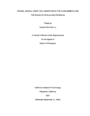
Tải xuống để đọc toàn bộCRANIAL NEURAL CREST CELL MIGRATION IN THE AVIAN EMBRYO AND THE ROLES OF EPH-A4 AND EPHRIN-AS Thesis by Carole Chih-Chen Lu In Partial Fulfillment of the Requirements for the degree of Doctor of Philosophy California Institute Of Technology Pasadena, California 2007 (Defended September 21, 2006) UMI Number: 3244121 INFORMATION TO USERS The quality of this reproduction is dependent upon the quality of the copy submitted. Broken or indistinct print, colored or poor quality illustrations and photographs, print bleed-through, substandard margins, and improper alignment can adversely affect reproduction. In the unlikely event that the author did not send a complete manuscript and there are missing pages, these will be noted. Also, if unauthorized copyright material had to be removed, a note will indicate the deletion.
® UMI UMI Microform 3244121 Copyright 2007 by ProQuest Information and Learning Company. All rights reserved. This microform edition is protected against unauthorized copying under Title 17, United States Code. ProQuest Information and Learning Company 300 North Zeeb Road P.
Box 1346 Ann Arbor, MI 48106-1346 Carole C. Lu © 2007 Carole Chih-Chen Lu All Rights Reserved Carole C. Lu iii ACKNOWLEDGEMENTS My relationship with developmental biology began in 1998, when | took my first developmental biology class. To me, it was love at first sight.
Since arriving at graduate school, the path has been mostly uphill, and there are many people who have supported, motivated, and kept me on the path. | thank my advisor and mentor, Scott E. Fraser, for his guidance and support all these years. Scott provides a unique research environment where graduate students are given freedom to pursue scientific questions.
He is also incredibly supportive and has (what must be) an infinite supply of optimistic advice. | thank the members of my committee, Marianne Bronner-Fraser, Paul Sternberg, and David Chan for scientific guidance and expertise. The Fraser lab is intimate because it is filled with friendly and helpful colleagues. | thank Paul Kulesa, who thoroughly and patiently showed me the ropes and provided me with a solid foundation.
| thank Sean Megason, Helen McBride, Elaine Bearer, and Andrew Ewald for invaluable scientific discussions and knowledge. | thank Michael Liebling for expertise with image analysis software, David Koos for advice in molecular biology and histology, Tatyana Demyanenko for advice in histology, and Aura Keeter for invaluable technical support. | thank Mary Flowers for being our lab mom, Gary Belford and Sonia Collazo for their skilled computer technical support. | thank Chris Waters and Dan Darcy for maintaining our confocal systems.
| also thank Julien Vermot, Le Trinh, Robia Pautler, Christie Canaria, Max Ezin, Mat Barnet, Larry Wade, Magdalena Bak-Maier, David Wu, Rusty Lansford, Mary Dickinson, Liz Jones, and Arian Forouhar for being awesome lab mates. | thank Marianne Bronner-Fraser for a wonderful teaching assistant experience and scientific discussions. The Bronner-Fraser lab next door graciously helps us with reagents and discussions. Specifically | thank Meghan Adams, Sujata Bhattacharyya, Vivian Lee, Martin Basch, Martin Garcia-Castro, Tatjana Sauka-Spengler, Ed Coles, Lisa Taneyhill, and Meyer Barembaum.
My fellow classmates were invaluable during graduate school. Specifically, | thank Gloria Choi, Pei-Yun Lee, and Magdalena Bak-Maier for the fun that we had when we studied for and passed our quails. Toby Rosen, Karli Watson, Xavier Ambroggio, and Johannes Graumann are great fun. | thank Sean Megason and Bernadine Tsung-Megason for adventures outside of lab.
| thank Stuart Freed for not throwing out my pots at the pottery studio and for years of therapeutic pottery. Lu iv | thank my dear sisters, Connie Lu and Cathy Lu, for providing me with many photographic opportunities and material for funny stories. | thank Attila Kovacs for feeding Connie. My parents, Kuen and Dong-Chih Lu, have consistently kept me on the path to graduation.
Lastly, | thank Alok J. Saldanha, my husband, for his love, brilliance, and enthusiasm. Alok is passionate about the world and is the best life partner that | could have ever hoped for. In memory of Eric Tse and Ben Edelson.
In memory of Molly. Lu Vv ABSTRACT The neural crest is a transient population of cells that migrate away from the dorsal neural tube in the vertebrate embryo. As the developing hindbrain constricts into rhombomeres, cranial neural crest cells migrate in three discrete streams adjacent to even-numbered rhombomeres, rhombomere 2 (r2), r4, and r6. To test the role of intrinsic versus extrinsic cues in influencing an individual cell’s trajectory, we implanted physical barriers in the chick mesoderm, distal to emerging neural crest cells (NCCs).
We analyzed spatio-temporal dynamics as NCCs encountered and responded to the barriers by using time-lapse confocal microscopy and cell tracking analysis. The majority of NCCs were able to overcome physical barriers. Even though the lead cells become temporarily blocked by a barrier, follower cells find a novel pathway around a barrier and become de novo leaders of a new stream. Quantitative analyses of cell trajectories find cells that encounter an r3 barrier migrate significantly faster but less directly than cells that encounter an r4 barrier, which migrate normally.
NCCs can also migrate into normally repulsive territory as they reroute. These results suggest that cranial neural crest cell trajectories are not intrinsically determined. NCCs can respond to minor alterations in the environment to retarget a peripheral destination. Both intrinsic and extrinsic cues are important in patterning.
We then tested the role of Eph/ephrin signaling on cranial neural crest migration by ectopically expressing full-length ephrin-A5 ligand; a truncated, Carole C. Lu vi constitutively active EphA4 receptor; and a truncated, kinase-dead EphA4 receptor within migratory neural crest cells. Ectopic expression of ephrin-A5 specifically causes the r6 subpopulation of neural crest cells to have truncated migration but does not affect directionality, suggesting that the r6 neural crest cells properly follow guidance cues. Our results support a role for ephrin-A5 in regulating the extent of migration.
Ectopic expression of constitutively active, truncated EphA4 causes NCCs to migrate aberrantly around the otic vesicle. Pathfinding errors are accompanied by changes in migratory behavior, with the NCCs migrating faster but with less directionality. Expression of a truncated, kinase-dead version of EphA4 also leads to pathfinding errors. Our results suggest Eph activity is involved in guidance and extent of migration.
Lu vii TABLE OF CONTENTS 10,9)160)0)08-i89. Vv TABLE OF CONTENTS1.ccccccccccccteecceceseeenecceeceecereensecneeeceeceecnee sees cneecaeeseecneensnesecesnessaeesaesnateniecnas vii LIST OF ïiic6 1. x CHAPTER 1: Introduction nh 6. 1 AVIAN CIDIYO oo.ccccccccecstcccntetesceneceaecneecneeceecneeeeesaeecaaesaaeceeseeseeteeceeeaaeecinecieetieesueesieenieetieenenesieatinees 1 Cranial neural crest cells: what are they and why are they ímporfanIf?.
2 Stereotypical pattern of mÍQFAflOH. HH HH KH ng TH nh kg 4 Possible intrinsic and @XtrinSiC mecha'liSIT. ánh HH HH HH HH HH rệt 5 Goal 0o. 14 CHAPTER 2: Time-lapse analysis reveals a series of events by which cranial neural crest cells reroute around physical 8-0-1.
22 Materials and MethOdlS. -- ác các HH HH KH HH HH KH KT cờ 27 0. 36 Ack'owledg6Im6FS.- cá HH HH HH HH HH HH T11 tá HT TH TC CC Cu crkt 41 San nẽnốố ố ốốốốốố. 45 CHAPTER 3: Time-lapse analysis of perturbations of ephrin-A5 and EphA4 during cranial neural crest Migration in the AVIAN embryO.
cu cece e cence ecee cece eeseseeecaeeecaececeeeeseeaeeseaeeeceeeeteeeeseaeteaiess 55 “Í>0ttPnnnn. 57 Materials And Method’ PS nhanh cố ốốố. 73 Tu i00 6s 1-7-0 ng cố ốốố. 88 CHAPTER 4: Summary and Future Direcfions.
HH hưu 108 SJ/////12a080PP0P7Ẽ7Ẽ7Ẽ85858Ẻ86 h6. 108 7//0/(29)/(-0/00/TT TT 0 no 3ẦÝÊŸỶỶ. Lu ix LIST OF FIGURES Figure 1.0: Stereotypical pattern OF miQrafÍOH. nen HH HT HH ng go Hà nh Hà ha 11 Figure 2.1: Foil barriers are ineffective at blocking Cranial neural cr@Sf.2: Leaders and followers change when a population of neural crest cells encounters a -/-EPPPSn.3: Neural crest cells that are blocked behind the barrier tend to migrate faster than cells Chat YO APOUNC UNC DAITIOL oo.
eee ccctccctscstcenscssecceeeeseesseccsessnecssecuseesasecsecaissssucssessaasessseseseaseceeessceensease 49 Figure 2.4: Neural crest cells are able to migrate into the r3 repulsive zone by migrating on top of CCH OMNOL eee eccecccneenceeececeeetetecsscansnecsaeeseseneesesesuesassauseassnssaesascuassascnsssasaaseasvasesatestcasecaterssatsaseatarasas 57 Figure 3.1: Differential expression of EphA4 and ephrin-A5 within the cranial region of an HH12 09165 PP®-.2: Ectopic ephrin-A& expression leads to fewer NCCs in BA3 inifially.3: Neural crest cells ectopically expressing ephrin-A5 do not migrate to BA3.4: Ephrin-A5+ r6 NCCs do not migrate tO BAS.5: EphA4(int) and EphA4(kd) causes mismigration along the otic vesicle.6: Cell morphology, temporal distribution, and Cell-traCkiNg.7: Eph/ephrin concentration and migrafÍOn. - - -- S121 HT HH Hệ, 98 Figure 3.8: Ectopic ephrin-A5 and EphA4 acfÍVÍẨW. ác c1 12 LH HH TY TH HH HH key 100 Figure 3.9: Summary of ephrin-A5 and EphA4 perturbations .cccccccccccessecssssessetesssenssessessenseees 102 Supplementary materials: cell death and Cell Proliferation .0: Distinct mechanisms guide neural crest migration at r4 and rồ. Lu x LIST OF TABLES Table 2.1: Mean velocity and directionality for barriers at r3 and r.
ST Hs he 53 Table 3.1: Cell tracking analysis: cranial neural Crest C@lÏS. -- - - cà HH HH HH Hệ, 103 Table 3.2: Cell tracking analysis: r8 subpopulation of cranial neural crest cellS.3: Rates of cell proliferation and deatÏh. HH HH HH HH HH Hệ, 105 Carole C. Lu xi KEYWORDS avian embryo cell migration cell-tracking confocal microscopy cranial neural crest electroporation EphA4 ephrin-A5 foil barrier particle-tracking time-lapse imaging whole-embryo explant culture Carole C.
Lu 1 CHAPTER 1: Introduction Introduction The process by which we develop out of a single fertilized egg is wonderfully complex. In theory, it is easy to understand— one cell divides into two daughter cells that too go on to divide until there is a population of cells that makes up an entire organism. How do we end up being a complex organism rather than a clump of cells? How do cells become patterned and coordinated into structures? The question of pattern formation is a global one. Cell division is but one aspect of development.
From the cell’s point of view, there are many different choices along the way. Not only can they divide, but also they can die, differentiate, migrate, respond to cues in the environment, secrete cues into the environment, or any combination of the above. All these actions by individual cells need to occur in an orchestrated fashion such that at the end, there is a complete and functional multi-cellular organism. During my tenure as a graduate student, | chose the migration of cranial neural crest cells within the avian embryo as the system in which to address how migration is involved in pattern formation.
Avian embryo The avian embryo has been a classic system for embryological studies since Aristotle (Aristotle, 350 B.E) for a number of reasons. Fresh, fertilized eggs are easy to obtain, available year round, relatively cheap, develop externally, and are easy to handle within a laboratory setting. The Hamburger Carole C. Lu 2 and Hamilton staging series (1951) allows one to conveniently set eggs for a certain amount of time to obtain embryos at the desired developmental stage.
A good anatomical understanding of the embryo is also available (Bellairs and Osmond, 2005). The accessibility and size of the embryo allows many types of microsurgical techniques such as ablation and grafting. Beside these more classic, embryological types of studies, new techniques have allowed us to take advantage of recent advances in molecular and cell biology. We can functionally test the roles of certain proteins or genes by implantation of protein-soaked beads; electroporation of DNA constructs, MRNA, or morpholinos; and viral transfection (Bronner-Fraser, 1996; Itasaki et al., 1999; Momose et al., 1999; Okada et al., 1999; Swartz et al., 2001; Thakur et al.
The chick genome has been sequenced and allows researchers to take advantage of newly available genomic resources (reviewed in Antin and Konieczka, 2005). The chick genome offers an interesting evolutionary perspective since it is positioned between lower vertebrates, such as fish, and higher vertebrates, such as humans. Lastly, since the avian embryo is a vertebrate embryo, many of the things we learn will be relevant to understanding human development. Cranial neural crest cells: what are they and why are they important?
Nội dung được bảo vệ bản quyền — Tải xuống đầy đủ
Trích dẫn luận án này
Carole Chih-Chen Lu (2007). Di cư tế bào neural crest sọ: vai trò EPH-A4 và Ephrin-A5 [Luận án tiến sĩ, California Institute of Technology]. LuanAn.net. https://luanan.net/sinh-hoc/sinh-hoc-phan-tu-te-bao/di-cu-te-bao-neural-crest-so-eph-a4-ephrin-a5
Từ khóa và chủ đề nghiên cứu
Từ khóa liên quan
Xem thêm luận án cùng lĩnh vực
Chủ đề nghiên cứu
Câu hỏi thường gặp
Luận án "Di cư tế bào neural crest sọ: vai trò EPH-A4 và Ephrin-A5" nghiên cứu về vấn đề gì?
Luận án tiến sĩ nghiên cứu di cư tế bào neural crest sọ trong phôi chim. Phân tích vai trò EPH-A4 và Ephrin-A5 trong điều hướng quỹ đạo tế bào.
Luận án "Di cư tế bào neural crest sọ: vai trò EPH-A4 và Ephrin-A5" được bảo vệ tại trường nào?
Luận án này được bảo vệ tại California Institute of Technology. Năm bảo vệ: 2007.
Luận án "Di cư tế bào neural crest sọ: vai trò EPH-A4 và Ephrin-A5" thuộc chuyên ngành gì?
Luận án "Di cư tế bào neural crest sọ: vai trò EPH-A4 và Ephrin-A5" thuộc chuyên ngành Sinh học phát triển. Danh mục: Sinh Học Phân Tử Tế Bào.
Luận án "Di cư tế bào neural crest sọ: vai trò EPH-A4 và Ephrin-A5" có bao nhiêu trang?
Luận án "Di cư tế bào neural crest sọ: vai trò EPH-A4 và Ephrin-A5" có 129 trang. Bạn có thể xem trước một phần tài liệu ngay trên trang web trước khi tải về.
Cách tải luận án "Di cư tế bào neural crest sọ: vai trò EPH-A4 và Ephrin-A5" về máy như thế nào?
Để tải luận án về máy, bạn nhấn nút "Tải xuống ngay" trên trang này, sau đó hoàn tất thanh toán phí lưu trữ. File sẽ được tải xuống ngay sau khi thanh toán thành công. Hỗ trợ qua Zalo: 0559 297 239.