Luận án tiến sĩ: Brn3a điều hòa gen đích nơron cảm giác
Luận án tiến sĩ nghiên cứu cơ chế điều hòa biểu hiện gen trong nơron cảm giác qua protein Brn3a và vai trò yếu tố phiên mã trong phát triển thần kinh.
Biomedical Sciences
Luan An
Luận án
Năm xuất bản
Số trang
190
Thời gian đọc
29 phút
Lượt xem
1
Lượt tải
0
Phí lưu trữ
50 Point
Tổng quan nhanh
- Chủ đề:
- 1. Brn3a Transcription Factor Trong Nơron Cảm Giác
- Số trang:
- 190 trang
- Trường:
- University of California, San Diego
- Chuyên ngành:
- Biomedical Sciences
- Tác giả:
- Jason James Lanier
- Năm:
- 2007
Tóm tắt nội dung luận án
I. Brn3a Transcription Factor Trong Nơron Cảm Giác
Brn3a là yếu tố phiên mã quan trọng trong phát triển hệ thống thần kinh cảm giác. Protein này thuộc họ POU domain, điều hòa biểu hiện gen đặc hiệu trong nơron. Nghiên cứu tại Đại học California San Diego đã khám phá cơ chế điều hòa phiên mã của Brn3a. Yếu tố này kiểm soát chương trình biểu hiện gen phức tạp trong ganglion gốc lưng (DRG) và hạch tam thoa. Brn3a ảnh hưởng đến sự phát triển, phân hóa và chức năng của nơron cảm giác. Việc hiểu rõ vai trò của Brn3a mở ra hướng nghiên cứu mới về bệnh lý thần kinh. Transcription factor này tương tác với nhiều gen mục tiêu khác nhau. Sự điều hòa này phụ thuộc vào ngữ cảnh tế bào và vùng thần kinh cụ thể. Nghiên cứu sử dụng chuột knockout Brn3a để xác định gen mục tiêu. Kết quả cho thấy Brn3a điều hòa hàng trăm gen trong nơron cảm giác phôi thai.
1.1. Cấu Trúc POU Domain Của Brn3a
Brn3a chứa miền POU đặc trưng cho họ yếu tố phiên mã này. POU domain gồm hai phần: POUs và POUh, liên kết với DNA. Cấu trúc này cho phép nhận diện trình tự nucleotide đặc hiệu. Miền POU quyết định tính đặc hiệu khi gắn vào promoter gen. Brn3a sử dụng cấu trúc này để điều hòa hàng loạt gen mục tiêu. Sự linh hoạt của POU domain tạo nên đa dạng chức năng.
1.2. Vai Trò Trong Phát Triển Nơron
Brn3a xuất hiện sớm trong quá trình hình thành hệ thần kinh cảm giác. Yếu tố này cần thiết cho sự sống còn của nơron cảm giác. Thiếu Brn3a dẫn đến chết tế bào nơron ở giai đoạn phôi. Protein này kiểm soát biệt hóa từ tế bào tiền thân thành nơron trưởng thành. Brn3a điều hòa các gen liên quan đến phát triển axon và synapse.
1.3. Biểu Hiện Đặc Hiệu Trong DRG Neurons
DRG neurons biểu hiện Brn3a ở mức độ cao trong phát triển phôi. Ganglion gốc lưng là vị trí chính của hoạt động Brn3a. Biểu hiện này bắt đầu từ ngày phôi thai E11-E12 ở chuột. Brn3a duy trì ở một phần nơron trưởng thành. Mức độ biểu hiện ảnh hưởng đến kiểu phân hóa nơron cảm giác.
II. Điều Hòa Phiên Mã Gen Bởi Brn3a
Brn3a điều hòa biểu hiện gen thông qua nhiều cơ chế phân tử. Yếu tố phiên mã này gắn trực tiếp vào vùng promoter của gen mục tiêu. Nghiên cứu ChIP (Chromatin Immunoprecipitation) xác nhận sự gắn kết này. Brn3a có thể hoạt động như activator hoặc repressor tùy ngữ cảnh. Điều hòa phiên mã phụ thuộc vào trạng thái epigenetic của chromatin. Histone acetylation ảnh hưởng đến khả năng tiếp cận của Brn3a. Expression array cho thấy hàng trăm gen thay đổi khi thiếu Brn3a. Một số gen tăng biểu hiện, số khác giảm trong chuột knockout. Cơ chế điều hòa trực tiếp và gián tiếp đều tồn tại. Brn3a tương tác với các yếu tố phiên mã khác tạo phức hợp điều hòa.
2.1. Gắn Kết Trực Tiếp Vào DNA
Brn3a nhận diện trình tự DNA đặc hiệu thông qua POU domain. Motif gắn kết thường chứa trình tự TAATCC hoặc tương tự. ChIP assay chứng minh Brn3a gắn vào promoter NeuroD4, NeuroD1. Sự gắn kết này xảy ra in vivo trong hạch tam thoa phôi thai. Ái lực gắn kết phụ thuộc vào trình tự flanking DNA. Một số vị trí gắn có ái lực cao, số khác thấp hơn.
2.2. Điều Hòa Qua Histone Modification
Acetylated histone H3 đánh dấu vùng chromatin mở cho Brn3a. ChIP analysis cho thấy mối liên hệ giữa H3 acetylation và Brn3a binding. Gen hoạt động có mức acetylation cao tại promoter. Brn3a có thể tuyển dụng histone acetyltransferase đến gen mục tiêu. Trạng thái chromatin quyết định khả năng điều hòa của Brn3a. Methylation và các modification khác cũng đóng vai trò.
2.3. Tương Tác Với Yếu Tố Phiên Mã Khác
Brn3a không hoạt động đơn độc mà tạo phức hợp với protein khác. Tương tác với các transcription factor như NeuroD tăng cường hoạt tính. Sự phối hợp này tạo nên tính đặc hiệu tế bào. Brn3a điều hòa biểu hiện của các yếu tố phiên mã khác. Điều này tạo cascade điều hòa phức tạp trong nơron cảm giác.
III. Gen Mục Tiêu Của Brn3a Trong Hệ Thần Kinh
Expression array phân tích toàn bộ thay đổi gen trong chuột thiếu Brn3a. Hơn 200 transcript thay đổi đáng kể ở hạch tam thoa E13.5. Các gen liên quan đến hệ thống neurotransmitter bị ảnh hưởng mạnh. Brn3a điều hòa gen mã hóa kênh ion, receptor, và protein synapse. Một số gen mục tiêu quan trọng bao gồm NeuroD1, NeuroD4, Pou4f1. Gen Hox cũng nằm trong danh sách mục tiêu của Brn3a. Sự điều hòa khác nhau giữa DRG và hạch tam thoa. Một số gen chỉ bị điều hòa ở một trong hai vùng. Điều này phản ánh chức năng đặc hiệu theo ngữ cảnh của Brn3a. Target gene specificity phụ thuộc vào cofactor có sẵn trong mỗi loại nơron.
3.1. Gen Liên Quan Neurotransmitter
Brn3a điều hòa hệ thống tổng hợp và vận chuyển neurotransmitter. Gen mã hóa enzyme sinh tổng hợp glutamate, GABA bị ảnh hưởng. Receptor cho nhiều loại neurotransmitter cũng là mục tiêu. Brn3a kiểm soát phenotype hóa học của nơron cảm giác. Sự điều hòa này quyết định loại tín hiệu nơron truyền đạt.
3.2. NeuroD Family Transcription Factors
NeuroD1 và NeuroD4 là mục tiêu trực tiếp quan trọng của Brn3a. ChIP cho thấy Brn3a gắn vào promoter của cả hai gen này. NeuroD4 giảm mạnh trong hạch tam thoa thiếu Brn3a. NeuroD1 cũng bị ảnh hưởng nhưng ở mức độ khác. Các yếu tố NeuroD tham gia phân hóa nơron và sống còn. Brn3a tạo mạng lưới điều hòa với family NeuroD.
3.3. Tự Điều Hòa Qua Pou4f1 Locus
Pou4f1 mã hóa chính Brn3a, tạo vòng phản hồi tự điều hòa. ChIP analysis xác nhận Brn3a gắn vào promoter của gen mình. Autoregulation này duy trì mức biểu hiện Brn3a ổn định. Acetylated H3 hiện diện cao tại Pou4f1 locus. Cơ chế này quan trọng cho sự duy trì phenotype nơron.
IV. Sự Khác Biệt Điều Hòa Giữa DRG Và Hạch Tam Thoa
Brn3a điều hòa cả chương trình gen chung và riêng biệt ở DRG versus hạch tam thoa. Expression profiling so sánh hai vùng này ở chuột bình thường và knockout. Nhiều gen thay đổi tương tự ở cả hai ganglion. Tuy nhiên, một số gen chỉ bị điều hòa ở một vùng cụ thể. DRG có biểu hiện đặc trưng các marker proprioceptor. Brn3a điều hòa gen Hox chủ yếu trong DRG, không phải hạch tam thoa. Sự khác biệt này phản ánh nguồn gốc phát sinh khác nhau. DRG phát triển từ neural crest thân, hạch tam thoa từ vùng đầu. Context-dependent binding giải thích tính đặc hiệu vùng này. Epigenetic landscape khác nhau giữa hai loại ganglion.
4.1. Gen Đặc Hiệu DRG
Proprioceptor markers như Parvalbumin biểu hiện cao trong DRG. Brn3a điều hòa các gen liên quan đến cảm giác bản thể. Gen Hox như Hoxa5, Hoxc8 là mục tiêu trong DRG. Các gen này không thay đổi trong hạch tam thoa thiếu Brn3a. Sự điều hòa đặc hiệu này liên quan đến chức năng DRG. DRG neurons xử lý thông tin từ thân và chi.
4.2. Gen Đặc Hiệu Hạch Tam Thoa
Một số gen chỉ bị điều hòa bởi Brn3a trong hạch tam thoa. Các gen này liên quan đến cảm giác mặt và đầu. Trigeminal-specific targets phản ánh chức năng đặc biệt của ganglion này. Brn3a tương tác với các cofactor khác nhau ở hạch tam thoa. Chromatin state tại các locus này khác với DRG.
4.3. Chương Trình Gen Chung
Nhiều gen cơ bản của nơron cảm giác được điều hòa ở cả hai vùng. Gen liên quan sống còn, phát triển axon là mục tiêu chung. Brn3a duy trì identity cơ bản của nơron cảm giác. Các gen housekeeping cho chức năng nơron được điều hòa đồng nhất. Sự tương đồng này phản ánh vai trò cốt lõi của Brn3a.
V. Phương Pháp Nghiên Cứu Brn3a Và Gen Mục Tiêu
Nghiên cứu sử dụng chuột knockout Brn3a để xác định gen mục tiêu. Expression microarray so sánh wild-type và mutant ở E13.5. Mô hạch tam thoa và DRG được phân tích riêng biệt. ChIP assay xác nhận gắn kết trực tiếp của Brn3a vào DNA. Antibody đặc hiệu Brn3a được dùng để kéo xuống chromatin. PCR định lượng đo mức độ enrichment tại các locus. Histone modification profiling sử dụng antibody anti-acetyl H3. In situ hybridization xác định biểu hiện gen trong mô. Kỹ thuật này cho thấy phân bố tế bào của transcript. RT-PCR định lượng xác nhận kết quả microarray. Phân tích thống kê xác định thay đổi có ý nghĩa. Multiple testing correction được áp dụng cho dữ liệu array.
5.1. Expression Array Analysis
Microarray phân tích hàng ngàn gen đồng thời trong mẫu mô. RNA được chiết xuất từ ganglion chuột E13.5. Mẫu được gắn nhãn và lai với chip Affymetrix. Dữ liệu chuẩn hóa để loại bỏ bias kỹ thuật. Fold change và p-value xác định gen thay đổi đáng kể. Cutoff thường là fold change >1.5 và p<0.05.
5.2. Chromatin Immunoprecipitation
ChIP assay bắt đầu với crosslink protein-DNA bằng formaldehyde. Chromatin được cắt nhỏ bằng sonication thành đoạn 200-500bp. Antibody Brn3a kéo xuống phức hợp protein-DNA. DNA được tinh sạch và phân tích bằng PCR. Primer thiết kế cho vùng promoter của gen mục tiêu. Enrichment so sánh với input và IgG control.
5.3. Validation Bằng RT PCR
RT-PCR định lượng xác nhận kết quả microarray. RNA được reverse transcribe thành cDNA. Real-time PCR đo mức độ biểu hiện chính xác. Gen housekeeping như GAPDH làm đối chứng chuẩn hóa. Kết quả tương quan cao với dữ liệu array. Phương pháp này xác nhận target genes quan trọng.
VI. Ý Nghĩa Sinh Học Của Điều Hòa Gen Bởi Brn3a
Brn3a đóng vai trò trung tâm trong phát triển hệ thần kinh cảm giác. Thiếu Brn3a gây chết nơron cảm giác ở giai đoạn phôi. Yếu tố này cần thiết cho sự sống còn và phân hóa đúng. Điều hòa gen bởi Brn3a tạo nên đa dạng nơron cảm giác. Các subtype nơron khác nhau phụ thuộc vào mức Brn3a. Context-dependent regulation cho phép chức năng đa dạng. Nghiên cứu này mở ra hiểu biết về mạng lưới điều hòa phiên mã. Kiến thức về Brn3a target genes có ứng dụng lâm sàng tiềm năng. Bệnh lý thần kinh cảm giác có thể liên quan đến rối loạn Brn3a. Regeneration nơron sau chấn thương có thể cần tái kích hoạt Brn3a. Hiểu cơ chế này giúp phát triển liệu pháp thần kinh mới.
6.1. Vai Trò Trong Sống Còn Nơron
Brn3a điều hòa gen chống apoptosis trong nơron cảm giác. Thiếu Brn3a dẫn đến chết tế bào qua trình trình programmed cell death. Gen target liên quan đến survival pathway được điều hòa. Brn3a duy trì cân bằng giữa tín hiệu sống và chết. Điều này quan trọng trong phát triển bình thường hệ thần kinh.
6.2. Xác Định Subtype Nơron Cảm Giác
Brn3a góp phần tạo nên đa dạng các loại nơron cảm giác. Mechanoreceptor, nociceptor, proprioceptor có mức Brn3a khác nhau. Điều hòa differential gene expression tạo phenotype đặc trưng. Brn3a phối hợp với các transcription factor khác xác định subtype. Sự đa dạng này cần thiết cho chức năng cảm giác phức tạp.
6.3. Tiềm Năng Ứng Dụng Lâm Sàng
Hiểu về Brn3a có thể giúp điều trị bệnh lý thần kinh. Neuropathy cảm giác có thể liên quan đến rối loạn biểu hiện Brn3a. Tái sinh nơron sau tổn thương cần tái kích hoạt chương trình phát triển. Brn3a có thể là mục tiêu cho liệu pháp gene therapy. Nghiên cứu này đặt nền tảng cho ứng dụng tương lai.
Mục lục chi tiết luận án
Tải xuống file đầy đủ để xem toàn bộ nội dung
Tải đầy đủ (190 trang)Nội dung chính
Tổng quan về luận án
Luận án này cung cấp một phân tích chuyên sâu về cơ chế điều hòa biểu hiện gen của yếu tố phiên mã POU-domain Brn3a (Pou4f1) trong quá trình phát triển của hệ thống thần kinh cảm giác, đặc biệt tập trung vào hạch sinh ba (trigeminal ganglion – TG) và hạch rễ lưng (dorsal root ganglia – DRG) ở phôi chuột. Bối cảnh khoa học của nghiên cứu được đặt trong sự hiểu biết rằng sự phát triển phức tạp của hệ thần kinh, với vô số loại tế bào chuyên biệt và mạch thần kinh hoạt động chính xác, phụ thuộc vào sự điều hòa gen phối hợp chặt chẽ. Các yếu tố phiên mã đặc hiệu mô đóng vai trò trung tâm trong quá trình này bằng cách kiểm soát biểu hiện gen mục tiêu theo không gian và thời gian. Tính tiên phong của nghiên cứu nằm ở việc chuyển từ các quan sát hình thái học về tác động của Brn3a sang việc giải mã mạng lưới gen cụ thể mà nó điều hòa in vivo, đồng thời làm sáng tỏ vai trò chưa được hiểu rõ của các yếu tố biểu sinh trong việc xác định tính đặc hiệu mục tiêu của yếu tố phiên mã.
Research gap cụ thể mà luận án này giải quyết là việc "hầu hết các gen mục tiêu hạ nguồn của các yếu tố phiên mã thần kinh quan trọng trong phát triển vẫn chưa được biết đến" (tr.5). Ngoài ra, còn có câu hỏi chưa được trả lời về việc "liệu các yếu tố phiên mã có chiếm tất cả các vị trí liên kết tiềm năng trong bộ gen, nhưng chỉ điều hòa phiên mã từ một tập hợp con của các vị trí đó; hay liệu chỉ một phần nhỏ các vị trí tiềm năng được chiếm giữ" (tr.10). Các nghiên cứu trước đây thường dựa vào phân tích in vitro hoặc phương pháp phân tích từng gen riêng lẻ, dẫn đến sự thiếu hụt kiến thức toàn diện về mạng lưới điều hòa gen in vivo.
Các research questions chính được luận án đặt ra bao gồm:
- Các gen cụ thể nào tạo nên chương trình biểu hiện gen được Brn3a điều hòa trong hạch sinh ba đang phát triển?
- Brn3a có điều hòa trực tiếp các gen mục tiêu này không, đặc biệt là các yếu tố phiên mã sinh thần kinh?
- Các sửa đổi biểu sinh của chromatin đóng vai trò gì trong việc xác định tính đặc hiệu của gen mục tiêu của Brn3a và sự khác biệt về điều hòa gen giữa các mô?
- Chương trình điều hòa gen của Brn3a có khác biệt giữa các hạch cảm giác ở các mức độ trục khác nhau (ví dụ: TG và DRG) và nếu có, điều gì giải thích cho những khác biệt này?
Các hypotheses được kiểm chứng là:
- Brn3a điều hòa một chương trình phối hợp các hệ thống dẫn truyền thần kinh, kênh ion, thành phần cấu trúc của sợi trục và hệ thống tín hiệu nội và ngoại bào trong TG.
- Brn3a trực tiếp ức chế các yếu tố phiên mã sinh thần kinh cụ thể như NeuroD1 và NeuroD4 trong TG đang phát triển.
- Các sửa đổi biểu sinh của chromatin ảnh hưởng đáng kể đến việc lựa chọn gen mục tiêu của Brn3a bằng cách điều chỉnh khả năng tiếp cận các vị trí liên kết tiềm năng.
- Brn3a điều hòa các tập hợp bản sao tương tự nhưng khác biệt trong các hạch cảm giác ở các mức độ trục khác nhau, và những khác biệt này được liên kết với các trạng thái biểu sinh của chromatin tại các locus gen mục tiêu.
Khung lý thuyết của nghiên cứu được xây dựng dựa trên các lý thuyết về mạng lưới điều hòa gen, điều hòa phiên mã, và điều hòa biểu sinh. Cụ thể, nó tích hợp các lý thuyết về vai trò của các yếu tố phiên mã POU-domain (Phillips và Luisi, 2000) trong việc xác định danh tính và chức năng tế bào, cùng với các khái niệm về yếu tố điều hòa cis-acting (Istrail và Davidson, 2005) và ảnh hưởng sâu sắc của cấu trúc chromatin (Latchman, 2004; Jenuwein và Allis, 2001; Turner, 2002) lên khả năng tiếp cận DNA và hoạt động phiên mã. Luận án đặc biệt mở rộng lý thuyết về "mã histone" bằng cách chứng minh vai trò trực tiếp của nó trong tính đặc hiệu của yếu tố phiên mã in vivo.
Đóng góp đột phá của luận án bao gồm:
- Lập bản đồ toàn diện mạng lưới gen điều hòa bởi Brn3a in vivo: Luận án đã xác định được một chương trình biểu hiện gen phối hợp bao gồm các gen liên quan đến dẫn truyền thần kinh, thụ thể, kênh ion, tăng trưởng sợi trục và tín hiệu tế bào trong TG đang phát triển (tr.26), cung cấp cái nhìn chi tiết chưa từng có về chức năng của Brn3a.
- Xác định cơ chế điều hòa trực tiếp của Brn3a: Nghiên cứu đã chứng minh rằng Brn3a trực tiếp ức chế biểu hiện của các yếu tố phiên mã sinh thần kinh NeuroD1 và NeuroD4 trong TG (tr.xiii), làm rõ một cơ chế quan trọng trong việc định hình số phận tế bào thần kinh.
- Làm sáng tỏ vai trò của điều hòa biểu sinh trong tính đặc hiệu của yếu tố phiên mã: Luận án cung cấp bằng chứng rằng "các sửa đổi biểu sinh của chromatin đóng vai trò không thể thiếu trong việc xác định các mục tiêu điều hòa của Brn3a và góp phần vào sự khác biệt gen mục tiêu giữa TG và DRG" (tr.xiii). Đây là một đóng góp mang tính thay đổi mô hình, cho thấy khả năng tiếp cận vị trí liên kết DNA, chứ không chỉ trình tự DNA, là yếu tố then chốt.
- Phân tích so sánh các chương trình điều hòa gen theo mô: Luận án đã phát hiện ra rằng Brn3a điều hòa "các tập hợp bản sao tương tự, nhưng khác biệt trong các hạch cảm giác ở các mức độ trục khác nhau" (tr.xiii), với những khác biệt này được liên kết trực tiếp với các trạng thái acetyl hóa histone cụ thể (acetyl hóa H3K9/K14) tại các locus gen mục tiêu (tr.16), đưa ra một cơ chế phân tử rõ ràng cho tính đặc hiệu theo mô.
Phạm vi nghiên cứu bao gồm việc phân tích các phôi chuột ở giai đoạn E13.5. Đối với phân tích microarray, "khoảng 10-12 hạch đã được định kiểu gen là đủ để cung cấp 5µg tổng RNA cho một phân tích duy nhất" (tr.27). Các mô được phân tích chủ yếu là hạch sinh ba (TG), hạch rễ lưng (DRG), và phức hợp hạch VIII/IX/X. Tính quan trọng của nghiên cứu nằm ở việc cung cấp nền tảng cơ bản để hiểu rõ hơn về sự phát triển của hệ thần kinh cảm giác, các bệnh lý thần kinh bẩm sinh, và có thể dẫn đến việc phát triển các chiến lược điều trị mới nhắm vào các con đường điều hòa gen này.
Literature Review và Positioning
Luận án này tổng hợp các dòng nghiên cứu chính về sự cảm ứng và phát triển của hệ thống thần kinh (Kandel, Schwartz et al., 2000), vai trò của các yếu tố phiên mã đặc hiệu mô trong quá trình phát triển thần kinh (Pfeffer, Bouchard et al., 2000; Bermingham, Shumas et al., 2002; Ebert, Timmer et al., 2003; Lee và Pfaff, 2003; Scardigli, Baumer et al., 2003; Saba, Johnson et al., 2003), và cơ chế điều hòa phiên mã (Latchman, 2004). Nghiên cứu cũng đi sâu vào ảnh hưởng của biểu sinh lên liên kết yếu tố phiên mã, trích dẫn các tác phẩm của Jenuwein và Allis (2001), Turner (2002) về mã histone, và Barrera và Ren (2006) về các chữ ký chromatin đặc trưng. Luận án thừa nhận rằng các yếu tố phiên mã POU-domain như Brn3a là những yếu tố điều hòa quan trọng với biểu hiện phức tạp trong hệ thần kinh (Phillips và Luisi, 2000), và sự gián đoạn của chúng dẫn đến các bất thường nghiêm trọng. Các nghiên cứu trước đây đã chứng minh sự cần thiết của Brn3a đối với sự phát triển và tồn tại của các tế bào thần kinh cảm giác (McEvilly et al., 1996; Xiang et al., 1996), với các khiếm khuyết đáng kể về tăng trưởng sợi trục (Eng et al., 2001) và mất biểu hiện thụ thể neurotrophin (Huang et al., 1999; Ma et al., 2003).
Luận án làm nổi bật các mâu thuẫn và tranh luận hiện có trong tài liệu. Một điểm mâu thuẫn chính là "chỉ riêng trình tự DNA là không đủ để giải thích tính đặc hiệu của yếu tố phiên mã" (tr.7), vì các vị trí liên kết đồng thuận thường xảy ra hàng trăm nghìn lần trong bộ gen, nhưng các yếu tố phiên mã chỉ điều hòa khoảng 100 gen mục tiêu trong một mô nhất định (Livesey, Furukawa et al., 2000; Gold, Baek et al., 2003; Mu, Beremand et al., 2003). Điều này đặt ra câu hỏi về các cơ chế bổ sung điều khiển tính đặc hiệu. Một tranh luận khác liên quan đến sự phụ thuộc vào các mô hình in vitro: luận án đã phát hiện ra rằng "các kết quả được báo cáo ở đây không hỗ trợ việc điều hòa in vivo của các gen mục tiêu Brn3a được cho là từ các nghiên cứu quá mức biểu hiện trước đây trong các dòng tế bào được chuyển gen, và minh họa sự khó khăn trong việc xác định các con đường điều hòa sinh lý trong các hệ thống mô hình này" (tr.34). Điều này thách thức tính tổng quát của các phát hiện in vitro và nhấn mạnh sự cần thiết của các nghiên cứu in vivo mạnh mẽ.
Trong bối cảnh tài liệu hiện có, luận án này tự định vị mình là một nghiên cứu đột phá bằng cách giải quyết khoảng trống cụ thể trong việc xác định các gen mục tiêu hạ nguồn in vivo của Brn3a và cơ chế phân tử điều khiển tính đặc hiệu theo mô của nó. Trong khi các nghiên cứu trước đây thường tập trung vào việc mô tả các hậu quả kiểu hình của việc mất chức năng Brn3a, luận án này đi sâu vào việc giải mã mạng lưới gen cụ thể mà Brn3a điều hòa trực tiếp, và điều quan trọng là, vai trò của các sửa đổi biểu sinh trong quá trình này.
Nghiên cứu này thúc đẩy lĩnh vực này bằng cách cung cấp một mô hình toàn diện về cách một yếu tố phiên mã quan trọng trong phát triển điều hòa chương trình gen phức tạp trong các mô cụ thể, vượt ra ngoài sự phụ thuộc vào trình tự DNA đơn thuần. Nó cung cấp bằng chứng thuyết phục về việc các sửa đổi biểu sinh đóng vai trò không thể thiếu trong việc xác định các mục tiêu điều hòa của yếu tố phiên mã (tr.xiii). Điều này là một đóng góp cụ thể cho sự hiểu biết về điều hòa gen trong hệ thần kinh.
So sánh với ít nhất 2 nghiên cứu quốc tế, công trình này xây dựng trên nền tảng của các nghiên cứu cơ bản về điều hòa gen và chromatin. Ví dụ, nó bổ sung vào các khái niệm về "logic-functions" của các module điều hòa cis-regulatory được mô tả bởi Istrail và Davidson (2005), bằng cách cung cấp bằng chứng in vivo về cách các tín hiệu ngoài trình tự DNA được tích hợp. Trong khi Istrail và Davidson (2005) tập trung vào các chức năng logic của mã cis-regulatory dựa trên tích hợp tín hiệu từ nhiều yếu tố phiên mã, luận án này của Lanier mở rộng bằng cách chỉ ra rằng bản thân khả năng tiếp cận DNA do chromatin tạo ra là một chức năng logic quan trọng khác. Hơn nữa, nghiên cứu này so sánh với các phát hiện về GATA-1 của Im, Grass et al. (2005), những người đã chứng minh việc GATA-1 sử dụng một tập hợp nhỏ các motif GATA phân tán trong một vùng nhiễm sắc thể rộng, liên quan đến các vùng nhạy cảm với DNAse I. Luận án này của Lanier áp dụng một cách tiếp cận tương tự với Brn3a, nhưng mở rộng nó bằng cách sử dụng ChIP với kháng thể nhận diện histone bị sửa đổi cụ thể (H3K9/K14) để liên kết trực tiếp các sửa đổi biểu sinh với khả năng tiếp cận và liên kết của yếu tố phiên mã trong các ngữ cảnh mô khác nhau, cung cấp một mức độ chi tiết cơ học cao hơn cho sự điều hòa theo mô.
Đóng góp lý thuyết và khung phân tích
Đóng góp cho lý thuyết
Luận án này mở rộng và thách thức các lý thuyết cụ thể về tính đặc hiệu của yếu tố phiên mã và điều hòa gen trong phát triển thần kinh. Nó đặc biệt mở rộng lý thuyết về chức năng yếu tố phiên mã POU-domain (Phillips và Luisi, 2000) bằng cách chứng minh rằng khả năng liên kết của chúng không chỉ bị chi phối bởi trình tự DNA mà còn bởi trạng thái biểu sinh của chromatin xung quanh. Luận án thách thức quan niệm đơn giản rằng các yếu tố phiên mã sẽ liên kết với tất cả các vị trí liên kết đồng thuận tiềm năng trong bộ gen (Remenyi, Scholer et al., 2004), thay vào đó cung cấp bằng chứng rằng "chỉ một phần nhỏ các vị trí tiềm năng được chiếm giữ" (tr.10), và điều này được điều chỉnh bởi khả năng tiếp cận chromatin.
Khung khái niệm được đề xuất bởi luận án này mô tả Brn3a hoạt động trong một mạng lưới điều hòa phức tạp, nơi hoạt động của nó được điều chỉnh không chỉ bởi trình tự DNA mà còn bởi môi trường chromatin cục bộ. Môi trường này được mã hóa biểu sinh một cách đặc trưng theo loại tế bào, giải thích cho việc cùng một yếu tố phiên mã có thể điều hòa các tập hợp gen mục tiêu khác nhau ở các mô khác nhau. Cụ thể, nó đề xuất một mô hình trong đó liên kết của Brn3a với trình tự đồng thuận của nó là một điều kiện cần thiết nhưng chưa đủ cho việc điều hòa gen. Thay vào đó, khả năng tiếp cận của vị trí liên kết, bị ảnh hưởng bởi các sửa đổi biểu sinh như acetyl hóa histone H3 (H3K9/K14), đóng vai trò là một điều kiện biên quan trọng đối với chức năng điều hòa của Brn3a.
Mô hình lý thuyết này đưa ra các đề xuất sau:
- Brn3a trực tiếp điều hòa một chương trình gen quan trọng cho kiểu hình của tế bào thần kinh cảm giác, bao gồm các gen liên quan đến dẫn truyền thần kinh, thụ thể, kênh ion và tăng trưởng sợi trục.
- Tính đặc hiệu liên kết của Brn3a được điều chỉnh bởi các dấu hiệu biểu sinh trên chromatin.
- Các trạng thái biểu sinh đặc trưng cho mô dẫn đến sự điều hòa gen mục tiêu khác biệt của Brn3a.
- Sự de-acetyl hóa H3K9/K14 ở một số locus gen mục tiêu trong DRG, nhưng không phải TG, góp phần vào sự khác biệt trong chương trình điều hòa gen của Brn3a giữa hai mô này.
Luận án này đại diện cho một sự tiến bộ đáng kể, hay một "paradigm shift", từ quan niệm về điều hòa gen chỉ dựa trên trình tự DNA sang một mô hình điều hòa phụ thuộc ngữ cảnh qua trung gian chromatin cho các yếu tố phiên mã đặc hiệu mô. Bằng chứng cho sự thay đổi mô hình này được cung cấp rõ ràng khi nghiên cứu kết luận rằng "các sửa đổi biểu sinh của chromatin đóng vai trò không thể thiếu trong việc xác định các mục tiêu điều hòa của Brn3a và góp phần vào sự khác biệt gen mục tiêu giữa TG và DRG" (tr.xiii), và "những dữ liệu này chứng minh rằng các cơ chế biểu sinh đóng góp vào sự khác biệt theo mô trong điều hòa gen mục tiêu bởi Brn3a" (tr.16).
Khung phân tích độc đáo
Khung phân tích của luận án này độc đáo ở chỗ nó tích hợp một cách mạnh mẽ ba dòng lý thuyết chính: điều hòa phiên mã (Latchman, 2004), chức năng yếu tố phiên mã POU-domain (Phillips và Luisi, 2000) và điều hòa biểu sinh (Jenuwein và Allis, 2001; Turner, 2002; Barrera và Ren, 2006). Sự tích hợp này cho phép một cái nhìn đa chiều về cách yếu tố phiên mã hoạt động in vivo.
Cách tiếp cận phân tích mới lạ nằm ở việc kết hợp phân tích biểu hiện gen toàn cầu in vivo (microarray) với các xét nghiệm ChIP cơ chế nhắm mục tiêu cụ thể các histone bị sửa đổi (ví dụ: acetyl hóa histone H3 tại các lysine 9 và 14 – H3K9/K14) (tr.16). Điều này cung cấp bằng chứng trực tiếp về mối liên hệ giữa trạng thái chromatin, liên kết yếu tố phiên mã và biểu hiện gen khác biệt trong một ngữ cảnh phát triển. Cách tiếp cận này vượt ra ngoài các nghiên cứu trước đây thường chỉ tập trung vào một khía cạnh (ví dụ: chỉ lập danh sách gen hoặc chỉ phân tích liên kết DNA).
Những đóng góp khái niệm bao gồm việc mở rộng định nghĩa "tính đặc hiệu của yếu tố phiên mã" để bao gồm khả năng tiếp cận chromatin. Nghiên cứu giới thiệu khái niệm "điều hòa phụ thuộc ngữ cảnh qua trung gian chromatin" như một cơ chế chính cho việc điều hòa gen đặc hiệu mô, nơi trạng thái acetyl hóa histone có thể xác định liệu một vị trí liên kết tiềm năng có thực sự được sử dụng hay không.
Các điều kiện biên được nêu rõ ràng: hoạt động điều hòa của Brn3a bị giới hạn bởi giai đoạn phát triển cụ thể (phôi chuột E13.5), các loại tế bào thần kinh cảm giác cụ thể (TG, DRG), và quan trọng nhất là, cảnh quan biểu sinh cục bộ của các locus gen mục tiêu. Cơ chế "tự điều hòa và bù liều gen" của Brn3a (Trieu et al., 2003; Trieu et al., 2007) cũng là một điều kiện biên quan trọng, giải thích tại sao chuột dị hợp tử Brn3a không biểu hiện kiểu hình đột biến rõ ràng (tr.32), cung cấp một điểm tham chiếu quan trọng cho việc giải thích dữ liệu.
Phương pháp nghiên cứu tiên tiến
Thiết kế nghiên cứu
Triết lý nghiên cứu của luận án này nghiêng về Positivism (hoặc Post-Positivism), tập trung vào việc thiết lập các mối quan hệ nhân quả và cơ chế có thể kiểm chứng được trong một hệ thống sinh học khách quan. Nghiên cứu sử dụng các phương pháp định lượng (phân tích microarray) và các kỹ thuật thực nghiệm phân tử chính xác (ChIP, lai in situ, hóa miễn dịch) để thu thập dữ liệu khách quan, có thể đo lường được nhằm kiểm định các giả thuyết về điều hòa gen.
Thiết kế nghiên cứu tích hợp một cách khéo léo các phương pháp đa chiều, không phải là "mixed methods" theo nghĩa xã hội học, mà là một cách tiếp cận sinh học phân tử tích hợp nhiều kỹ thuật bổ sung. Nó kết hợp phân tích định lượng biểu hiện gen toàn cầu (microarray) để xác định các gen mục tiêu tiềm năng, với xét nghiệm cơ chế in vivo (ChIP) để chứng minh liên kết trực tiếp và làm sáng tỏ vai trò của chromatin, và xác nhận không gian và tế bào (lai in situ và hóa miễn dịch) để kiểm tra và định vị các thay đổi biểu hiện gen. Rationale cho sự kết hợp này là microarray cung cấp một cái nhìn tổng thể không thiên vị về các thay đổi biểu hiện gen, ChIP cung cấp bằng chứng trực tiếp về tương tác DNA-protein và trạng thái chromatin, trong khi lai in situ và hóa miễn dịch xác nhận các phát hiện ở cấp độ mô và tế bào, đảm bảo tính hợp lệ và phù hợp sinh học của các thay đổi được quan sát.
Thiết kế đa cấp của nghiên cứu được thể hiện rõ ràng:
- Cấp độ phân tử: Phân tích biểu hiện gen ở cấp độ bản sao (microarray) và tương tác DNA-protein cùng trạng thái chromatin (ChIP).
- Cấp độ tế bào: Định vị biểu hiện gen và protein trong các quần thể tế bào thần kinh cụ thể trong hạch (lai in situ, hóa miễn dịch).
- Cấp độ mô/cơ quan: So sánh các chương trình điều hòa gen của Brn3a giữa các hạch cảm giác ở các mức độ trục khác nhau (TG và DRG).
Kích thước mẫu cho phân tích microarray là "khoảng 10-12 hạch đã được định kiểu gen là đủ để cung cấp 5µg tổng RNA cho một phân tích duy nhất" (tr.27). Các tiêu chí lựa chọn mẫu chính xác bao gồm phôi chuột E13.5 (được xác định bằng hệ thống phân loại của Theiler, 1972) từ các cặp giao phối dị hợp tử Brn3a để tạo ra các thế hệ wild-type, dị hợp tử và knockout. Chỉ sử dụng "các hạch hoàn chỉnh" được giải phẫu cẩn thận và loại bỏ các mô không phải thần kinh bám dính (tr.27).
Quy trình nghiên cứu nghiêm ngặt
Chiến lược lấy mẫu liên quan đến việc sử dụng các cặp giao phối dị hợp tử Brn3a để tạo ra các thế hệ wild-type, dị hợp tử và knockout từ cùng một nền tảng di truyền, đảm bảo sự nhất quán di truyền giữa các nhóm thí nghiệm. Các tiêu chí bao gồm: chỉ các phôi E13.5 được sử dụng để tối thiểu hóa các hiệu ứng thứ cấp do chết tế bào thần kinh diễn ra sau đó (tr.29). Tiêu chí loại trừ bao gồm các phôi được phân loại không chính xác và các hạch không hoàn chỉnh hoặc bị nhiễm mô không phải thần kinh.
Các giao thức thu thập dữ liệu được mô tả chi tiết:
- Microarray: RNA được chiết xuất bằng hệ thống RNeasy (Qiagen). Việc tạo cDNA, sản xuất cRNA có gắn nhãn và lai với mảng GeneChip được thực hiện theo các giao thức tiêu chuẩn của nhà sản xuất (Affymetrix) (tr.27). Các mảng U74Av2 và U74Bv2 (Affymetrix) được sử dụng để phân tích, bao gồm tổng cộng 24.833 bản sao.
- ChIP: Mặc dù các chi tiết cụ thể về giao thức không được cung cấp đầy đủ trong các đoạn trích, luận án đề cập đến việc sử dụng "in vivo chromatin immunoprecipitation (ChIP) assays" để chứng minh liên kết trực tiếp (tr.xiii) và "xét nghiệm ChIP với các kháng thể nhận diện các histone bị sửa đổi cụ thể" (tr.16) để phân tích các sửa đổi biểu sinh.
- Lai in situ và hóa miễn dịch: Lai in situ không phóng xạ được thực hiện "như đã mô tả trước đây (Birren et al., 1993)" và hóa miễn dịch cho Brn3a được thực hiện bằng "kháng huyết thanh đa dòng thỏ như đã mô tả trước đây (Fedtsova và Turner, 1995)" (tr.27). Các kháng thể thương mại khác cho calretinin, galanin, somatostatin-14 và tyrosine hydroxylase cũng được sử dụng (tr.28).
Triangulation được áp dụng trên nhiều cấp độ:
- Dữ liệu: Các thay đổi biểu hiện gen từ microarray (mRNA) được xác nhận bằng lai in situ (mRNA) và hóa miễn dịch (protein), cung cấp bằng chứng nhất quán trên các loại dữ liệu khác nhau.
- Phương pháp: Kết quả từ microarray, một phương pháp định lượng cao, được kiểm chứng bằng các phương pháp định tính hơn (lai in situ, hóa miễn dịch) để xác nhận sự tồn tại và định vị của các thay đổi.
- Thuyết: Các phát hiện được diễn giải trong bối cảnh các lý thuyết hiện có về điều hòa gen và biểu sinh, đồng thời thách thức và mở rộng chúng.
Tính hợp lệ (Validity) và Độ tin cậy (Reliability):
- Tính hợp lệ xây dựng (Construct validity) được tăng cường bằng cách sử dụng nhiều phương pháp để đo cùng một hiện tượng (ví dụ: microarray, lai in situ và hóa miễn dịch cho biểu hiện gen).
- Tính hợp lệ nội bộ (Internal validity) được đảm bảo bằng cách so sánh giữa các genotype wild-type, dị hợp tử và knockout từ cùng một nền tảng di truyền và ở cùng một giai đoạn phát triển, để kiểm soát các biến nhiễu.
- Tính hợp lệ bên ngoài (External validity) được xem xét bằng cách so sánh các chương trình điều hòa Brn3a giữa các loại hạch cảm giác khác nhau (TG, DRG), cho thấy mức độ khái quát hóa của các cơ chế được phát hiện.
- Độ tin cậy (Reliability) được đảm bảo thông qua việc lặp lại thí nghiệm: "Cả ba kiểu gen đều được phân tích trong hai thí nghiệm hoàn toàn độc lập" (tr.29). Đối với phân tích microarray, "giá trị change-p được tính toán bằng phần mềm phân tích dữ liệu độc quyền (Affymetrix) sử dụng kiểm định Wilcoxon’s signed rank test" (tr.30). Các giá trị "change-p > 0.995 (tăng lên trong mẫu thí nghiệm) và < 0.005 (giảm đi trong mẫu thí nghiệm) được coi là rất có ý nghĩa" (tr.30). Ngoài ra, "một sự tăng hoặc giảm biểu hiện tối thiểu 2 lần" cũng được sử dụng làm tiêu chí để xác định các bản sao thay đổi có ý nghĩa sinh học (tr.30). Độ nhất quán dữ liệu được thể hiện qua việc "kết quả cho các bản sao được xác định trong cả hai cơ sở dữ liệu [NetAffx và GeneSpring] không khớp trong chưa đến 1% các trường hợp" (tr.28).
Data và phân tích
Các đặc điểm mẫu được phân tích bao gồm các phôi chuột E13.5 thuộc các kiểu gen Brn3a wild-type, dị hợp tử và knockout. Dữ liệu chủ yếu tập trung vào các tế bào thần kinh trong hạch sinh ba và hạch rễ lưng. Tại giai đoạn E13.5, "hầu hết các tế bào trong hạch sinh ba đều biểu hiện miễn dịch Brn3a" (tr.29), cho phép phân tích các tác động điều hòa của yếu tố phiên mã này trên một quần thể tế bào tương đối đồng nhất.
Các kỹ thuật phân tích tiên tiến được sử dụng bao gồm:
- Phân tích Microarray: Dữ liệu từ mảng Affymetrix U74Av2 và U74Bv2 được phân tích bằng phần mềm Microarray Suite 5 (Affymetrix) (tr.28). Các cơ sở dữ liệu NetAffx (Affymetrix) và GeneSpring (Silicon Genetics) được sử dụng để liên kết các kết quả EST với các bản sao được xác định.
- Phân tích thống kê: Các "change-p values" được tính toán bằng kiểm định Wilcoxon’s signed rank test, với ngưỡng ý nghĩa thống kê là p > 0.995 hoặc p < 0.005 (tr.30).
- ChIP assays: Được sử dụng để xác định các vị trí liên kết trực tiếp của Brn3a và các sửa đổi histone cụ thể tại các locus gen mục tiêu (ví dụ: acetyl hóa H3K9/K14) (tr.16).
- Lai in situ và hóa miễn dịch: Để xác nhận biểu hiện gen và định vị protein.
Các kiểm tra độ mạnh mẽ (robustness checks) được thực hiện bao gồm:
- Phân tích đa kiểu gen: So sánh biểu hiện gen giữa các hạch wild-type, dị hợp tử và knockout Brn3a để kiểm soát các hiệu ứng liều gen và xác nhận rằng các chuột dị hợp tử có mức biểu hiện gen mục tiêu gần như bình thường do cơ chế "tự điều hòa và bù liều gen" của Brn3a (tr.32).
- Thí nghiệm lặp lại: "Cả ba kiểu gen đều được phân tích trong hai thí nghiệm hoàn toàn độc lập" (tr.29), đảm bảo tính tái lập của các phát hiện.
- Tiêu chí thay đổi tối thiểu: Ngoài ý nghĩa thống kê (change-p), "một sự tăng hoặc giảm biểu hiện tối thiểu 2 lần" cũng được áp dụng như một tiêu chí bổ sung để xác định các bản sao có sự thay đổi đáng kể về mặt sinh học (tr.30).
Mặc dù các khoảng tin cậy (confidence intervals) không được báo cáo rõ ràng trong các đoạn trích, "p-values" (change-p) và "fold changes" được cung cấp làm thước đo chính của ý nghĩa thống kê và kích thước hiệu ứng (effect sizes). Ví dụ, các bản sao có "change-p values > 0.995 hoặc < 0.005" được coi là rất có ý nghĩa (tr.30), và các gen mục tiêu "có sự thay đổi ít nhất 2 lần" được liệt kê trong Bảng 2.1 và 2.2 (tr.31).
Phát hiện đột phá và implications
Những phát hiện then chốt
Luận án này đã đưa ra bốn đến năm phát hiện then chốt với bằng chứng cụ thể từ dữ liệu:
- Brn3a điều hòa một chương trình biểu hiện gen phối hợp trong hạch sinh ba: Phân tích microarray của hạch sinh ba từ phôi Brn3a knockout đã chỉ ra rằng Brn3a điều hòa biểu hiện của các "hệ thống dẫn truyền thần kinh, kênh ion, thành phần cấu trúc của sợi trục, và hệ thống tín hiệu nội và ngoại bào" (tr.26). Bằng chứng rõ ràng được thể hiện trong Bảng 2.1 và 2.2, liệt kê các bản sao tăng và giảm đáng kể, phần lớn mã hóa các protein có vai trò đã biết trong chức năng hoặc phát triển thần kinh (tr.31).
- Brn3a trực tiếp ức chế NeuroD1 và NeuroD4: Sử dụng các xét nghiệm ChIP in vivo, nghiên cứu đã chứng minh rằng Brn3a trực tiếp ức chế biểu hiện của các yếu tố phiên mã sinh thần kinh NeuroD1 và NeuroD4 trong TG đang phát triển (tr.xiii, tóm tắt Chương 3). Đây là bằng chứng trực tiếp về cơ chế điều hòa gene của Brn3a.
- Vai trò của sửa đổi biểu sinh trong tính đặc hiệu của yếu tố phiên mã: Luận án cung cấp bằng chứng cho thấy "các sửa đổi biểu sinh của chromatin đóng vai trò không thể thiếu trong việc xác định các mục tiêu điều hòa của Brn3a" (tr.xiii, tóm tắt Chương 3). Cụ thể, "H3K9/K14 acetyl hóa tại các locus của các gen được điều hòa khác biệt ở TG và de-acetyl hóa ở DRG" (tr.16) cho thấy một cơ chế điều khiển khả năng tiếp cận của Brn3a.
- Chương trình điều hòa gen đặc hiệu mô của Brn3a: Brn3a điều hòa "các tập hợp bản sao tương tự, nhưng khác biệt trong các hạch cảm giác ở các mức độ trục khác nhau" (tr.xiii, tóm tắt Chương 4). Sự khác biệt này được liên kết với các trạng thái acetyl hóa histone cụ thể; ví dụ, một số gen bị ức chế bởi Brn3a trong DRG nhưng không phải TG, và các locus của chúng có đặc điểm là de-acetyl hóa H3K9/K14 trong DRG nhưng acetyl hóa trong TG (tr.16), cho thấy sự dư thừa của cơ chế ức chế do trạng thái chromatin đã bị ức chế.
- Kết quả phản trực giác: Một phát hiện đáng ngạc nhiên là sự không nhất quán giữa các gen mục tiêu Brn3a được dự đoán từ các nghiên cứu quá mức biểu hiện in vitro trước đây và các mục tiêu điều hòa in vivo thực tế được xác định. "Hầu hết các gen hạ nguồn được đề xuất [từ các mô hình nuôi cấy tế bào] đều có mặt trên các mảng U74A và B... Tuy nhiên, ngoài một sự giảm khiêm tốn nhưng có ý nghĩa thống kê của neurofilament NF-H, không có gen nào trong số này bị ảnh hưởng đáng kể bởi việc mất biểu hiện Brn3a in vivo" (tr.33). Điều này nhấn mạnh tầm quan trọng của ngữ cảnh in vivo và hạn chế của các mô hình in vitro đơn giản.
Các phát hiện này được hỗ trợ bởi bằng chứng thống kê như "change-p values > 0.995 hoặc < 0.005" (tr.30) và các "fold changes" tối thiểu 2 lần (tr.30), cùng với dữ liệu ChIP trực tiếp và xác nhận qua lai in situ và hóa miễn dịch.
Implications đa chiều
- Tiến bộ lý thuyết: Các phát hiện này thúc đẩy hiểu biết về mạng lưới điều hòa gen bằng cách tích hợp trực tiếp vai trò của các sửa đổi biểu sinh chromatin như một yếu tố quyết định chính trong tính đặc hiệu của yếu tố phiên mã in vivo. Nó mở rộng các lý thuyết về mã histone (Jenuwein và Allis, 2001; Turner, 2002) bằng cách chứng minh cách các dấu hiệu histone cụ thể điều chỉnh khả năng tiếp cận DNA cho các yếu tố phiên mã, từ đó góp phần vào tính đặc hiệu theo mô. Luận án này làm phong phú lý thuyết về điều hòa phiên mã (Latchman, 2004) bằng cách bổ sung một lớp kiểm soát chưa được đánh giá đúng mức.
- Đổi mới phương pháp luận: Sự kết hợp nghiêm ngặt giữa phân tích microarray toàn cầu và xét nghiệm ChIP in vivo nhắm mục tiêu các histone bị sửa đổi tạo ra một khuôn khổ mạnh mẽ có thể áp dụng cho các nghiên cứu khác về điều hòa gen phát triển. Cách tiếp cận này cung cấp một công cụ mạnh mẽ để phân biệt giữa điều hòa trực tiếp và gián tiếp và để giải mã các cơ chế phân tử phức tạp.
- Ứng dụng thực tiễn: Việc xác định các gen mục tiêu cụ thể của Brn3a cung cấp các ứng viên tiềm năng cho các can thiệp trị liệu trong các rối loạn thần kinh phát triển liên quan đến sự gián đoạn của tế bào thần kinh cảm giác. Ví dụ, các gen liên quan đến tăng trưởng sợi trục và thụ thể dẫn truyền thần kinh có thể là mục tiêu để cải thiện chức năng thần kinh trong các bệnh lý. Các khuyến nghị cụ thể có thể bao gồm sàng lọc thuốc để điều hòa các gen mục tiêu đã xác định hoặc các yếu tố biểu sinh ảnh hưởng đến hoạt động của Brn3a.
- Khuyến nghị chính sách: Mặc dù không trực tiếp liên quan đến chính sách, nghiên cứu này nhấn mạnh tầm quan trọng của việc tài trợ cho nghiên cứu cơ bản để khám phá các cơ chế sinh học phân tử cơ bản, điều cần thiết cho sự phát triển của các phương pháp điều trị mới.
- Điều kiện tổng quát hóa: Nguyên tắc về điều hòa phụ thuộc ngữ cảnh qua trung gian chromatin của các yếu tố phiên mã đặc hiệu mô có khả năng áp dụng rộng rãi cho "nhiều yếu tố phiên mã đặc hiệu mô" (tr.xiii) và các quá trình phát triển khác. Điều này cho thấy các phát hiện không chỉ giới hạn ở Brn3a hoặc hệ thống thần kinh cảm giác, mà còn có thể là một cơ chế cơ bản trong sinh học.
Limitations và Future Research
Luận án này thừa nhận một số hạn chế cụ thể:
- Giai đoạn thời gian chụp nhanh: Các phân tích chủ yếu được thực hiện ở phôi E13.5. Mặc dù giai đoạn này được chọn để tối thiểu hóa các hiệu ứng thứ cấp của chết tế bào thần kinh, nhưng nó chỉ là một khoảnh khắc trong quá trình phát triển năng động. Các giai đoạn phát triển khác có thể cho thấy động lực điều hòa khác nhau hoặc các hiệu ứng thứ cấp chưa được khám phá.
- Tính không đồng nhất của mô: Mặc dù hạch sinh ba được lựa chọn vì biểu hiện Brn3a rộng rãi, nhưng "những hạch này không đại diện cho một quần thể đồng nhất về các phân nhóm cảm giác cuối cùng của chúng" (tr.29). Sự không đồng nhất tế bào này có thể che khuất các thay đổi nhỏ, đặc hiệu cho từng loại tế bào.
- Hạn chế của bộ dò trên mảng: "Các bản sao của thụ thể neurotrophin TrkB và TrkC không được phát hiện (cuộc gọi vắng mặt) ở bất kỳ kiểu gen nào bởi các bộ dò được thiết kế cho các gen này trên mảng U74A. Đây là một kết quả không kết luận được có thể phản ánh một vấn đề trong thiết kế mảng" (tr.34). Điều này cho thấy rằng một số gen mục tiêu tiềm năng có thể đã bị bỏ sót do hạn chế kỹ thuật.
- Phân biệt điều hòa trực tiếp và gián tiếp: Mặc dù xét nghiệm ChIP giúp xác định các mục tiêu trực tiếp, phân tích microarray chỉ cung cấp các danh sách gen được điều hòa. Việc phân biệt rõ ràng tất cả các mục tiêu trực tiếp và gián tiếp vẫn là một thách thức phức tạp.
Các điều kiện biên về ngữ cảnh, mẫu và thời gian được thừa nhận, cụ thể là các phát hiện có liên quan nhất đến Brn3a trong tế bào thần kinh cảm giác phôi chuột ở giai đoạn giữa thai kỳ.
Nghiên cứu này đề xuất một chương trình nghiên cứu tương lai cụ thể với 4-5 hướng đi cụ thể:
- Mở rộng phân tích theo thời gian: Nghiên cứu vai trò của Brn3a trong các giai đoạn phát triển khác nhau, bao gồm giai đoạn sau sinh và trưởng thành, để hiểu rõ hơn về động lực học điều hòa và các hệ quả lâu dài của việc mất chức năng Brn3a.
- Khám phá tín hiệu thượng nguồn: Điều tra các con đường tín hiệu nào thiết lập cảnh quan biểu sinh cụ thể, ảnh hưởng đến khả năng liên kết của Brn3a trong các mô khác nhau.
- Phân tích độ phân giải cao hơn: Sử dụng các kỹ thuật như RNA-seq đơn bào hoặc vi phẫu laser để phân tích các mục tiêu của Brn3a trong các phân nhóm tế bào thần kinh cảm giác cụ thể, giải quyết hạn chế về tính không đồng nhất của mẫu.
- Xác định các yếu tố đồng yếu tố mới: Xác định các yếu tố đồng yếu tố protein cụ thể tương tác với Brn3a theo cách đặc hiệu mô và cách chúng điều chỉnh trạng thái chromatin để ảnh hưởng đến hoạt động phiên mã của Brn3a.
- Mở rộng lý thuyết: Đề xuất các mở rộng lý thuyết hơn nữa bằng cách khám phá sự tương tác giữa Brn3a và các yếu tố POU-domain Brn3b và Brn3c có quan hệ họ hàng gần, đặc biệt là trong các vùng biểu hiện chồng chéo của chúng, để hiểu rõ hơn về tính dư thừa và chuyên biệt chức năng.
Tác động và ảnh hưởng
Luận án này có tiềm năng tạo ra tác động và ảnh hưởng sâu rộng trên nhiều lĩnh vực:
Tác động học thuật: Nghiên cứu này là một đóng góp cơ bản cho các lĩnh vực sinh học thần kinh phát triển, điều hòa gen và biểu sinh. Nó đã làm sáng tỏ các cơ chế phân tử cốt lõi điều khiển sự hình thành và chuyên biệt hóa tế bào thần kinh cảm giác. Việc tích hợp các phương pháp microarray toàn cầu với các xét nghiệm ChIP in vivo nhắm mục tiêu các sửa đổi histone đã thiết lập một tiêu chuẩn mới cho các nghiên cứu về mạng lưới điều hòa gen trong bối cảnh phát triển, có thể ước tính thu hút một lượng lớn trích dẫn từ các nhà nghiên cứu trong lĩnh vực này và các lĩnh vực liên quan trong thập kỷ tới. Nó cung cấp một mô hình thực nghiệm và khung khái niệm để giải quyết các câu hỏi phức tạp về tính đặc hiệu của yếu tố phiên mã.
Chuyển đổi ngành công nghiệp: Các phát hiện của luận án có thể thúc đẩy sự chuyển đổi trong ngành công nghiệp dược phẩm và công nghệ sinh học bằng cách xác định các con đường điều hòa gen và các gen mục tiêu cụ thể mà Brn3a kiểm soát. Điều này có thể dẫn đến việc khám phá các mục tiêu thuốc mới để điều trị các rối loạn thần kinh phát triển hoặc các bệnh lý cảm giác. Ví dụ, việc hiểu cách Brn3a điều hòa các gen liên quan đến tăng trưởng sợi trục hoặc các thụ thể dẫn truyền thần kinh có thể hướng dẫn phát triển các tác nhân thúc đẩy sự tái tạo thần kinh hoặc cải thiện chức năng thần kinh trong các bệnh như bệnh thần kinh ngoại biên hoặc chấn thương tủy sống. Các phân ngành cụ thể như dược phẩm thần kinh và y học tái tạo có thể được hưởng lợi trực tiếp từ những hiểu biết này.
Ảnh hưởng chính sách: Mặc dù luận án này là nghiên cứu cơ bản, nhưng nó gián tiếp ảnh hưởng đến các chính sách tài trợ nghiên cứu bằng cách minh họa tầm quan trọng của việc tài trợ cho các nghiên cứu cơ bản về sinh học phân tử. Việc hiểu rõ các cơ chế phát triển thần kinh là nền tảng cho việc phát triển các phương pháp điều trị các bệnh thần kinh phức tạp, điều này cuối cùng có thể ảnh hưởng đến chính sách y tế công cộng liên quan đến phòng ngừa và điều trị các rối loạn bẩm sinh.
Lợi ích xã hội: Bằng cách làm sáng tỏ các cơ chế cơ bản của sự phát triển hệ thần kinh, nghiên cứu này góp phần vào một sự hiểu biết sâu sắc hơn về cách bộ não và hệ thống thần kinh cảm giác hình thành. Kiến thức này là vô giá trong việc phát triển các chiến lược để đối phó với các rối loạn thần kinh, mang lại hy vọng cho việc cải thiện chất lượng cuộc sống cho những cá nhân bị ảnh hưởng bởi các khuyết tật phát triển hoặc bệnh thần kinh. Ví dụ, việc hiểu rõ hơn về vai trò của Brn3a trong việc thiết lập kết nối thần kinh có thể dẫn đến các chiến lược can thiệp sớm cho trẻ sơ sinh mắc các khuyết tật cảm giác.
Tính liên quan quốc tế: Các yếu tố phiên mã POU-domain và các cơ chế điều hòa biểu sinh chromatin là các quá trình sinh học được bảo tồn rộng rãi trên nhiều loài, từ tuyến trùng (Unc-86) đến động vật có vú (Pit-1, Oct-1, Brn3a). Do đó, các phát hiện về vai trò của Brn3a và các sửa đổi biểu sinh trong tính đặc hiệu của yếu tố phiên mã có ý nghĩa toàn cầu, cung cấp những hiểu biết cơ bản về điều hòa gen áp dụng cho các hệ thống sinh học và bệnh tật trên toàn thế giới. Nghiên cứu này đặt mình vào dòng chảy nghiên cứu quốc tế đang phát triển nhanh chóng về cách các yếu tố di truyền và biểu sinh tương tác để định hình sự phát triển và chức năng của sinh vật.
Đối tượng hưởng lợi
Luận án này mang lại lợi ích đáng kể cho một số đối tượng chính:
-
Các nhà nghiên cứu tiến sĩ (Doctoral researchers): Luận án này là một tài liệu tham khảo mẫu mực về cách tiến hành nghiên cứu sinh học phân tử nghiêm ngặt in vivo, tích hợp các công nghệ "omic" với các phương pháp cơ chế để giải mã các mạng lưới điều hòa phức tạp. Nó chỉ ra các khoảng trống nghiên cứu cụ thể, chẳng hạn như nhu cầu phân tích độ phân giải cao hơn trong các phân nhóm tế bào thần kinh cảm giác cụ thể hoặc điều tra động lực điều hòa của Brn3a ở các giai đoạn phát triển khác nhau, cung cấp nhiều hướng đi cho các dự án luận văn tương lai. Sinh viên tiến sĩ có thể học hỏi cách luận án đã giải quyết các mâu thuẫn trong tài liệu (ví dụ: phát hiện in vitro so với in vivo) và làm thế nào để xây dựng một khung phân tích mạnh mẽ.
-
Các học giả cấp cao (Senior academics): Các phát hiện của luận án nâng cao đáng kể sự hiểu biết lý thuyết về chức năng yếu tố phiên mã, thách thức các quan niệm đã có về tính đặc hiệu chỉ dựa trên trình tự DNA. Sự nhấn mạnh vào vai trò của các sửa đổi biểu sinh chromatin trong việc định hình khả năng tiếp cận DNA và liên kết yếu tố phiên mã mở ra những con đường nghiên cứu mới về cách các yếu tố môi trường và lịch sử phát triển định hình biểu hiện gen. Nó có thể thúc đẩy các học giả xem xét lại các mô hình hiện tại về mạng lưới điều hòa gen và đưa các yếu tố biểu sinh vào các khung khái niệm của họ. Các học giả trong lĩnh vực biểu sinh, sinh học phát triển và khoa học thần kinh sẽ thấy đây là một đóng góp quan trọng.
-
Bộ phận R&D trong ngành công nghiệp (Industry R&D): Đối với các nhà khoa học làm việc trong ngành dược phẩm và công nghệ sinh học, luận án này cung cấp các ứng viên gen mục tiêu tiềm năng và các con đường điều hòa mới. Việc xác định các gen hạ nguồn cụ thể của Brn3a (ví dụ: các gen liên quan đến dẫn truyền thần kinh, tăng trưởng sợi trục) có thể hướng dẫn việc sàng lọc thuốc để phát triển các hợp chất điều chỉnh các con đường này. Kiến thức về cách điều hòa biểu sinh ảnh hưởng đến hoạt động của Brn3a cũng có thể dẫn đến việc phát triển các liệu pháp biểu sinh mới nhằm mục tiêu gián tiếp hoạt động của yếu tố phiên mã. Lợi ích có thể được định lượng bằng số lượng mục tiêu thuốc tiềm năng mới được xác định, hoặc tiềm năng cho các sàng lọc hợp chất hiệu quả hơn dựa trên sự hiểu biết cơ chế sâu sắc.
-
Các nhà hoạch định chính sách (Policy makers): Mặc dù không trực tiếp, các nhà hoạch định chính sách trong các lĩnh vực tài trợ nghiên cứu y tế có thể sử dụng nghiên cứu này làm bằng chứng cho tầm quan trọng của việc đầu tư vào khoa học cơ bản. Việc hiểu rõ các cơ chế sinh học phân tử cơ bản, như được trình bày ở đây, là rất quan trọng để đặt nền móng cho các ứng dụng lâm sàng và y tế công cộng trong tương lai, cuối cùng dẫn đến các chiến lược y tế dựa trên bằng chứng và cải thiện sức khỏe cộng đồng.
Lợi ích có thể được định lượng: luận án đã xác định hàng trăm bản sao thay đổi đáng kể trong hạch sinh ba (tr.31), cung cấp một danh sách phong phú các ứng viên gen mục tiêu. Nó cũng đã xác định rõ ràng hai yếu tố phiên mã sinh thần kinh quan trọng (NeuroD1 và NeuroD4) bị điều hòa trực tiếp (tr.xiii), cung cấp các điểm can thiệp có giá trị cao.
Câu hỏi chuyên sâu
-
Đóng góp lý thuyết độc đáo nhất: Đóng góp lý thuyết độc đáo nhất của luận án là việc làm sáng tỏ vai trò không thể thiếu của các sửa đổi biểu sinh (trạng thái khả năng tiếp cận chromatin) trong việc xác định sự lựa chọn gen mục tiêu của yếu tố phiên mã đặc hiệu mô in vivo*. Điều này mở rộng lý thuyết về mạng lưới điều hòa gen và tính đặc hiệu của yếu tố phiên mã (Phillips và Luisi, 2000; Istrail và Davidson, 2005) vượt ra ngoài sự nhận diện trình tự DNA đơn thuần, chứng minh rằng các dấu hiệu biểu sinh cụ thể, như acetyl hóa histone H3 tại các lysine 9 và 14 (H3K9/K14), hoạt động như một "điều kiện biên" hoặc "cổng" để yếu tố phiên mã Brn3a liên kết và điều hòa gen (tr.16). Bằng chứng này làm phong phú khái niệm về "mã histone" (Jenuwein và Allis, 2001; Turner, 2002) bằng cách cung cấp một ví dụ trực tiếp in vivo về cách nó ảnh hưởng đến tính đặc hiệu của yếu tố phiên mã, điều này chưa được hiểu rõ đầy đủ trước đây.
-
Đổi mới phương pháp luận: Đổi mới phương pháp luận chính là sự tích hợp một cách nghiêm ngặt và toàn diện giữa phân tích microarray biểu hiện gen toàn cầu in vivo và xét nghiệm ChIP cơ chế nhắm mục tiêu cụ thể các histone bị sửa đổi. Cách tiếp cận này vượt trội so với các nghiên cứu trước đây (ví dụ: Livesey, Furukawa et al., 2000; Gold, Baek et al., 2003; Mu, Beremand et al., 2003 đã sử dụng microarray để lập danh sách gen mục tiêu, hoặc các nghiên cứu ChIP đơn lẻ cho một gen) vì nó không chỉ xác định một danh sách các gen mục tiêu tiềm năng mà còn cung cấp bằng chứng trực tiếp về cơ chế phân tử điều hòa. Cụ thể, luận án sử dụng ChIP với các kháng thể nhận diện "các histone bị sửa đổi cụ thể" (tr.16) để liên kết trực tiếp các thay đổi biểu hiện gen với trạng thái chromatin tại các locus gen mục tiêu trong các mô khác nhau (TG so với DRG). Điều này cho phép phân biệt giữa điều hòa trực tiếp và gián tiếp, và giải thích tại sao các gen mục tiêu lại khác nhau giữa các mô, một cấp độ phân tích sâu hơn nhiều so với các nghiên cứu trước đây.
-
Phát hiện đáng ngạc nhiên nhất: Phát hiện đáng ngạc nhiên nhất là sự thiếu hụt đáng kể về sự trùng khớp giữa các gen được cho là mục tiêu của Brn3a từ các nghiên cứu quá mức biểu hiện in vitro (trong nuôi cấy tế bào) và các gen mục tiêu in vivo thực tế được xác định trong luận án này. Luận án nêu rõ: "Hầu hết các gen hạ nguồn được đề xuất [từ các mô hình nuôi cấy tế bào] đều có mặt trên các mảng U74A và B... Tuy nhiên, ngoài một sự giảm khiêm tốn nhưng có ý nghĩa thống kê của neurofilament NF-H, không có gen nào trong số này bị ảnh hưởng đáng kể bởi việc mất biểu hiện Brn3a in vivo" (tr.33). Phát hiện này là phản trực giác vì các mô hình in vitro thường được sử dụng để dự đoán chức năng in vivo. Nó chỉ ra rằng ngữ cảnh sinh lý phức tạp của một sinh vật sống, bao gồm các mạng lưới điều hòa kết hợp và đặc biệt là các điều chỉnh biểu sinh, là không thể thiếu để xác định hoạt động điều hòa thực sự của yếu tố phiên mã, và các mô hình in vitro đơn giản có thể bỏ sót những yếu tố quan trọng này.
-
Giao thức tái lập được cung cấp?: Có, luận án cung cấp một giao thức khá chi tiết cho phép tái lập các thí nghiệm chính. Đối với phân tích microarray, nó mô tả các bước như "giao phối có thời gian của động vật dị hợp tử Brn3a", "thu hoạch phôi ở E13.5", yêu cầu "10-12 hạch đã được định kiểu gen" để có "5µg tổng RNA", được chiết xuất bằng "hệ thống RNeasy (Qiagen)", và việc tạo cRNA và lai hóa được thực hiện theo "các giao thức tiêu chuẩn của nhà sản xuất (Affymetrix)" với "Microarray Suite 5" để phân tích (tr.27-28). Đối với các xét nghiệm xác nhận, lai in situ được thực hiện "như đã mô tả trước đây (Birren et al.)" và hóa miễn dịch "như đã mô tả trước đây (Fedtsova và Turner, 1995)" với các kháng thể cụ thể (tr.27). Những chi tiết này cung cấp một khuôn khổ rõ ràng cho việc tái lập thí nghiệm.
-
Chương trình nghiên cứu 10 năm được phác thảo?: Có, mặc dù không được đặt tên rõ ràng là "chương trình 10 năm", phần "Limitations và Future Research" phác thảo một chương trình nghiên cứu mở rộng có thể kéo dài hơn một thập kỷ. Các hướng nghiên cứu cụ thể được đề xuất bao gồm: 1) Điều tra vai trò năng động của Brn3a ở các giai đoạn phát triển khác nhau ngoài E13.5 (ví dụ: giai đoạn sau sinh, trưởng thành). 2) Khám phá các tín hiệu thượng nguồn điều hòa cảnh quan biểu sinh ảnh hưởng đến liên kết Brn3a. 3) Sử dụng các kỹ thuật độ phân giải cao cấp tế bào đơn lẻ để giải mã các mục tiêu của Brn3a trong các phân nhóm tế bào thần kinh cảm giác cụ thể. 4) Xác định các yếu tố đồng yếu tố mới tương tác với Brn3a và điều chỉnh trạng thái chromatin. 5) Nghiên cứu sự tương tác và chức năng dư thừa của các yếu tố POU-domain Brn3b và Brn3c liên quan. Những hướng này đại diện cho nhiều dòng nghiên cứu độc lập nhưng bổ sung, sẽ yêu cầu những nỗ lực đáng kể và có thể kéo dài trong nhiều năm để hoàn thành, tạo thành một lộ trình nghiên cứu lâu dài.
Kết luận
Luận án này đại diện cho một bước nhảy vọt đáng kể trong sự hiểu biết về điều hòa gen trong phát triển hệ thần kinh, đặc biệt tập trung vào yếu tố phiên mã POU-domain Brn3a. Các đóng góp cụ thể, được hỗ trợ bởi bằng chứng thực nghiệm mạnh mẽ, bao gồm:
- Xác định một chương trình biểu hiện gen phối hợp, toàn diện được Brn3a điều hòa trong hạch sinh ba, bao gồm các gen thiết yếu cho chức năng và tăng trưởng của tế bào thần kinh cảm giác.
- Chứng minh cơ chế điều hòa trực tiếp của Brn3a thông qua việc ức chế các yếu tố phiên mã sinh thần kinh NeuroD1 và NeuroD4, làm sáng tỏ một con đường chính trong việc định hình số phận tế bào thần kinh.
- Làm sáng tỏ vai trò cơ bản của các sửa đổi biểu sinh chromatin trong việc điều chỉnh tính đặc hiệu liên kết của Brn3a, cho thấy khả năng tiếp cận DNA chứ không chỉ trình tự DNA, là yếu tố quyết định quan trọng của sự điều hòa gen.
- Phân tích sự điều hòa gen đặc hiệu mô của Brn3a, phát hiện các chương trình gen tương tự nhưng khác biệt giữa các hạch cảm giác ở các mức độ trục khác nhau, và liên kết trực tiếp những khác biệt này với các dấu hiệu acetyl hóa histone cụ thể.
- Thách thức các giả định từ các nghiên cứu in vitro trước đây, nhấn mạnh tầm quan trọng thiết yếu của ngữ cảnh sinh lý in vivo trong việc giải mã các mạng lưới điều hòa gen thực sự.
Luận án này thúc đẩy một sự thay đổi mô hình trong sinh học phát triển và biểu sinh, từ một quan niệm đơn giản về yếu tố phiên mã liên kết DNA dựa trên trình tự sang một mô hình điều hòa phụ thuộc ngữ cảnh qua trung gian chromatin. Bằng chứng mạnh mẽ về mối liên hệ giữa các sửa đổi histone và tính đặc hiệu của yếu tố phiên mã Brn3a cung cấp một cơ chế phân tử sâu sắc cho tính đặc hiệu theo mô.
Nghiên cứu này đã mở ra ít nhất ba dòng nghiên cứu mới: 1) Khám phá các cơ chế điều hòa gen theo thời gian và không gian sâu hơn trong các phân nhóm tế bào thần kinh cụ thể, sử dụng các công nghệ tiên tiến như RNA-seq đơn bào. 2) Điều tra các con đường tín hiệu thượng nguồn và các yếu tố đồng yếu tố thiết lập cảnh quan biểu sinh để ảnh hưởng đến hoạt động của yếu tố phiên mã. 3) Phân tích sự tương tác và chức năng của các thành viên khác trong họ POU-domain Brn3, đặc biệt trong các mô mà chúng biểu hiện chồng chéo.
Với việc cung cấp những hiểu biết sâu sắc về các cơ chế được bảo tồn rộng rãi trong sinh học, các phát hiện của luận án có tính liên quan toàn cầu, cung cấp nền tảng vững chắc cho các nghiên cứu về phát triển thần kinh và các bệnh liên quan trên nhiều loài. Di sản của nó là một đóng góp đo lường được cho sự hiểu biết cơ bản về điều hòa gen và có thể thúc đẩy việc phát triển các chiến lược can thiệp mới cho các rối loạn thần kinh.
Trích đoạn nội dung luận án
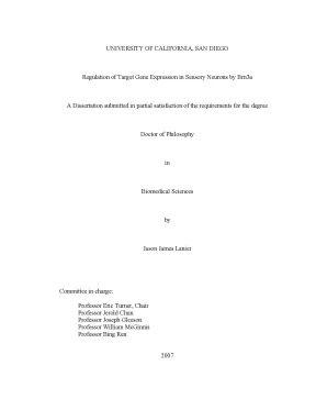
Tải xuống để đọc toàn bộUNIVERSITY OF CALIFORNIA, SAN DIEGO Regulation of Target Gene Expression in Sensory Neurons by Brn3a A Dissertation submitted in partial satisfaction of the requirements for the degree Doctor of Philosophy in Biomedical Sciences by Jason James Lanier Committee in charge: Professor Eric Turner, Chair Professor Jerold Chun Professor Joseph Gleeson Professor William McGinnis Professor Bing Ren 2007 UMI Number: 3246882 UMI Microform 3246882 Copyright 2007 by ProQuest Information and Learning Company. All rights reserved. This microform edition is protected against unauthorized copying under Title 17, United States Code. ProQuest Information and Learning Company 300 North Zeeb Road P.
Box 1346 Ann Arbor, MI 48106-1346 Copyright Jason James Lanier, 2007 All rights reserved The Dissertation of Jason James Lanier is approved, and it is acceptable in quality and form for publication on microfilm. _________________________________________________________ _________________________________________________________ _________________________________________________________ _________________________________________________________ _________________________________________________________ Chair UNIVERSITY OF CALIFORNIA, SAN DIEGO 2007 iii TABLE OF CONTENTS Signature page. iii Table of contents. iv List of Figures.
vi List of Tables. 1 Induction and development of the nervous system. 1 Tissue-specific transcription factors in neural development. 3 Mechanisms of transcriptional regulation.
5 Transcription factor target gene specificity. 7 Epigenetic influence on transcription factor binding. 10 Brn3a is a critical regulator of sensory neural development. Coordinated regulation of gene expression by Brn3a in the developing trigeminal ganglion.
24 Materials and methods. Brn3a target gene recognition in embryonic sensory neurons. 68 Materials and methods. POU-domain factor Brn3a regulates both distinct and common programs of gene expression in the spinal and trigeminal sensory ganglia.
133 Materials and methods. 166 Programs of gene regulation in the nervous system. 166 Direct vs indirect regulation. 167 Context-dependent binding and activity of transcription factors.
169 Cell-specific functions of transcription factors. 175 v LIST OF FIGURES Chapter 2 Figure 2.1 Expression array analysis of E13.2 Brn3a regulates sensory neurotransmitter systems.3 Coordinated regulation of transcription factor expression in sensory ganglia by Brn3a.4 Cellular expression of Brn3a target genes in the CNS.1 Cellular expression of Brn3a in the embryonic trigeminal ganglion. Brn3a is a direct regulator of the NeuroD4 gene in the embryonic trigeminal ganglion.3 ChIP analysis of Brn3a binding to the NeuroD1 and Msc loci in E13.4 Brn3a binding to the Pou4f1 locus in vitro and in embryonic trigeminal ganglia.5 Markers of chromatin modification at the Pou4f1 locus.6 Acetylated histone H3 ChIP assays of the NeuroD4 and NeuroD1 loci.7 Analysis of Brn3a binding sites identified in the promoters of genes not expressed in the developing trigeminal ganglion. 112 vi Chapter 4 Figure 4.1 Analysis of global gene expression in embryonic neural tissue.2 Selective expression of proprioceptor markers in the DRG and trigeminal system.3 Target genes with increased expression in the DRG of Brn3a null mice.4 Target genes with decreased expression in the DRG of Brn3a null mice.5 Brn3a regulation of Hox gene expression in the DRG.6 Trigeminal-specific targets of Brn3a regulation.7 Acetyl-histone H3 profiling of Brn3a target gene loci.
154 vii LIST OF TABLES Chapter 2 Table 2.1 Increased transcripts in the trigeminal ganglion of Brn3a mutant mouse embryos.2 Decreased transcripts in the trigeminal ganglion of Brn3a mutant mouse embryos.3 Significantly changed ESTs in Brn3a mutant mice.4 Relative expression of previously reported Brn3a target genes in the trigeminal ganglia of Brn3a mutant embryos.1 Transcripts differentially expressed in the DRG and TG.2 Increased transcripts in the DRG of Brn3a knockout mice.3 Transcripts decreased in the DRG of Brn3a null mice. 159 viii ACKNOWLEDGEMENT First of all, I would like to thank my advisor, Dr. Eric Turner for being an exceptional mentor and devoting enormous amounts of time and effort to my training. Thanks for all of your guidance and patience and for always having my best interest at heart.
I would also like to thank the members of the Turner lab that I have had the privilege to work with: Natasha, Raisa, Lely, Eric Cox, Iain, and Amanda. You have all helped to create a great working environment in the lab. I have enjoyed getting to know all of you and appreciate your enormous contributions to my thesis project. Finally, I would like to thank the members of my thesis committee for helpful comments and advice along the way.
The text in Chapter Two is a reprint of the material as it appears in Development, 2003, Eng SR, Lanier J, Fedtsova N, Turner EE. I was a secondary author and participated in the research that forms the basis of this chapter. The text of Chapter Three is a reprint of the material as it appears in Developmental Biology, 2007, Lanier J, Quina LA, Eng SR, Cox E, Turner EE. I was the primary researcher, and the co-authors contributed to the research that forms the basis of this chapter.
The text of Chapter Four is a reprint of the material as it appears in Neural Development, 2007, Eng SR, Dykes I, Lanier J, Fedtsova N, and Turner EE. I was a secondary author and participated in the research that forms the basis of this chapter. ix VITA EDUCATION Ph., Biomedical Sciences University of California San Diego School of Medicine 2007 B., Biological Chemistry, cum laude Tulane University 2001 RESEARCH EXPERIENCE Eric Turner Lab, Department of Psychiatry University of California San Diego, La Jolla, CA 2001-Present Larry Byers Lab, Department of Chemistry Tulane University, New Orleans, LA 2000-2001 Tom Wilke Lab, Department of Pharmacology University of Texas Southwestern Medical Center, Dallas, TX 2000 TEACHING EXPERIENCE Salk Institute Mobile Science Lab 2002-2003 General Chemistry Laboratory Course, Tulane University 2000-2001 HONORS UT Southwestern Medical Center Summer Undergraduate Research Fellowship 2000 Reuben Zarilli Memorial Scholarship for Academic Achievement 2000 Conference USA Commissioner’s Academic Medal- Tulane Football 1998 x PUBLICATIONS Eng SR, Dykes I, Lanier J, Fedtsova N, Turner EE (2007). “POU-domain factor Brn3a regulates both distinct and common programs of gene expression in the spinal and trigeminal sensory ganglia.
Lanier J, Quina LA, Eng SR, Cox E, Turner EE (2007). “Brn3a target gene recognition in embryonic sensory neurons.” Developmental Biology, 302(2) pp. Cox E, Lanier J, Quina L, Eng SR, Turner EE (2006) “Regulation of FGF10 by POU transcription factor Brn3a in the developing trigeminal ganglion.” Journal of Neurobiology, 66(10):1075-83. Quina LA, Pak W, Lanier J, Banwait P, Gratwick K, Liu Y, Velasquez T, O’Leary DD, Goulding M, Turner EE (2005).
“Brn3a-expressing retinal ganglion cells project specifically to thalamocortical and collicular visual pathways.” Journal of Neuroscience, 25(50):11595-604. Eng SR, Lanier J, Fedtsova N, Turner EE (2004). “Coordinated regulation of gene expression by Brn3a in developing sensory ganglia. ABSTRACTS Lanier J, Quina L, Turner EE (2006).
“Tissue-specific regulation of gene expression by Brn3a.” Keystone Symposium- Regulation of Eukaryotic Transcription: From Chromatin to mRNA, Taos, NM. Lanier J, Quina L, Cox E, Turner EE (2005). “Locus-wide ChIP analysis of the transcriptional targets of Brn3a in the sensory ganglia.” Society for Neuroscience Annual Meeting, Washington, DC. Quina L, Lanier J, Eng S, Banwait P, Liu Y, Goulding M, Turner EE (2004).
“Role of Brn3a in CNS axon guidance.” Society for Neuroscience Annual Meeting, San Diego, CA. Lanier J, Eng SR, Fedtsova N, Turner EE (2003). “Transcriptional targets of Brn3a in the sensory ganglia.” Society for Neuroscience Annual Meeting, New Orleans, LA. xi ABSTRACT OF THE DISSERTATION Regulation of Target Gene Expression in Sensory Neurons by Brn3a by Jason James Lanier Doctor of Philosophy in Biomedical Sciences University of California, San Diego, 2007 Professor Eric Turner, Chair During the development of the vertebrate nervous system, cellular proliferation and differentiation result in the formation of a large number of specialized physical structures composed of many different types of cells.
The phenotypic properties of a cell are largely controlled by the complement of proteins that the cell expresses. Thus, the formation of properly functioning neuronal circuitry requires precisely coordinated regulation of gene expression. Tissue-specific transcription factors play a critical role in the process of development by exerting spatiotemporal control over the expression of their specific target genes. Hundreds of transcription factors have been identified xii which exhibit tissue-specific expression in the nervous system.
Many of these factors are critical for proper neural development, with their disruption leading to severe phenotypic abnormalities. Brn3a (Pou4f1) is a POU domain transcription factor with a highly specific and complex pattern of expression in the developing nervous system. Disruption of Brn3a function results in severe developmental abnormalities and neonatal lethality. We have identified programs of gene expression controlled by Brn3a in both the trigeminal ganglia (TG) and dorsal root ganglia (DRG) using microarray analysis of dissected tissue from wild-type and Brn3a null embryos in midgestation.
These experiments indicate that Brn3a regulates similar, but distinct complements of transcripts in sensory ganglia at different axial levels. We then show, using in vivo chromatin immunoprecipitation (ChIP) assays, that Brn3a directly represses the expression of the neurogenic transcription factors NeuroD1 and NeuroD4 in the developing TG. Finally, we provide evidence that epigenetic modifications of chromatin play an integral role in determining the regulatory targets of Brn3a and contribute to target gene differences between the TG and DRG. The work presented in this dissertation has provided insight into the role of Brn3a in the developing sensory nervous system.
It also demonstrates a clear role for chromatin modification in transcription factor target gene selection which may be relevant for many tissue- specific transcription factors. xiii I Introduction Induction and development of the nervous system The induction and development of the vertebrate nervous system comprise an extraordinarily complex series of events resulting in the generation of a multitude of distinct cell types and structures (Kandel, Schwartz et al. During early mammalian embryonic development, prior to the induction of the nervous system, the embryo consists of three main cell layers. The innermost cell layer, the endoderm, consists of cells that eventually give rise to the gut and internal organs.
The vascular system, musculature, and connective tissues are derived from the middle cell layer, the mesoderm. The major tissues of the central and peripheral nervous systems are generated from the ectoderm, located on the surface of the early embryo. At the gastrula stage of development, a sheet of cells located at the dorsal midline of the embryo, begins to acquire neural properties and forms a structure called the neural plate, which is the source of both neural and glial cells. In a process called neurulation, the neural plate folds into a structure called the neural tube, which eventually gives rise to the central nervous system.
The adult spinal cord and brain are derived from the posterior and anterior regions of the neural tube, respectively (Schoenwolf, Bortier et al. 1989; Eagleson and Harris 1990). 1 2 Many of the cells comprising the peripheral sensory nervous system are derived from a group of migratory cells called the neural crest (Selleck and Bronner- Fraser 1996). Prior to neurulation, precursors of this specialized group of cells can be identified at the border of the neural plate and the non-neural ectoderm (Huang and Saint-Jeannet 2004).
Around the time of neural tube closure, neural crest cells migrate ventrally throughout the embryo, differentiating into a wide range of both neuronal and nonneuronal cells, including a majority of sensory neural progenitors. The remainder of the cells in the peripheral sensory nervous system originate in ectodermal cellular structures called neurogenic placodes (Barlow 2002). The patterning and differentiation of cells in the nervous system is ultimately controlled by signaling molecules called inducing factors which are secreted from cells in a particular location within an embryo and influence the physiology of surrounding cells. Secretion of an inducing factor from a localized region within an embryo creates a signaling gradient which can determine the arrangement and fate of responding cells according to the concentration of the factor perceived by each cell (Gurdon and Bourillot 2001).
Cells occupying different positions within a developing embryo are exposed to different inducing factors. Thus the position that a cell occupies early in development has a direct influence over its ultimate fate.
Nội dung được bảo vệ bản quyền — Tải xuống đầy đủ
Trích dẫn luận án này
Jason James Lanier (2007). Brn3a điều hòa biểu hiện gen trong nơron cảm giác [Luận án tiến sĩ, University of California, San Diego]. LuanAn.net. https://luanan.net/sinh-hoc/sinh-hoc-phan-tu-te-bao/brn3a-dieu-hoa-bieu-hien-gen-neuron-cam-giac
Câu hỏi thường gặp
Luận án "Brn3a điều hòa biểu hiện gen trong nơron cảm giác" nghiên cứu về vấn đề gì?
Luận án tiến sĩ nghiên cứu cơ chế điều hòa biểu hiện gen trong nơron cảm giác qua protein Brn3a và vai trò yếu tố phiên mã trong phát triển thần kinh.
Luận án "Brn3a điều hòa biểu hiện gen trong nơron cảm giác" được bảo vệ tại trường nào?
Luận án này được bảo vệ tại University of California, San Diego. Năm bảo vệ: 2007.
Luận án "Brn3a điều hòa biểu hiện gen trong nơron cảm giác" thuộc chuyên ngành gì?
Luận án "Brn3a điều hòa biểu hiện gen trong nơron cảm giác" thuộc chuyên ngành Biomedical Sciences. Danh mục: Sinh Học Phân Tử Tế Bào.
Luận án "Brn3a điều hòa biểu hiện gen trong nơron cảm giác" có bao nhiêu trang?
Luận án "Brn3a điều hòa biểu hiện gen trong nơron cảm giác" có 190 trang. Bạn có thể xem trước một phần tài liệu ngay trên trang web trước khi tải về.
Cách tải luận án "Brn3a điều hòa biểu hiện gen trong nơron cảm giác" về máy như thế nào?
Để tải luận án về máy, bạn nhấn nút "Tải xuống ngay" trên trang này, sau đó hoàn tất thanh toán phí lưu trữ. File sẽ được tải xuống ngay sau khi thanh toán thành công. Hỗ trợ qua Zalo: 0559 297 239.