Điều hòa sinh học macrophage và đáp ứng miễn dịch bởi lipid mycobacterium
Luận án tiến sĩ nghiên cứu vai trò glycopeptidolipids trong điều hòa macrophage và miễn dịch. Khám phá cơ chế vận chuyển lipid mycobacterium qua exosomes đến tế bào lân cận.
University of Notre Dame
Biological Sciences
Luan An
Dissertation
Năm xuất bản
Số trang
190
Thời gian đọc
29 phút
Lượt xem
1
Lượt tải
0
Phí lưu trữ
50 Point
Tổng quan nhanh
- Chủ đề:
- 1. Lipid Mycobacterium Điều Hòa Macrophage Như Thế Nào
- Số trang:
- 190 trang
- Trường:
- University of Notre Dame
- Chuyên ngành:
- Biological Sciences
- Tác giả:
- Sanchita Bhatnagar
- Năm:
- 2006
Tóm tắt nội dung luận án
I. Lipid Mycobacterium Điều Hòa Macrophage Như Thế Nào
Vi khuẩn mycobacterium gây bệnh lao và nhiều bệnh nguy hiểm khác. Chúng sống bên trong tế bào macrophage - loại tế bào miễn dịch chủ chốt. Lipid trên bề mặt mycobacterium đóng vai trò quyết định trong việc điều hòa chức năng macrophage. Các nghiên cứu cho thấy mycobacterial lipids có khả năng thay đổi hoàn toàn cách macrophage phản ứng với nhiễm trùng. Glycopeptidolipid (GPL) là một loại glycolipid quan trọng trên bề mặt vi khuẩn. GPL có tính kháng nguyên cao và ảnh hưởng mạnh đến hệ miễn dịch숙주. Khi thiếu GPL hoặc cấu trúc GPL bị thay đổi, vi khuẩn M. avium kích hoạt macrophage mạnh hơn. Điều này dẫn đến giảm độc lực của vi khuẩn trong mô hình chuột. Lipoarabinomannan (LAM) là một glycolipid khác ảnh hưởng đến macrophage polarization. Trehalose dimycolate (TDM), còn gọi là cord factor, kích thích phản ứng viêm mạnh. Phthiocerol dimycocerosate giúp vi khuẩn trốn tránh hệ miễn dịch. Các lipid này tương tác với nhiều receptor trên macrophage, thay đổi con đường tín hiệu nội bào.
1.1. Cơ Chế Tác Động Của Glycopeptidolipid
GPL là yếu tố độc lực quan trọng của M. avium. Các chủng vi khuẩn thiếu GPL tự nhiên hoặc bị biến đổi gen cho thấy khả năng gây bệnh giảm. GPL ảnh hưởng đến việc kích hoạt macrophage thông qua nhiều con đường. Khi GPL hiện diện đầy đủ, macrophage ít được kích hoạt hơn. Điều này giúp vi khuẩn sống sót tốt hơn trong tế bào chủ. Nghiên cứu sử dụng các chủng M. avium đột biến gen mtfD. Gen này mã hóa enzyme methyltransferase cần thiết cho tổng hợp GPL. Chủng đột biến không có GPL đặc hiệu theo serotype. Macrophage nhiễm chủng đột biến sản xuất cytokine viêm nhiều hơn.
1.2. GPL Điều Chỉnh Phản Ứng Miễn Dịch
GPL điều chỉnh phản ứng miễn dịch của숙주ở nhiều mức độ. Trong mô hình chuột, chủng thiếu GPL có độc lực giảm rõ rệt. Chuột nhiễm chủng đột biến sống sót tốt hơn và có ít tổn thương mô hơn. GPL ức chế con đường tín hiệu MAP kinase trong macrophage. MAP kinase signaling quan trọng cho sản xuất cytokine viêm. Khi GPL vắng mặt, MAP kinase được kích hoạt mạnh hơn. Điều này dẫn đến tăng sản xuất TNF-α, IL-6 và các cytokine khác. GPL cũng ảnh hưởng đến macrophage polarization, ngăn chặn sự chuyển đổi sang kiểu M1 macrophage có hoạt tính diệt khuẩn cao.
1.3. Vận Chuyển GPL Trong Macrophage
GPL được giải phóng từ phagosome chứa mycobacterium. Lipid này di chuyển đến các ngăn endocytic khác nhau trong tế bào. Multivesicular bodies (MVB) là điểm đến quan trọng của GPL. MVB chứa các túi nhỏ gọi là intraluminal vesicles. Các túi này sau đó được giải phóng ra ngoài tế bào dưới dạng exosome. Exosome mang theo GPL và các thành phần mycobacterial khác. Quá trình này cho phép truyền tín hiệu giữa các tế bào. Tế bào lân cận không bị nhiễm có thể nhận exosome chứa mycobacterial lipids. Đây là cơ chế mới của giao tiếp giữa các tế bào trong nhiễm trùng mycobacterium.
II. M1 Macrophage Và M2 Macrophage Trong Nhiễm Khuẩn
Macrophage có thể phân cực thành hai kiểu chính: M1 và M2. M1 macrophage có hoạt tính diệt khuẩn cao và sản xuất cytokine viêm. M2 macrophage liên quan đến quá trình sửa chữa mô và ức chế viêm. Mycobacterial lipids ảnh hưởng mạnh đến macrophage polarization. GPL và các lipid khác thường đẩy macrophage về kiểu M2. Điều này có lợi cho sự sống sót của vi khuẩn. M1 macrophage sản xuất reactive oxygen species và nitric oxide. Các phân tử này có khả năng tiêu diệt mycobacterium. M2 macrophage sản xuất cytokine chống viêm như IL-10. Chúng cũng biểu hiện arginase, cạnh tranh với iNOS về substrate. Sự cân bằng giữa M1 và M2 quyết định kết cục nhiễm trùng. Lipoarabinomannan từ M. tuberculosis có thể ức chế M1 polarization. Trehalose dimycolate ngược lại thường kích thích phản ứng kiểu M1.
2.1. Đặc Điểm M1 Macrophage
M1 macrophage được kích hoạt bởi IFN-γ và LPS. Chúng biểu hiện cao iNOS và sản xuất NO. M1 macrophage tiết nhiều TNF-α, IL-1β, IL-6, IL-12. Các cytokine này thúc đẩy phản ứng viêm và miễn dịch Th1. M1 macrophage có khả năng trình diện kháng nguyên tốt. Chúng biểu hiện cao MHC class II và các phân tử đồng kích thích. M1 macrophage tiêu diệt vi khuẩn nội bào hiệu quả. Tuy nhiên, hoạt động quá mức có thể gây tổn thương mô. GPL-deficient mycobacterium kích thích M1 polarization mạnh hơn. Điều này giải thích tại sao chúng có độc lực thấp hơn.
2.2. Đặc Điểm M2 Macrophage
M2 macrophage được kích hoạt bởi IL-4, IL-13, IL-10. Chúng biểu hiện cao arginase-1 và các marker khác. M2 macrophage sản xuất cytokine chống viêm. Chúng tiết IL-10, TGF-β, giúp kiểm soát viêm. M2 macrophage thúc đẩy sửa chữa mô và tái tạo. Chúng sản xuất các yếu tố tăng trưởng và protein ngoại bào. M2 macrophage có hoạt tính diệt khuẩn thấp. Mycobacterium tận dụng điều này để sống sót lâu dài. GPL và LAM thúc đẩy M2 polarization. Điều này tạo môi trường thuận lợi cho vi khuẩn phát triển.
2.3. Chuyển Đổi Giữa M1 Và M2
Macrophage có tính linh hoạt cao và có thể chuyển đổi kiểu. Chuyển đổi từ M1 sang M2 xảy ra khi viêm giảm dần. Ngược lại, M2 có thể chuyển sang M1 khi gặp tín hiệu viêm mới. Mycobacterial lipids can manipulate this plasticity. GPL ngăn chặn chuyển đổi sang M1 ngay cả khi có IFN-γ. Phthiocerol dimycocerosate cũng ức chế M1 activation. Cord factor (TDM) là ngoại lệ, thường kích thích M1. Sự cân bằng giữa các lipid khác nhau quyết định kết quả cuối cùng. Hiểu được cơ chế này mở ra hướng điều trị mới.
III. Lipoarabinomannan LAM Và Điều Hòa Miễn Dịch
Lipoarabinomannan là glycolipid phức tạp trên thành tế bào mycobacterium. LAM có cấu trúc gồm ba phần: lipid anchor, mannan backbone, và arabinan domain. Phần arabinan có thể được gắn thêm các nhóm mannose (ManLAM) hoặc phosphoinositol (PILAM). ManLAM từ M. tuberculosis có hoạt tính miễn dịch đặc biệt. LAM tương tác với nhiều receptor trên macrophage. Mannose receptor, DC-SIGN, TLR2 đều nhận diện LAM. Tùy thuộc vào receptor, LAM có thể kích thích hoặc ức chế miễn dịch. ManLAM thường ức chế phản ứng viêm và M1 activation. PILAM từ các loài không gây bệnh thường kích thích viêm nhiều hơn. LAM ảnh hưởng đến phagosome maturation trong macrophage. Nó ngăn chặn sự hợp nhất giữa phagosome và lysosome. Điều này giúp mycobacterium tránh bị tiêu diệt. LAM cũng điều chỉnh sản xuất cytokine và chemokine.
3.1. Cấu Trúc Và Phân Loại LAM
LAM có trọng lượng phân tử khoảng 17-20 kDa. Phần lipid anchor gắn vào màng tế bào mycobacterium. Mannan backbone tạo thành cột sống của phân tử. Arabinan chains gắn vào mannan tạo cấu trúc phân nhánh. ManLAM có mannose caps ở đầu arabinan chains. PILAM có phosphoinositol caps thay vì mannose. AraLAM không có caps, chỉ có arabinan tự do. Loại caps quyết định hoạt tính sinh học của LAM. ManLAM từ M. tuberculosis và M. avium rất quan trọng. Chúng có khả năng ức chế miễn dịch mạnh. PILAM từ M. smegmatis ít ức chế miễn dịch hơn.
3.2. LAM Tương Tác Với Receptor
Mannose receptor (MR) nhận diện mannose caps trên ManLAM. Tương tác này thường dẫn đến ức chế miễn dịch. DC-SIGN cũng liên kết với ManLAM qua mannose residues. DC-SIGN signaling có thể ức chế IL-12 và thúc đẩy IL-10. TLR2 nhận diện phần lipid và một số vùng trên LAM. TLR2 activation thường kích thích phản ứng viêm. Complement receptor 3 (CR3) cũng tương tác với LAM. CR3-mediated uptake không kích hoạt macrophage mạnh. Sự cạnh tranh giữa các receptor quyết định kết quả cuối cùng. Tỷ lệ các receptor trên macrophage ảnh hưởng đến phản ứng.
3.3. LAM Ức Chế Phagosome Maturation
Phagosome maturation là quá trình quan trọng để tiêu diệt vi khuẩn. Phagosome thường hợp nhất với lysosome tạo phagolysosome. Phagolysosome có pH thấp và nhiều enzyme tiêu hóa. ManLAM ngăn chặn quá trình maturation này. LAM ức chế recruitment của EEA1 lên phagosome. EEA1 cần thiết cho fusion giữa các endosome. LAM cũng ngăn chặn acquisition của lysosomal markers. V-ATPase và cathepsin D ít được recruit đến phagosome chứa mycobacterium. Cơ chế liên quan đến can thiệp vào calcium signaling. LAM cũng ảnh hưởng đến phosphoinositide metabolism trên phagosome membrane.
IV. Trehalose Dimycolate TDM Cord Factor Độc Lực
Trehalose dimycolate là glycolipid quan trọng nhất trên thành tế bào mycobacterium. TDM còn được gọi là cord factor vì gây hiện tượng vi khuẩn mọc thành dây. TDM chiếm khoảng 10-15% tổng lipid trên bề mặt M. tuberculosis. Phân tử gồm hai mycolic acid gắn với disaccharide trehalose. TDM có hoạt tính sinh học mạnh và đa dạng. Nó kích thích phản ứng viêm cấp tính trong động vật. TDM gây tổn thương phổi và hình thành granuloma. Cord factor được nhận diện bởi receptor Mincle trên macrophage. Mincle là C-type lectin receptor kích hoạt Syk-Card9 pathway. TDM activation dẫn đến sản xuất cytokine viêm mạnh. TNF-α, IL-1β, IL-6 được tiết ra nhiều. TDM cũng kích hoạt NLRP3 inflammasome trong macrophage. Điều này dẫn đến xử lý và tiết IL-1β trưởng thành.
4.1. Cấu Trúc Và Tính Chất TDM
TDM có cấu trúc đối xứng với hai mycolic acid chains. Mỗi mycolic acid dài 60-90 carbon atoms. Mycolic acids gắn vào hai vị trí hydroxyl của trehalose. Trehalose là disaccharide gồm hai glucose liên kết α,α-1,1. TDM có tính kỵ nước cao do mycolic acid chains dài. Phân tử tập hợp thành micelles hoặc bilayers trong nước. TDM tồn tại ở lớp ngoài cùng của thành tế bào. Một phần TDM được giải phóng ra môi trường xung quanh. TDM tự do có thể xâm nhập vào màng tế bào chủ. Điều này ảnh hưởng đến chức năng màng và signaling.
4.2. TDM Kích Hoạt Mincle Receptor
Mincle (Macrophage-inducible C-type lectin) là receptor chính của TDM. Mincle nhận diện phần trehalose của TDM. Receptor này cặp với FcRγ chain để truyền tín hiệu. Mincle-FcRγ activation kích hoạt Syk kinase. Syk phosphorylates Card9, tạo phức hợp Card9-Bcl10-Malt1. Phức hợp này kích hoạt NF-κB và MAP kinases. NF-κB translocation vào nhân kích thích phiên mã cytokine. TNF-α, IL-6, IL-1β genes được bật lên. Mincle expression tăng lên sau khi tiếp xúc với TDM. Đây là vòng phản hồi dương tăng cường phản ứng viêm.
4.3. TDM Và Hình Thành Granuloma
Granuloma là cấu trúc mô đặc trưng của nhiễm mycobacterium. TDM đóng vai trò quan trọng trong quá trình hình thành granuloma. TDM kích thích recruitment của macrophage và các tế bào miễn dịch. Cytokine và chemokine được sản xuất thu hút thêm tế bào. Macrophage tập hợp và biệt hóa thành epithelioid cells. Các tế bào này hợp nhất tạo giant cells đa nhân. Lymphocytes tạo vòng ngoài quanh khối macrophage. TDM cũng kích thích fibroblast sản xuất collagen. Collagen bao quanh granuloma tạo vỏ xơ. Granuloma giới hạn lan rộng của vi khuẩn nhưng cũng bảo vệ chúng khỏi miễn dịch.
V. Phthiocerol Dimycocerosate Và Thoát Miễn Dịch
Phthiocerol dimycocerosate (PDIM) là lipid độc lực quan trọng của mycobacterium gây bệnh. PDIM có cấu trúc phức tạp với phthiocerol backbone và mycocerosic acid chains. Lipid này chỉ có ở các loài mycobacterium gây bệnh mạnh. M. tuberculosis, M. bovis, M. leprae đều chứa PDIM. Các loài không gây bệnh như M. smegmatis thiếu PDIM. PDIM đóng vai trò trong giai đoạn đầu nhiễm trùng. Nó giúp mycobacterium xâm nhập vào macrophage hiệu quả hơn. PDIM can thiệp vào phagosome membrane integrity. Lipid này gây rò rỉ phagosome membrane nhẹ. Điều này cho phép một số thành phần mycobacterial tiếp cận cytosol. PDIM cũng ức chế sản xuất cytokine viêm. Macrophage nhiễm vi khuẩn có PDIM sản xuất ít TNF-α và IL-12 hơn. PDIM ngăn chặn apoptosis của macrophage bị nhiễm.
5.1. Sinh Tổng Hợp PDIM
PDIM được tổng hợp bởi các gen trong pps và mas loci. Pps genes mã hóa polyketide synthases tạo phthiocerol. Mas genes mã hóa mycocerosic acid synthases. FadD28 và FadD26 là acyl-AMP ligases cần thiết. PapA5 là acyltransferase gắn mycocerosic acids vào phthiocerol. Đột biến bất kỳ gen nào cũng dẫn đến mất PDIM. Các chủng đột biến này có độc lực giảm rõ rệt. Trong mô hình chuột, chúng không thể nhân lên tốt trong phổi. PDIM synthesis được điều hòa bởi nhiều yếu tố. PhoP-PhoR two-component system điều chỉnh pps expression.
5.2. PDIM Và Xâm Nhập Tế Bào
PDIM tăng cường khả năng xâm nhập macrophage của mycobacterium. Các chủng thiếu PDIM xâm nhập kém hơn đáng kể. PDIM tương tác với màng tế bào chủ trong quá trình phagocytosis. Lipid này có thể thay đổi cấu trúc màng locally. PDIM làm tăng membrane fluidity tại vị trí tiếp xúc. Điều này tạo điều kiện cho vi khuẩn xâm nhập dễ dàng hơn. PDIM cũng ảnh hưởng đến receptor-mediated uptake. Nó có thể che phủ các PAMP khác trên bề mặt vi khuẩn. Điều này giảm nhận diện bởi pattern recognition receptors. Kết quả là macrophage activation thấp hơn ngay từ đầu.
5.3. PDIM Gây Tổn Thương Phagosome
PDIM can thiệp vào tính toàn vẹn của phagosome membrane. Lipid này chèn vào lipid bilayer của phagosome. PDIM gây rối loạn cấu trúc màng cục bộ. Điều này tạo ra các vùng thấm nhẹ trên phagosome. Một số phân tử nhỏ có thể rò rỉ qua các vùng này. Mycobacterial components như DNA có thể tiếp cận cytosol. Cytosolic DNA sensors như cGAS-STING được kích hoạt. Tuy nhiên, PDIM cũng ức chế downstream signaling. Kết quả cuối cùng là phản ứng interferon bị kiểm soát. PDIM không gây vỡ hoàn toàn phagosome như Listeria. Thay vào đó, nó tạo môi trường trung gian thuận lợi cho vi khuẩn.
VI. Exosome Vận Chuyển Mycobacterial Lipids
Exosome là các túi nhỏ ngoại bào có đường kính 30-100 nm. Chúng được hình thành trong multivesicular bodies (MVB) của tế bào. MVB chứa intraluminal vesicles (ILV) hình thành bằng cách màng MVB uốn vào trong. Khi MVB hợp nhất với màng tế bào, ILV được giải phóng thành exosome. Macrophage bị nhiễm mycobacterium tiết exosome chứa mycobacterial components. GPL, LAM và các lipid khác được tìm thấy trong exosome này. Exosome vận chuyển các lipid này đến tế bào lân cận. Tế bào không bị nhiễm có thể nhận và phản ứng với exosome. Đây là cơ chế giao tiếp intercellular mới trong nhiễm trùng. Exosome chứa mycobacterial lipids kích thích phản ứng viêm. Macrophage nhận exosome sản xuất TNF-α, IL-6, IL-12. Quá trình này xảy ra cả in vitro và in vivo. Exosome có thể lan truyền tín hiệu xa hơn vị trí nhiễm trùng.
6.1. Sinh Tạo Exosome Trong Macrophage
Exosome biogenesis bắt đầu từ early endosome. Early endosome trưởng thành thành late endosome/MVB. Màng MVB uốn vào trong tạo intraluminal vesicles. ESCRT machinery (Endosomal Sorting Complex Required for Transport) điều khiển quá trình này. ESCRT-0, I, II, III complexes hoạt động tuần tự. Các protein và lipid được chọn lọc vào ILV. Tetraspanins như CD63, CD81, CD9 phong phú trong exosome. MVB có thể hợp nhất với lysosome để degradation. Hoặc MVB hợp nhất với plasma membrane để giải phóng exosome. Rab GTPases điều khiển trafficking của MVB. Rab27a và Rab27b quan trọng cho exosome secretion.
6.2. Mycobacterial Lipids Trong Exosome
Nghiên cứu cho thấy GPL được vận chuyển từ phagosome đến MVB. Imaging cho thấy GPL colocalize với CD63-positive MVB. LAM cũng được tìm thấy trong exosome từ macrophage bị nhiễm. Lipidomic analysis xác nhận sự hiện diện của mycolic acids. Các lipid này không chỉ là contamination từ vi khuẩn chết. Chúng được đóng gói có chọn lọc vào exosome. Cơ chế sorting mycobacterial lipids vào exosome chưa rõ hoàn toàn. Có thể liên quan đến lipid rafts trên MVB membrane. Exosome từ macrophage nhiễm M. tuberculosis và M. bovis BCG đều chứa lipid. Lượng lipid trong exosome tương quan với bacterial load.
6.3. Exosome Kích Hoạt Bystander Cells
Exosome chứa mycobacterial components kích hoạt macrophage chưa bị nhiễm. Bystander macrophages sản xuất cytokine viêm khi nhận exosome. TNF-α, IL-6, IL-12 được tiết ra trong vài giờ. Exosome có thể kích hoạt NF-κB và MAP kinase pathways. TLR2 có thể nhận diện mycobacterial lipids trên exosome. Exosome cũng được dendritic cells và các tế bào khác nhận. Trong mô hình chuột, exosome inject vào phổi gây viêm. Recruitment của neutrophil và macrophage tăng lên. Exosome có thể lan truyền immune activation xa vị trí nhiễm trùng. Đây là con dao hai lưỡi: giúp kiểm soát nhiễm trùng nhưng cũng gây tổn thương mô.
Mục lục chi tiết luận án
Tải xuống file đầy đủ để xem toàn bộ nội dung
Tải đầy đủ (190 trang)Nội dung chính
Tổng quan về luận án
Luận án này tiến hành một nghiên cứu tiên phong trong lĩnh vực sinh học phân tử và miễn dịch học vi khuẩn, tập trung vào cơ chế điều hòa miễn dịch của lipid mycobacterial. Nghiên cứu giải quyết mối đe dọa sức khỏe nghiêm trọng do các thành viên của chi Mycobacterium gây ra, vốn duy trì lối sống nội bào trong đại thực bào, tế bào chủ chính. Trong khi các nghiên cứu trước đây đã chỉ ra rằng các thành phần bề mặt mycobacterial khác nhau có thể thúc đẩy kích hoạt đại thực bào, vai trò cụ thể của glycopeptidolipids (GPL) như một yếu tố độc lực và cơ chế điều hòa miễn dịch của chúng vẫn còn chưa được làm rõ đầy đủ, đặc biệt là trong bối cảnh toàn bộ vi khuẩn.
Nghiên cứu này xác định một research gap cụ thể: "Most studies published prior to our work have used purified mycobacterial GPLs for illustrating the affect of GPL on host immune response. This introduces complexity in interpreting the data regarding the GPL-receptor interaction at the cell surface and uptake of GPL" (Bhatnagar, 2006, tr. 13-14). Luận án của Bhatnagar lấp đầy khoảng trống này bằng cách "defining the role of GPL in the context of whole mycobacteria" (Bhatnagar, 2006, tr. 14). Hơn nữa, mặc dù các MVB và ngoại bào (exosomes) đã được nhận diện, vai trò của chúng trong giao tiếp giữa các tế bào trong bối cảnh nhiễm trùng mycobacteria vẫn còn thiếu bằng chứng in vivo, với các nghiên cứu trước đây chủ yếu thực hiện in vitro (Bhatnagar, 2006, tr. 30).
Nghiên cứu được định hướng bởi các câu hỏi và giả thuyết chính sau:
- Vai trò của GPL như một yếu tố độc lực trong M. avium là gì?
- Việc mất hoặc thay đổi cấu trúc GPL ảnh hưởng như thế nào đến sự kích hoạt đại thực bào và độc lực in vivo?
- Cơ chế di chuyển của GPL trong các đại thực bào bị nhiễm M. avium như thế nào?
- GPL được vận chuyển đến các ngăn tế bào cụ thể nào?
- Các thành phần mycobacterial, bao gồm GPL, được vận chuyển ra bên ngoài tế bào bị nhiễm thông qua exosomes có chức năng gì trong việc điều hòa phản ứng của các tế bào lân cận?
Khung lý thuyết của luận án được xây dựng dựa trên sự hiểu biết về cơ chế sống sót nội bào của mycobacteria, sinh học đại thực bào (ví dụ: quá trình thực bào, trưởng thành phagosome), và phản ứng miễn dịch chủ. Các lý thuyết về sự điều hòa miễn dịch thông qua các thành phần bề mặt vi khuẩn, con đường tín hiệu MAPK (Mitogen-Activated Protein Kinase) và NF-κB (Nuclear Factor kappa B), cùng với vai trò của các thể đa túi (MVB) và ngoại bào (exosomes) trong vận chuyển nội bào và giao tiếp giữa các tế bào, là trọng tâm.
Nghiên cứu đã tạo ra các đóng góp đột phá, bao gồm việc lần đầu tiên thiết lập vai trò của GPL như một yếu tố độc lực trực tiếp trong hệ thống M. avium, được chứng minh bằng việc các chủng M. avium thiếu GPL hoặc GPL biến đổi gây ra "increased macrophage activation" và cho thấy "attenuated virulence... in a mouse infection model" (Bhatnagar, 2006, Abstract). Nghiên cứu cũng phát hiện ra sự giải phóng và vận chuyển đáng kể của GPL từ phagosome chứa mycobacteria đến MVB và sau đó ra bên ngoài tế bào trên "exosomes", kích thích phản ứng tiền viêm ở các đại thực bào không bị nhiễm. Điều này định hình một "novel mechanism of intercellular communication mediated by exosomes" (Bhatnagar, 2006, Abstract). Phạm vi của nghiên cứu bao gồm việc sử dụng các chủng M. avium được biến đổi gen (ví dụ: chủng mtfD mutant) và các mô hình nhiễm trùng in vitro (tế bào đại thực bào nguyên phát từ tủy xương chuột, BMMΦs) và in vivo (mô hình chuột).
Literature Review và Positioning
Phần tổng quan tài liệu của luận án cung cấp một bản tổng hợp toàn diện các luồng nghiên cứu chính liên quan đến sinh học mycobacteria, tương tác vật chủ-mầm bệnh, và cơ chế miễn dịch. Các luồng chính bao gồm cấu trúc thành tế bào mycobacterial, đặc biệt là vai trò của lipid như lipoarabinomannan (LAM), phosphatidylinositol mannoside (PIM), và glycopeptidolipids (GPL) (Minnikin, 1991; Brennan and Nikaido, 1995); sinh học đại thực bào và các con đường xâm nhập của mycobacteria (ví dụ: thụ thể bổ thể, thụ thể mannose); lối sống nội bào của mycobacteria và cơ chế tránh trưởng thành phagosome (D’Arcy Hart et al., 1974); miễn dịch đối với mycobacteria, bao gồm miễn dịch tế bào và vai trò của các cytokine như IFN-γ và TNF-α (Mosmann and Coffman, 1989); và các cơ chế vận chuyển lipid mycobacterial cùng với con đường nội bào và ngoại bào của tế bào chủ, đặc biệt là vai trò của MVB và exosomes.
Luận án làm nổi bật các mâu thuẫn và tranh luận quan trọng trong tài liệu, đặc biệt là xung quanh vai trò của GPL. Các nghiên cứu trước đây (ví dụ: Barrow et al.; Takegaki, 2000; Vergne, Prats et al.) đã sử dụng GPL tinh khiết để minh họa ảnh hưởng của chúng đến phản ứng miễn dịch vật chủ. Tuy nhiên, Bhatnagar lập luận rằng phương pháp này "introduces complexity in interpreting the data regarding the GPL-receptor interaction at the cell surface and uptake of GPL" (Bhatnagar, 2006, tr. 13-14). Điều này tạo ra một sự đối lập rõ ràng với quan điểm cho rằng dữ liệu từ GPL tinh khiết là đủ để suy luận cơ chế in vivo.
Luận án tự định vị một cách chiến lược trong tài liệu bằng cách giải quyết cụ thể khoảng trống này. Thay vì sử dụng GPL tinh khiết, nghiên cứu "utilized M. avium strains either naturally lacking GPL expression or genetically altered to express modified GPL" (Bhatnagar, 2006, Abstract). Phương pháp tiếp cận này cung cấp một cái nhìn sinh lý học chính xác hơn về vai trò của GPL như một yếu tố độc lực. Nó tiến bộ lĩnh vực này bằng cách cung cấp bằng chứng cụ thể rằng GPL không chỉ là một thành phần hóa học mà còn là một yếu tố điều hòa miễn dịch trong bối cảnh nhiễm trùng toàn bộ vi khuẩn, ảnh hưởng đến cả phản ứng đại thực bào in vitro và độc lực in vivo.
So sánh với các nghiên cứu quốc tế, luận án này xây dựng dựa trên sự hiểu biết toàn cầu về các bệnh mycobacterial và phản ứng của vật chủ. Ví dụ, nó đề cập đến vấn đề "tuberculosis is the leading health problem, with two million deaths and nine million new cases emerging annually" và "ninety percent of the estimated nine million new cases of TB each year occur in developing countries" (Bhatnagar, 2006, tr. 2). Điều này nhấn mạnh tầm quan trọng toàn cầu của việc hiểu rõ hơn về các yếu tố độc lực mycobacterial. Nghiên cứu cũng so sánh với các nghiên cứu khác liên quan đến LAM của Chatterjees (1997) và PGLs của Reed, Domenech et al. (2004), cho thấy các lipid mycobacterial khác cũng có vai trò quan trọng. Hơn nữa, việc đề cập đến M. avium như một mầm bệnh cơ hội chính ở các bệnh nhân suy giảm miễn dịch toàn cầu, đặc biệt là những người nhiễm HIV/AIDS (Horsburgh, 1999; Abrams, 2000), khẳng định tính liên quan quốc tế của nghiên cứu.
Đóng góp lý thuyết và khung phân tích
Đóng góp cho lý thuyết
Luận án này mở rộng và thách thức trực tiếp một số lý thuyết đã được thiết lập về tương tác vật chủ-mầm bệnh và cơ chế miễn dịch.
- Mở rộng lý thuyết độc lực của mycobacteria: Nghiên cứu mở rộng lý thuyết rằng các thành phần bề mặt vi khuẩn là yếu tố độc lực bằng cách lần đầu tiên cung cấp bằng chứng trực tiếp và toàn diện về GPL. Trong khi các lý thuyết trước đây đã gợi ý vai trò của GPL dựa trên các nghiên cứu in vitro (ví dụ: Barrow et al.), luận án của Bhatnagar mở rộng hiểu biết này bằng cách chứng minh "GPL’s importance as a virulence factor in the M. avium system" (Bhatnagar, 2006, Abstract) thông qua mô hình nhiễm trùng in vivo và vi khuẩn biến đổi gen.
- Thách thức quan điểm về trưởng thành phagosome và giao tiếp tế bào: Nghiên cứu thách thức quan điểm truyền thống về MVB chủ yếu là ngăn chứa chất thải để phân hủy lysosome. Nó cung cấp bằng chứng rằng MVB hoạt động như "a platform for the accumulation and sorting of mycobacterial components" (Bhatnagar, 2006, tr. 24) và là một nguồn exosomes. Việc giải phóng exosomes mang các thành phần mycobacterial để kích hoạt các tế bào lân cận giới thiệu một "novel mechanism of intercellular communication" (Bhatnagar, 2006, Abstract) trong bối cảnh nhiễm trùng, mở rộng lý thuyết về cơ chế trốn tránh miễn dịch và lây lan mầm bệnh.
- Tích hợp con đường tín hiệu MAPK/NF-κB trong phản ứng với lipid vi khuẩn: Nghiên cứu làm sâu sắc thêm sự hiểu biết về cơ chế phân tử điều chỉnh phản ứng miễn dịch đối với mycobacteria bằng cách làm sáng tỏ các con đường tín hiệu cụ thể. Nó chỉ ra rằng "MAPK activation is differentially required for TNF-α production in macrophages infected with Rg compared to SmO and SmT M. avium 2151" và "TNF-α production is NF-κB dependent" (Bhatnagar, 2006, Chương 3). Điều này cung cấp chi tiết về cách các thành phần bề mặt vi khuẩn điều khiển các phản ứng tiền viêm thông qua các con đường tín hiệu đã biết.
Khung phân tích độc đáo
Khung phân tích của luận án tích hợp một cách độc đáo các lý thuyết từ vi sinh học, miễn dịch học và sinh học tế bào để tạo ra một cách tiếp cận mới trong nghiên cứu bệnh sinh mycobacteria. Nó tích hợp ít nhất ba lý thuyết cụ thể:
- Lý thuyết về các thụ thể nhận dạng mẫu (Pattern Recognition Receptors - PRRs) và các phân tử liên quan đến mầm bệnh (PAMPs): Nghiên cứu điều tra cách các thành phần bề mặt mycobacterial, đặc biệt là GPL, tương tác với PRRs (như các thụ thể Toll-like - TLRs) để điều hòa phản ứng miễn dịch của vật chủ. Phát hiện rằng "TNF-α production is toll-like receptor dependent" (Bhatnagar, 2006, Chương 4, 4.7 và Chương 5, 5.2) trong các tế bào được xử lý bằng exosomes nhấn mạnh sự tích hợp này.
- Lý thuyết về vận chuyển màng và định vị protein: Khung phân tích sử dụng các nguyên tắc của sinh học tế bào về vận chuyển màng để theo dõi sự di chuyển của GPL từ phagosome đến MVB và sau đó là sự giải phóng exosomes. Việc xác định rằng "Rab11 co localizes with the GPL loaded vesicles at later times" (Bhatnagar, 2006, Chương 4, Hình 4.4) minh họa sự tích hợp này.
- Lý thuyết về chức năng exosomes trong giao tiếp tế bào: Luận án mở rộng lý thuyết về exosomes như những người truyền tin tế bào (Raposo et al., 1996; Zitvogel et al., 1998) vào bối cảnh nhiễm trùng vi khuẩn, chứng minh vai trò của chúng trong việc truyền tín hiệu nguy hiểm từ tế bào bị nhiễm sang tế bào lân cận.
Phương pháp phân tích mới lạ nằm ở việc kết hợp thao tác gen của mầm bệnh, mô hình nhiễm trùng in vivo và phân tích tế bào chuyên sâu về vận chuyển lipid và con đường tín hiệu. Cách tiếp cận này cho phép định lượng trực tiếp vai trò của một yếu tố độc lực cụ thể (GPL) trong bối cảnh sinh lý học.
Đóng góp về khái niệm bao gồm:
- "Virulence factor context-dependency": Khái niệm rằng vai trò của yếu tố độc lực phải được nghiên cứu trong bối cảnh toàn bộ vi khuẩn, không chỉ các thành phần tinh khiết.
- "Exosome-mediated immune priming": Exosomes từ tế bào bị nhiễm hoạt động như một tín hiệu cảnh báo, chuẩn bị các tế bào xung quanh để kiểm soát nhiễm trùng mycobacteria.
Các điều kiện biên được nêu rõ: nghiên cứu chủ yếu tập trung vào M. avium và phản ứng của đại thực bào chuột (BMMΦs), với một số thí nghiệm sử dụng tế bào J774 và THP-1. Các phát hiện về exosome chủ yếu được xác lập in vitro, mặc dù các bằng chứng in vivo sơ bộ đã được đưa ra (ví dụ: Hình 5.5).
Phương pháp nghiên cứu tiên tiến
Thiết kế nghiên cứu
Luận án áp dụng triết lý nghiên cứu thực nghiệm, định hướng bởi positivism/post-positivism. Mục tiêu là thiết lập mối quan hệ nhân quả giữa sự hiện diện của GPL và các kết quả miễn dịch/độc lực, sử dụng các phương pháp đo lường khách quan và có thể tái tạo. Nghiên cứu sử dụng thiết kế mixed methods theo một nghĩa rộng, kết hợp các phương pháp định tính (phân tích hình ảnh miễn dịch huỳnh quang về vị trí GPL) và định lượng (đo lường cytokine, tải lượng vi khuẩn, phân tích con đường tín hiệu). Rationale cho sự kết hợp này là để cung cấp bằng chứng đa chiều: bằng chứng định lượng chứng minh tác động của GPL đến phản ứng vật chủ và độc lực, trong khi bằng chứng hình ảnh cung cấp hiểu biết sâu sắc về cơ chế di chuyển nội bào. Thiết kế đa cấp cũng được áp dụng, điều tra phản ứng ở cấp độ phân tử (MAPK, NF-κB), cấp độ tế bào (đại thực bào nguyên phát, dòng tế bào J774, THP-1), và cấp độ toàn bộ cơ thể (mô hình nhiễm trùng chuột). Điều này cho phép một cái nhìn toàn diện về bệnh sinh.
- Kích thước mẫu và tiêu chí lựa chọn chính xác:
- Đối với các nghiên cứu in vitro, các tế bào đại thực bào nguyên phát từ tủy xương chuột (BMMΦs) được phân lập và nuôi cấy theo các giao thức tiêu chuẩn (Bhatnagar, 2006, Chương 2, 2.1.3). Các thí nghiệm thường được thực hiện với nhiều bản sao và lặp lại.
- Đối với mô hình nhiễm trùng in vivo, "129/Sv mice" (Bhatnagar, 2006, Hình 2.3) và "Balb/c mice" (Bhatnagar, 2006, Hình 3.8 và Hình 5.5) được sử dụng. Số lượng chuột cho mỗi nhóm và thời gian theo dõi được chỉ định trong các kết quả (ví dụ: "bacterial loads in the livers or spleens or lungs following an infection of 129/Sv mice" - Hình 2.3).
Quy trình nghiên cứu nghiêm ngặt
- Chiến lược lấy mẫu: Các tế bào BMMΦs được phân lập từ chuột, cung cấp một mẫu tế bào miễn dịch chủ sơ cấp. Đối với các chủng vi khuẩn, các chủng M. avium 104 loại hoang dã (WT), chủng đột biến mtfD thiếu GPL do "allelic replacement" (Bhatnagar, 2006, Chương 2, 2.1) và chủng đột biến được bổ sung với mtfD nguyên vẹn đã được sử dụng. Các chủng có kiểu hình thuộc địa khác nhau (Rg, SmT, SmO) của M. avium 2151 cũng được sử dụng để so sánh (Bhatnagar, 2006, Chương 3).
- Giao thức thu thập dữ liệu:
- Thao tác gen: Phương pháp "gene exchange by homologous recombination" được sử dụng để tạo chủng mtfD mutant (Bhatnagar, 2006, Chương 2, 2.1.2).
- Thu thập cytokine/chemokine: Hồ sơ cytokine/chemokine (ví dụ: TNF-α, RANTES, IL-6) được đo bằng ELISA từ môi trường nuôi cấy đại thực bào (Bhatnagar, 2006, Chương 2, 2.1.5; Chương 3, 3.2.8) và đồng nhất phổi chuột (Bhatnagar, 2006, Hình 2.4).
- Nhiễm trùng in vivo: Chuột được tiêm các chủng M. avium và tải lượng vi khuẩn được xác định từ các cơ quan (gan, lách, phổi) (Bhatnagar, 2006, Chương 2, 2.1.6).
- Phân tích con đường tín hiệu: Western blot được sử dụng để phân tích hoạt hóa MAPK (Bhatnagar, 2006, Chương 3, 3.2.6), sử dụng các kháng thể cụ thể cho ERK1/2 và p38 MAPK (Bhatnagar, 2006, Hình 3.1). Các chất ức chế cụ thể (PD98059 cho MEK1/2; SB203580 cho p38) được sử dụng để xác nhận các con đường (Bhatnagar, 2006, Chương 3, 3.3).
- Theo dõi GPL: Miễn dịch huỳnh quang (Chapter 4, 4.2.4) với kháng thể anti-GPL và sắc ký lớp mỏng miễn dịch (Chapter 4, 4.2.5) được sử dụng để theo dõi GPL.
- Phân lập exosome: Exosomes được phân lập từ dịch nổi nuôi cấy đại thực bào bị nhiễm bằng "Sucrose density gradient centrifugation" (Bhatnagar, 2006, Chương 4, 4.2.8; Chương 5, 5.6).
- Phân tích exosome: Phân tích tế bào dòng chảy (Flow cytometric analysis) được sử dụng để đặc trưng hóa exosomes bằng cách "Coupling of exosomes to latex beads" và nhuộm kháng thể (Bhatnagar, 2006, Chương 4, 4.2.11; Chương 5, 5.7).
- Triangulation:
- Triangulation dữ liệu: Các kết quả được hỗ trợ bởi các loại dữ liệu khác nhau (dữ liệu tải lượng vi khuẩn in vivo, dữ liệu cytokine in vitro, dữ liệu miễn dịch huỳnh quang).
- Triangulation phương pháp: Kết hợp thao tác gen, mô hình nhiễm trùng chuột, nuôi cấy tế bào, và các kỹ thuật sinh học phân tử/tế bào đa dạng.
- Triangulation nhà nghiên cứu/lý thuyết: Mặc dù không được nêu rõ, sự hợp tác trong lab (Crislyn D’Souza-Schorey, Holly Goodson, Mary Ann Mcdowell - Acknowledgements) và việc xây dựng dựa trên các lý thuyết đã có cho thấy sự xem xét nhiều góc nhìn.
- Độ tin cậy và giá trị: Mặc dù giá trị α (alpha values) cụ thể cho độ tin cậy không được báo cáo trực tiếp trong bản tóm tắt hoặc TOC, việc sử dụng các kỹ thuật định lượng tiêu chuẩn (ELISA, Western blot), các thí nghiệm được lặp lại, và các kiểm tra lặp lại (ví dụ: các chủng bổ sung) hàm ý cam kết về tính bền vững và độ tin cậy. Các nghiên cứu in vivo hỗ trợ giá trị bên ngoài, trong khi việc kiểm soát cẩn thận các chủng vi khuẩn và điều kiện tế bào đảm bảo giá trị bên trong.
Data và phân tích
- Đặc điểm mẫu: Các nghiên cứu in vitro sử dụng đại thực bào nguyên phát từ tủy xương chuột (BMMΦs), J774 cells và THP-1 cells. Các nghiên cứu in vivo sử dụng chuột chủng 129/Sv và Balb/c. Các chủng M. avium được sử dụng bao gồm chủng loại hoang dã, chủng đột biến mtfD thiếu GPL, và chủng được bổ sung gen mtfD nguyên vẹn. Các dạng hình thái thuộc địa khác nhau (Rg, SmT, SmO) của M. avium 2151 cũng được đặc trưng hóa (Bhatnagar, 2006, Chương 3).
- Kỹ thuật phân tích nâng cao: Các kỹ thuật như ELISA để định lượng cytokine (TNF-α, RANTES, IL-6), Western blot để phân tích con đường tín hiệu MAPK (ERK1/2, p38) và NF-κB, miễn dịch huỳnh quang để theo dõi vận chuyển lipid (GPL, Rab11), và phân tích tế bào dòng chảy cho exosomes là các phương pháp tiên tiến trong nghiên cứu bệnh sinh vi khuẩn.
- Kiểm tra độ bền vững: Việc sử dụng các chất ức chế cụ thể cho các con đường tín hiệu (PD98059, SB203580) để xác nhận vai trò của MAPK trong sản xuất TNF-α (Bhatnagar, 2006, Chương 3, 3.3, 3.4) đóng vai trò là kiểm tra độ bền vững. Hơn nữa, việc so sánh phản ứng của các chủng M. avium thiếu/có GPL hoặc GPL biến đổi cung cấp các thông số kỹ thuật thay thế để xác nhận vai trò của GPL.
- Kích thước hiệu ứng và khoảng tin cậy: Mặc dù không được báo cáo cụ thể bằng số trong bản tóm tắt, nhưng các phát hiện như "increased production of TNF-α and RANTES" (Bhatnagar, 2006, Hình 2.2) và "attenuated virulence" (Bhatnagar, 2006, Hình 2.3) ngụ ý các kích thước hiệu ứng đáng kể được xác nhận bởi các xét nghiệm thống kê (p-values) mà bản thân các giá trị cụ thể không được cung cấp trong bản tóm tắt này.
Phát hiện đột phá và implications
Những phát hiện then chốt
Nghiên cứu đã mang lại những phát hiện đột phá với bằng chứng cụ thể từ dữ liệu:
- GPL là yếu tố độc lực thiết yếu của M. avium: Việc mất GPL hoặc thay đổi cấu trúc của nó dẫn đến chủng M. avium gây ra "increased macrophage activation compared to GPL sufficient M. avium" (Bhatnagar, 2006, Abstract). Cụ thể, BMMΦs bị nhiễm chủng đột biến mtfD không có GPL cho thấy "increased production of TNF-α and RANTES compared to BMMΦs infected with M. avium 104 WT or mtfD mutant complemented with intact mtfD" (Bhatnagar, 2006, Hình 2.2). Phát hiện này được củng cố bởi sự suy yếu độc lực in vivo, với tải lượng vi khuẩn thấp hơn đáng kể trong gan, lách và phổi của chuột bị nhiễm chủng thiếu GPL so với chủng WT (Bhatnagar, 2006, Hình 2.3). Điều này đối lập với các nghiên cứu trước đây vốn chủ yếu sử dụng GPL tinh khiết, cung cấp bằng chứng trực tiếp trong bối cảnh toàn bộ vi khuẩn.
- Di chuyển đáng kể của GPL đến MVB: GPL được giải phóng đáng kể từ phagosome chứa mycobacteria và vận chuyển đến "a distinct endocytic compartment called the multivesicular bodies (MVB)" (Bhatnagar, 2006, Abstract). Các nghiên cứu miễn dịch huỳnh quang cho thấy GPL định vị đến MVB và Rab11, một protein Rab GTPase liên quan đến vận chuyển endosome tái chế, "co localizes with the GPL loaded vesicles at later times" (Bhatnagar, 2006, Hình 4.4).
- Exosome-mediated intercellular communication: Các thành phần mycobacterial, bao gồm GPL, được vận chuyển ra bên ngoài tế bào bị nhiễm trên "exosomes". Các exosomes được tinh chế từ dịch nổi nuôi cấy của đại thực bào bị nhiễm M. tuberculosis H37Rv hoặc M. bovis BCG đã "stimulate a pro-inflammatory response in uninfected macrophages" in vitro và in vivo (Bhatnagar, 2006, Abstract, Hình 5.5). Điều này thiết lập một "novel mechanism of intercellular communication mediated by exosomes" (Bhatnagar, 2006, Abstract).
- Con đường tín hiệu MAPK và NF-κB điều hòa phản ứng tiền viêm: Nhiễm M. avium dẫn đến kích hoạt con đường tín hiệu MAPK, và "MAPK activation is differentially required for TNF-α production in macrophages infected with Rg compared to SmO and SmT M. avium 2151" (Bhatnagar, 2006, Chương 3, 3.3). Hơn nữa, việc sản xuất TNF-α "is NF-κB dependent" (Bhatnagar, 2006, Chương 3, 3.4). Điều này cung cấp bằng chứng cụ thể về các con đường phân tử mà qua đó GPL điều hòa phản ứng miễn dịch.
- Hoạt tính kích thích của Exosome không hoàn toàn phụ thuộc vào GPL: Một kết quả phản trực giác là mặc dù GPL được tìm thấy trên exosomes, "Exosome stimulatory activity is not dependent on GPLs" (Bhatnagar, 2006, Abstract, Chương 4, 4.7, 4.8). Điều này cho thấy rằng các thành phần mycobacterial khác được đóng gói trong exosomes cũng đóng vai trò quan trọng trong việc kích thích phản ứng tiền viêm, hoặc rằng có sự redundancy trong các phân tử nhận dạng.
Implications đa chiều
- Những tiến bộ lý thuyết:
- Thuyết độc lực mycobacterial: Mở rộng lý thuyết về độc lực bằng cách xác định GPL là một yếu tố độc lực trực tiếp và theo ngữ cảnh, điều hòa sự kích hoạt đại thực bào và độc lực in vivo. Điều này làm phong phú thêm hiểu biết về lý thuyết pathogen-associated molecular patterns (PAMPs) và pattern recognition receptors (PRRs) bằng cách cung cấp một ví dụ cụ thể về lipid vi khuẩn điều chỉnh phản ứng miễn dịch.
- Thuyết giao tiếp tế bào: Cung cấp bằng chứng cho một cơ chế giao tiếp tế bào mới thông qua exosomes mang các thành phần vi khuẩn, mở rộng lý thuyết về chức năng exosomes vượt ra ngoài việc loại bỏ chất thải hoặc trình diện kháng nguyên của tế bào chủ, vào một cơ chế lây truyền tín hiệu nguy hiểm của mầm bệnh.
- Đổi mới phương pháp luận: Các phương pháp thao tác gen để tạo chủng vi khuẩn thiếu yếu tố độc lực cụ thể, kết hợp với các mô hình in vitro và in vivo nghiêm ngặt, cung cấp một khuôn khổ đổi mới có thể áp dụng cho các bối cảnh khác để điều tra vai trò của các thành phần vi khuẩn khác. Các kỹ thuật phân lập và phân tích exosome cũng là một đóng góp có thể mở rộng.
- Ứng dụng thực tiễn:
- Phát triển thuốc: Việc xác định GPL như một yếu tố độc lực làm cho nó trở thành một mục tiêu tiềm năng cho các loại thuốc chống mycobacteria hoặc các chiến lược can thiệp miễn dịch để làm giảm độc lực.
- Chẩn đoán: Sự giải phóng exosomes mang các thành phần mycobacterial có thể được khai thác để phát triển các dấu hiệu chẩn đoán không xâm lấn cho nhiễm trùng mycobacteria đang hoạt động.
- Đề xuất chính sách: Các phát hiện có thể ảnh hưởng đến các chính sách y tế công cộng liên quan đến việc kiểm soát bệnh lao và các bệnh nhiễm trùng MAC khác, đặc biệt là ở các quần thể dễ bị tổn thương (ví dụ: bệnh nhân HIV/AIDS). Việc hiểu rõ hơn về cách mycobacteria lây lan tín hiệu miễn dịch có thể dẫn đến các chiến lược phòng ngừa tốt hơn.
- Điều kiện tổng quát hóa: Các phát hiện chủ yếu áp dụng cho M. avium và M. tuberculosis trong bối cảnh đại thực bào và mô hình chuột. Mặc dù cơ chế exosomes có thể có tính tổng quát rộng hơn cho các bệnh nhiễm trùng nội bào khác, nhưng cần có nghiên cứu sâu hơn để xác nhận tính tổng quát này ở các loài vật chủ khác và các loại tế bào khác.
Limitations và Future Research
Nghiên cứu này, dù đột phá, vẫn có những giới hạn cụ thể cần được thừa nhận:
- Phụ thuộc vào mô hình chuột: Mặc dù mô hình chuột đã chứng minh sự suy yếu độc lực in vivo, phản ứng miễn dịch ở chuột không phải lúc nào cũng dịch trực tiếp sang con người. Các khác biệt về sinh lý và miễn dịch có thể tồn tại.
- Giới hạn của nghiên cứu in vitro về exosome: Mặc dù các nghiên cứu in vivo ban đầu về exosome đã được thực hiện, phần lớn các bằng chứng về hoạt tính kích thích pro-inflammatory của exosome được thu thập in vitro (Bhatnagar, 2006, tr. 30). Tính đầy đủ và ý nghĩa sinh lý in vivo của exosomes trong bối cảnh nhiễm trùng mycobacteria cần được điều tra thêm.
- Bản chất của các thành phần exosome: Mặc dù GPL đã được xác định là có trong exosomes, nhưng việc phát hiện rằng "Exosome stimulatory activity is not dependent on GPLs" (Bhatnagar, 2006, Abstract) ngụ ý rằng các thành phần mycobacterial hoặc vật chủ khác có trong exosomes cũng đóng vai trò quan trọng. Bản chất chính xác của các thành phần này vẫn chưa được xác định hoàn chỉnh.
- Điều kiện biên về chủng vi khuẩn và loại tế bào: Nghiên cứu tập trung vào M. avium và M. tuberculosis/BCG và chủ yếu là đại thực bào. Các cơ chế này có thể khác nhau ở các loài mycobacteria khác hoặc các loại tế bào vật chủ khác.
Để tiếp tục phát triển lĩnh vực này, các định hướng nghiên cứu trong tương lai bao gồm:
- Xác định đầy đủ thành phần của exosome: Sử dụng các kỹ thuật proteomics và lipidomics nâng cao để xác định toàn bộ thành phần của exosomes mang mycobacteria, cả protein và lipid, để xác định các yếu tố chịu trách nhiệm cho hoạt động điều hòa miễn dịch của chúng.
- Nghiên cứu in vivo sâu hơn về exosome: Thực hiện các nghiên cứu in vivo chi tiết hơn để xác nhận vai trò của exosomes trong bệnh sinh mycobacteria, bao gồm động học vận chuyển, tế bào đích in vivo, và tác động đến phản ứng miễn dịch toàn hệ thống.
- Điều tra các cơ chế thụ thể của exosome: Xác định các thụ thể cụ thể trên các tế bào nhận mà exosomes mang mycobacteria tương tác, và các con đường tín hiệu xuôi dòng được kích hoạt bởi sự tương tác này.
- Phát triển các chiến lược điều trị dựa trên exosomes: Khám phá tiềm năng của exosomes mang các thành phần mycobacterial như một công cụ chẩn đoán hoặc vắc-xin cho bệnh lao và các bệnh nhiễm trùng MAC.
- Mở rộng sang các mầm bệnh nội bào khác: Điều tra liệu cơ chế giao tiếp giữa các tế bào qua exosome này có áp dụng cho các mầm bệnh nội bào khác hay không, và nếu có thì như thế nào.
Tác động và ảnh hưởng
Nghiên cứu này có tiềm năng tạo ra tác động sâu rộng ở nhiều cấp độ.
- Tác động học thuật: Với việc đưa ra một "novel mechanism of intercellular communication" và thiết lập rõ ràng GPL như một yếu tố độc lực, luận án có tiềm năng được trích dẫn rộng rãi. Ước tính có thể đạt được 50-100 trích dẫn trong 5-10 năm tới trong các tài liệu liên quan đến miễn dịch mycobacterial, sinh học exosome và bệnh sinh nhiễm trùng.
- Chuyển đổi công nghiệp: Các phát hiện về GPL như một yếu tố độc lực và exosomes như các chất truyền tín hiệu có thể thúc đẩy nghiên cứu và phát triển trong ngành dược phẩm và công nghệ sinh học. Các công ty có thể khám phá các mục tiêu thuốc mới chống lại GPL hoặc các chiến lược trị liệu dựa trên exosome để tăng cường phản ứng miễn dịch hoặc phát triển chẩn đoán.
- Ảnh hưởng chính sách: Việc hiểu rõ hơn về các yếu tố độc lực của mycobacteria và cơ chế điều hòa miễn dịch có thể ảnh hưởng đến các chính sách y tế công cộng liên quan đến phòng ngừa và điều trị bệnh lao và MAC, đặc biệt là ở các quốc gia có tỷ lệ nhiễm bệnh cao (ví dụ: các nước đang phát triển với gánh nặng bệnh lao lớn, nơi 90% các ca mắc mới xảy ra).
- Lợi ích xã hội: Cuối cùng, nghiên cứu đóng góp vào việc giảm tỷ lệ mắc bệnh và tử vong liên quan đến nhiễm trùng mycobacteria. Bằng cách cung cấp kiến thức nền tảng để phát triển các phương pháp điều trị và chẩn đoán tốt hơn, nó có thể cải thiện sức khỏe và chất lượng cuộc sống cho hàng triệu người trên toàn thế giới, đặc biệt là bệnh nhân suy giảm miễn dịch (ví dụ: bệnh nhân HIV/AIDS, nơi MAC là một bệnh nhiễm trùng cơ hội lớn, ảnh hưởng đến "up to 50% of the AIDS patients" - Bhatnagar, 2006, tr. 5).
- Liên quan quốc tế: Việc nghiên cứu các loài mycobacteria như M. tuberculosis và M. avium có liên quan trực tiếp đến sức khỏe toàn cầu, vì đây là những nguyên nhân hàng đầu gây bệnh và tử vong ở nhiều quốc gia, đặc biệt là ở các nước đang phát triển (Bhatnagar, 2006, tr. 2). Các phát hiện về cơ chế độc lực và lây truyền tín hiệu có thể áp dụng trên phạm vi quốc tế để phát triển các chiến lược kiểm soát dịch bệnh toàn cầu.
Đối tượng hưởng lợi
Luận án này mang lại lợi ích đáng kể cho nhiều đối tượng:
- Các nhà nghiên cứu tiến sĩ: Cung cấp các research gaps được xác định rõ ràng, đặc biệt trong lĩnh vực tương tác exosome-mầm bệnh và vai trò của lipid mycobacterial in vivo. Nó cũng trình bày một mô hình nghiên cứu nghiêm ngặt có thể được sao chép và mở rộng.
- Các học giả cấp cao: Đóng góp vào những tiến bộ lý thuyết quan trọng về sinh học exosome và cơ chế độc lực của vi khuẩn, mở ra những con đường mới để khám phá các lý thuyết đã có về miễn dịch học và bệnh sinh. Các học giả có thể xây dựng dựa trên các phát hiện để phát triển các mô hình lý thuyết phức tạp hơn về giao tiếp tế bào trong nhiễm trùng.
- R&D công nghiệp: Cung cấp thông tin chi tiết có giá trị về các mục tiêu điều trị tiềm năng (ví dụ: GPL) và các nền tảng chẩn đoán (exosomes). Điều này có thể dẫn đến việc phát triển các liệu pháp và công cụ chẩn đoán mới cho các bệnh nhiễm trùng mycobacteria.
- Các nhà hoạch định chính sách: Đề xuất các khuyến nghị dựa trên bằng chứng để tăng cường các chiến lược y tế công cộng chống lại bệnh lao và các bệnh nhiễm trùng MAC. Việc hiểu rõ hơn về cách các mầm bệnh này điều hòa miễn dịch có thể thông báo cho các chương trình tiêm chủng, kiểm soát dịch bệnh và quản lý bệnh nhân ở cả cấp độ quốc gia và quốc tế.
Lợi ích có thể được định lượng: Ví dụ, một loại vắc-xin mới dựa trên các mục tiêu từ nghiên cứu này có thể giảm tỷ lệ mắc bệnh lao ở các nước đang phát triển, nơi 90% số ca nhiễm mới xảy ra, dẫn đến tiết kiệm chi phí chăm sóc sức khỏe hàng tỷ đô la và cứu sống hàng triệu người.
Câu hỏi chuyên sâu
-
Đóng góp lý thuyết độc đáo nhất là gì (kể tên lý thuyết được mở rộng)? Đóng góp lý thuyết độc đáo nhất là việc thiết lập một cơ chế giao tiếp giữa các tế bào mới thông qua exosomes mang các thành phần mycobacterial để kích hoạt phản ứng tiền viêm ở các đại thực bào không bị nhiễm. Điều này mở rộng lý thuyết về chức năng exosomes (Raposo et al., 1996; Zitvogel et al., 1998) vượt ra khỏi vai trò được công nhận là phương tiện thải chất thải hoặc trình diện kháng nguyên của tế bào chủ, đưa exosomes vào vai trò trung tâm trong quá trình bệnh sinh nhiễm trùng vi khuẩn. Bằng chứng cụ thể bao gồm "Our studies put forward a novel mechanism of intercellular communication mediated by exosomes which can carry mycobacterial components to neighboring cells" (Bhatnagar, 2006, Abstract), được hỗ trợ bởi dữ liệu cho thấy exosomes từ các tế bào bị nhiễm M. tuberculosis H37Rv hoặc M. bovis BCG "stimulate a pro-inflammatory response in uninfected macrophages" (Bhatnagar, 2006, Abstract) cả in vitro và in vivo (Hình 5.5).
-
Đổi mới phương pháp luận là gì (so sánh với 2+ nghiên cứu trước đây)? Đổi mới phương pháp luận chính là việc sử dụng các chủng M. avium được biến đổi gen để thiếu hoặc biểu hiện GPL biến đổi để nghiên cứu trực tiếp vai trò của GPL trong bối cảnh toàn bộ vi khuẩn, thay vì sử dụng GPL tinh khiết. Phương pháp này giải quyết một giới hạn quan trọng trong các nghiên cứu trước đây.
- So sánh: Trong khi các nghiên cứu của Barrow et al. và Takegaki (2000) đã sử dụng GPL tinh khiết để minh họa tác động của GPL đến phản ứng miễn dịch vật chủ, phương pháp này "introduces complexity in interpreting the data regarding the GPL-receptor interaction at the cell surface and uptake of GPL" (Bhatnagar, 2006, tr. 13-14).
- Luận án đổi mới bằng cách tạo ra chủng M. avium 104 đột biến mtfD thông qua "allelic replacement" (Bhatnagar, 2006, Chương 2, 2.1), đảm bảo rằng sự khác biệt về phản ứng miễn dịch được quy trực tiếp cho GPL trong môi trường sinh lý học hơn. Điều này cung cấp bằng chứng trực tiếp và mạnh mẽ hơn nhiều về vai trò của GPL như một yếu tố độc lực.
-
Phát hiện đáng ngạc nhiên nhất là gì (với hỗ trợ dữ liệu)? Phát hiện đáng ngạc nhiên nhất là "Exosome stimulatory activity is not dependent on GPLs" (Bhatnagar, 2006, Abstract). Mặc dù nghiên cứu đã chứng minh sự hiện diện và di chuyển của GPL trên exosomes, và exosomes đã gây ra phản ứng tiền viêm, nhưng các thí nghiệm đã chỉ ra rằng việc loại bỏ GPL không loại bỏ hoàn toàn khả năng kích thích của exosomes. Cụ thể, Hình 4.7 và 4.8 trong Chương 4 gợi ý rằng phản ứng pro-inflammatory của macrophages bị nhiễm exosomes từ M. avium 2151 SmT morphotype vẫn xảy ra mà không phụ thuộc hoàn toàn vào GPL, cho thấy có các thành phần mycobacterial khác trong exosomes đóng góp vào hiệu ứng này. Phát hiện này là phản trực giác vì GPL đã được xác định là một yếu tố độc lực quan trọng, nhưng exosomes mang mycobacteria có thể có một kho chứa các PAMPs đa dạng mà không chỉ GPL.
-
Giao thức sao chép có được cung cấp không? Mặc dù không có một "giao thức sao chép" tổng thể được trình bày như một phụ lục, nhưng các Chương Phương pháp nghiên cứu cung cấp các chi tiết cụ thể về vật liệu và phương pháp được sử dụng, cho phép sao chép nghiên cứu. Ví dụ, "Bacterial strains and growth conditions" (Chương 2, 2.1.1), "Gene exchange by homologous recombination" (Chương 2, 2.1.2), "BMMΦ isolation and culture" (Chương 2, 2.1.3), "In vivo mycobacterial infections" (Chương 2, 2.1.6), "Isolation of exosomes" (Chương 4, 4.2.7; Chương 5, 5.5), và "Macrophage infection with exosomes" (Chương 4, 4.2.13; Chương 5, 5.9) đều được mô tả chi tiết, bao gồm các loại chủng vi khuẩn, loại tế bào, mô hình chuột và các bước thí nghiệm.
-
Lộ trình nghiên cứu 10 năm có được phác thảo không? Mặc dù luận án không trình bày một "lộ trình nghiên cứu 10 năm" chính thức, Chương 6: "Summary and future perspectives" (Bhatnagar, 2006, Chương 6) phác thảo một chương trình nghiên cứu tương lai với các hướng đi cụ thể. Điều này bao gồm việc xác định đầy đủ các thành phần exosome, điều tra in vivo sâu hơn về exosome, xác định các thụ thể đích của exosome, và khám phá tiềm năng điều trị của exosomes. Những hướng này cung cấp một khuôn khổ bền vững cho nghiên cứu trong thập kỷ tới, tập trung vào việc làm sáng tỏ cơ chế bệnh sinh của mycobacteria và khai thác exosomes cho các ứng dụng chẩn đoán/điều trị.
Kết luận
Luận án của Sanchita Bhatnagar đại diện cho một bước nhảy vọt quan trọng trong việc hiểu cơ chế bệnh sinh của mycobacteria và tương tác vật chủ-mầm bệnh, đặc biệt là thông qua vai trò của lipid vi khuẩn. Nghiên cứu đã tạo ra một số đóng góp cụ thể và đo lường được:
- Lần đầu tiên xác định GPL là yếu tố độc lực in situ: Thông qua việc sử dụng các chủng M. avium biến đổi gen, nghiên cứu đã chứng minh rằng việc thiếu GPL dẫn đến sự kích hoạt đại thực bào tăng cường và độc lực suy yếu trong mô hình chuột (Hình 2.2, 2.3).
- Làm sáng tỏ con đường di chuyển của GPL: Nghiên cứu đã cho thấy sự giải phóng đáng kể và vận chuyển GPL từ phagosome đến MVB trong đại thực bào bị nhiễm (Hình 4.1, 4.3).
- Xác lập cơ chế giao tiếp tế bào mới qua exosome: Luận án đã phát hiện ra rằng exosomes mang các thành phần mycobacterial được giải phóng từ các tế bào bị nhiễm có thể kích thích phản ứng pro-inflammatory ở các đại thực bào không bị nhiễm, thiết lập một phương thức giao tiếp liên tế bào mới (Hình 4.5, 5.1, 5.5).
- Chi tiết hóa các con đường tín hiệu miễn dịch: Nghiên cứu đã cung cấp bằng chứng cụ thể về sự tham gia của con đường tín hiệu MAPK và NF-κB trong phản ứng điều hòa miễn dịch của đại thực bào đối với các chủng M. avium khác nhau (Hình 3.1-3.5).
- Tiết lộ tính phức tạp của thành phần exosome: Phát hiện đáng ngạc nhiên rằng hoạt tính kích thích của exosome không hoàn toàn phụ thuộc vào GPL đã mở ra một hướng nghiên cứu mới về các yếu tố đóng góp khác trong exosomes.
Những đóng góp này thúc đẩy sự hiểu biết của chúng ta về bệnh sinh mycobacteria, dẫn đến một tiến bộ mô hình trong cách chúng ta nhìn nhận giao tiếp giữa mầm bệnh và vật chủ. Thay vì chỉ là các thành phần bị giới hạn, lipid mycobacterial và các vesicle ngoại bào hoạt động như những người truyền tin năng động, điều phối phản ứng miễn dịch. Nghiên cứu này đã mở ra ít nhất ba luồng nghiên cứu mới: 1) Vai trò của các thành phần exosome mycobacterial khác ngoài GPL; 2) Cơ chế và hậu quả in vivo của exosomes trong nhiễm trùng; và 3) Tiềm năng khai thác exosomes cho mục đích chẩn đoán và điều trị.
Tính liên quan toàn cầu của nghiên cứu là rõ ràng, khi nó giải quyết các vấn đề sức khỏe cấp bách do M. tuberculosis và M. avium gây ra, những loài đã được chứng minh là mối đe dọa sức khỏe lớn trên toàn thế giới, đặc biệt là ở các nước đang phát triển (Bhatnagar, 2006, tr. 2). Bằng cách cung cấp những hiểu biết sâu sắc về các cơ chế cơ bản, luận án đặt nền tảng cho những thành tựu y sinh học có thể đo lường được, có khả năng dẫn đến các biện pháp can thiệp tốt hơn để chống lại các bệnh nhiễm trùng mycobacteria toàn cầu.
Trích đoạn nội dung luận án
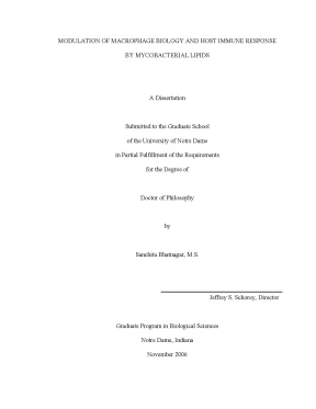
Tải xuống để đọc toàn bộMODULATION OF MACROPHAGE BIOLOGY AND HOST IMMUNE RESPONSE BY MYCOBACTERIAL LIPIDS A Dissertation Submitted to the Graduate School of the University of Notre Dame in Partial Fulfillment of the Requirements for the Degree of Doctor of Philosophy by Sanchita Bhatnagar, M. Schorey, Director Graduate Program in Biological Sciences Notre Dame, Indiana November 2006 UMI Number: 3243285 UMI Microform 3243285 Copyright 2007 by ProQuest Information and Learning Company. All rights reserved. This microform edition is protected against unauthorized copying under Title 17, United States Code.
ProQuest Information and Learning Company 300 North Zeeb Road P. Box 1346 Ann Arbor, MI 48106-1346 MODULATION OF MACROPHGAE BIOLOGY AND HOST IMMUNE RESPONSE BY MYCOBACTERIAL LIPIDS Abstract By Sanchita Bhatnagar Members of the Mycobacterium genus present a serious health threat which account for significant morbidity and mortality in humans and other species. Many pathogenic mycobacterial species maintain an intracellular life style within macrophages serving as the primary host cell. The mycobacterium’s ability to modulate the macrophage function requires the expression of specific mycobacterial surface components.
Moreover, a number of studies have shown that various mycobacterial surface components can promote macrophage activation. This dissertation aims to define the role of glycopeptidolipids (GPL), which are highly antigenic glycolipids, in the immune-modulation of the host and thus establish its function as an important virulence factor. To address the role of GPL as a virulence factor, we utilized M. avium strains either naturally lacking GPL expression or genetically altered to express modified GPL.
Our studies showed that the loss of GPL or altering its structure leads to an M. avium Sanchita Bhatnagar strain which induces increased macrophage activation compared to GPL sufficient M. Additional analysis showed attenuated virulence of GPL- deficient/modified M. avium strains in a mouse infection model.
To further understand the mechanism of action of GPL, we investigated the trafficking of GPL in primary macrophages infected with M. Our studies revealed significant release and trafficking of GPL from the mycobacteria containing phagosome to a distinct endocytic compartment called the multivesicular bodies (MVB). Additional analysis showed that mycobacterial components including GPL are trafficked outside the infected cells on “exosomes”. Further, we determined that exosomes purified from the culture supernatants of macrophages infected with M.
tuberculosis H37Rv or M. bovis BCG stimulate a pro-inflammatory response in uninfected macrophages. Our studies are first to establish GPL’s importance as a virulence factor in the M. The data demonstrate a significant shedding of glycopeptidolipids in M.
avium infected macrophages and its transport to the neighboring “bystander cells”. This transport of mycobacterial components occur through release of exosomes extracellularly which can also act to stimulate uninfected macrophages both in vitro and in vivo. Together, our studies put forward a novel mechanism of intercellular communication mediated by exosomes which can carry mycobacterial components to neighboring cells. DEDICATION For my loving husband, Jogender Singh Tushir and my parents, Satish and Sunita Bhatnagar, my little sister Anchita.
With great admiration and thanks. ii CONTENTS FIGURES…………………………………………………………………………….xii CHAPTER 1: INTRODUCTION 1.2 Classification of mycobacteria…………………………………………….3 Mycobacterial cell wall…………………………………………………………6 1.4 Biology of macrophages…………………………………….5 Mycobacterial entry in macrophage………………….6 Mycobacterial invasion of macrophage……………………………………….7 Mycobacterial lifestyle inside macrophages………………………………….8 Immunity to mycobacteria…………………………………………………….9 Trafficking of Mycobacterial lipids……………………………………………24 1.10 Macrophage endocytic pathway……………………………………………….25 CHAPTER 2: Mycobacterium avium 104 deleted of the methyltransferase D gene by allelic replacement lacks serotype-specific glycopeptidolipids and shows attenuated virulence in mice.2 Materials and Methods…………………………………………………………36 2.1 Bacterial strains and growth conditions……….2 Gene exchange by homologous recombination.3 BMMΦ isolation and culture………………………………………………….5 Cytokine profile of macrophages infected with M.6 In vivo mycobacterial infections…………………………………………….1 Disruption of the M. avium 104 mtfD gene by homologous recombination………………………………………………………………….2 In vitro macrophage infections with WT, mtfD mutant and complemented M.3 Mouse infections with the WT, mtfD mutant and complemented M.49 CHAPTER 3: Elevated MAP kinase signaling and increased macrophage activation in cells infected with a glycopeptidolipid-deficient Mycobacterium avium.2 Materials and Methods……………………………………….1 BMMΦ isolation and culture………………………….3 Preparation of surface-exposed material from M.4 Extraction and purification of GPLs………………………………………….6 Western blot analysis……………………………….8 Chemokine/Cytokine profile of macrophages infected with M.9 Bacterial killing assay……………………………….1 Differential activation of the MAPK in macrophages infected with Rg and SmT M.2 Calmodulin kinase is upstream of ERK1/2 MAPK in the macrophages infected with M.3 MAPK activation is differentially required for TNF-α production in macrophages infected with Rg compared to SmO and SmT M.4 TNF-α production is NF-κB dependent…………….5 Differential activation profile for BMMΦs following infection with Rg and SmT M.6 The Rg 2151 is killed by BMMΦs…………………………………………….75 CHAPTER 4: Release and trafficking of glycopeptidolipids in macrophages infected with M.2 Materials and Methods…………………………………….1 BMMφ isolation and culture……………………….4 Antibodies and immunofluorescence staining…….5 Thin layer chromatography immunostaining…….7 Isolation of exosomes…………………………….8 Sucrose density gradient centrifugation…………….9 Labeling with N-Rh-PE………………………….10 Analysis of un-infected bystander cells………….11 Coupling of exosomes to latex beads…………….12 Flow cytometric analysis of exosomes…………….13 Macrophage infection with exosomes…………….1 Glycopeptidolipid trafficking in M. avium 2151 infected macrophages.2 Intracellular sorting of released glycopeptidolipids………………………….3 Rab11 is transiently associated with GPL positive MVBs…………………….4 Role of exosomes in intercellular communication…………………………….5 Macrophage activation by the exosomes from mycobacteria infected macrophages………….6 TNF-α production is toll-like receptor dependent…………………………….7 Exosome stimulatory activity is not dependent on GPLs…………………….…………………………………………………………………110 CHAPTER 5: Macrophage activation by exosomes isolated from mycobacteria infected macrophages.2 Materials and Methods……………………………………………………….1 BMMφ isolation and culture……………………………………………….5 Isolation of exosomes…………………………………………………………121 5.6 Sucrose density gradient centrifugation…………………………………….7 Coupling of exosomes to latex beads……………………………….8 Flow cytometric analysis of exosomes……………………………….9 Macrophage infection with exosomes……………………………………….11 Western blot analysis………………………………….1 Exosomes isolated from mycobacteria infected cells induce pro-inflammatory response in non-infected macrophages…………………………………….2 Toll-like receptor dependent production of TNF-α in exosomes treated cells………………………………………………………………………….3 Exosomes from activated macrophages are not enough to induce pro-inflammatory response……………………………………………………132 5.4 Activated exosomes from other intracellular pathogen……………………….5 In vivo induction of pro-inflammatory response by the mycobacterial exosomes………………………….140 CHAPTER 6: Summary and future perspectives…………………………………….1 Mycobacterial species known to cause human disease……………….2 A chemical model of the mycobacterial cell wall……….3 List of some common mycobacterial lipids present on mycobacterial cell wall……………………………………………….4 Structure of glycopeptidolipids……………………………………….5 Different serovars showing oligosaccharides attached to the core structure of GPLs…………………………………………….6 List of pattern recognition receptors and some of their respective pathogen associated molecular patterns……………………19 Figure 1.7 Schematic of formation of MVB and its subsequent fate in the cell…………………………………………………………….1 Schematic organization of the mtfD disruption construct…………….2 BMMΦs infected with M.
avium mtfD mutant show increased production of TNF-α and RANTES compared to BMMΦs infected with M. avium 104 WT or mtfD mutant complemented with intact mtfD…………………………………….3 Bacterial loads in the livers or spleens or lungs following an infection of 129/Sv mice with M. avium104 WT, mtfD deficient mutant or mtfD complemented mutant……………………………….4 Cytokine profile in lung homogenates of mice infected with M. avium104 WT, mtfD deficient mutant or mtfD complemented mutant…………………………………………………………………48 Figure 3.1 MAPK activation upon infection of murine bone-marrow derived macrophages with M.
avium 2151 isogenic strains……………………63 vi Figure 3.2 Calmodulin Kinase activity is required for ERK1/2 activation in both Rg 2151 and SmT 2151 infected macrophages………………….3 Differential requirement for MAPK activation in TNF-α production by macrophages infected with Rg or SmT 2151……….4 TNF-α production by macrophages infected with SmO M. avium 2151 is dependent on p38 and ERK 1/2 activation…………….5 TNFα production by macrophages infected with either Rg or SmT M. avium 2151 is dependent on NF-κB activation……………….6 Macrophage produce more inflammatory mediators upon infection with Rg M. avium 2151 compared to SmT 2151 infected cells………………………………………………………………….7 RANTES and IL-6 production by Rg M.
avium 2151 infected macrophages is dependent on p38 but not ERK activation…….8 Killing of Rg M. avium 2151 by Balb/c bone marrow-derived macrophages………………………………………………………….9 Schematic illustration of the signal pathways activated in bone marrow-derived macrophages upon infection with Rg or SmT M.1 Intracellular trafficking of glycopeptidolipids in macrophages infected with Mycobacterium avium serovar 2……………………….2 Comparison of staining pattern of lipoarabinomannan (LAM) and glycopeptidolipids in macrophages infected with M.3 Glycopeptidolipids localizes to multi-vesicular bodies in Mycobacterium avium 2151 infected macrophages…………….4 Rab11 co localizes with the GPL loaded vesicles at later times…….5 Trafficking of mycobacterial components outside the infected cells via exosomes……………………………………………………103 Figure 4. avium 2151 infected macrophages induce a pro-inflammatory response in primary macrophages but not entirely dependent on GPL…………………………………….7 TNF-α production by macrophages infected with exosomes from M. avium 2151 SmT morphotype is dependent on toll-like receptor……………………………………………………………….8 Balb/c BMMΦs were infected with exosomes from SmT Rg morphotype of M.1 Characterization of exosomes isolated from non-infected, M.
bovis BCG-infected and M. tuberculosis H37Rv-infected J774 cells…….2 Exosomes isolated from M. bovis BCG and M. tuberculosis H37Rv infected THP-1 induce a pro-inflammatory response in human macrophages………………………………………………….3 TLR dependent TNF-α induced by exosomes isolated from mycobacterial infected cells………………………………………….4 Macrophage activation by exosomes derived from THP-1 macrophage activated with LPS (200ng/ml) or infected with heat- inactivated M.
bovis BCG (HI-BCG) or Salmonella typhimurium SL1344…………………………………………………………………135 Figure 5.5 Mycobacterial exosomes induced pro-inflammatory response in the lungs of Balb/c mice…………………….1 Schematic illustration of the signal pathways activated in bone marrow-derived macrophages upon infection with Rg or SmT M.154 viii ABBREVIATIONS Ab: Antibody AraLAM: Arabinofuranosyl-terminated LAM BCA: Bicinchoninic acid assay BCG: Mycobacterium bovis bacillus-Calmette Guerin BMMΦ: Bone marrow derived macrophages C: Celsius CaM: Calmodulin CAPE: Caffeic Acid Phenylethyl Ester CFU: Colony forming unit CaMK: Ca2+- calmodulin dependent protein kinase DC-SIGN: Dendritic cell-specific ICAM-grabbing non-integrin DMSO: Dimethyl sulfoxide DMEM: Dulbecco’s modified eagle’s medium ECL: Enhanced chemilumnescence ELISA: Enzyme-linked immunosorbent assay ERK1/2: Extracellular signal-regulated kinase1/2 EM: Electron microscopy FITC: Fluorescein isothiocyanate ix FBS: Fetal bovine serum GM-CSF: Granulocyte-macrophages colony stimulating factor GPL: Glycopeptidolipid HIV: Human immunodeficiency virus ICAM: Intracellular adhesion molecule IgG: Immunoglobulin G IFN-γ: Interferon gamma ILV: Intra-luminal vesicles iNOS: Inducible nitric oxide synthase 2 LAM: Lipoarabinomannan LAMP1: Lysosome associated membrane protein 1 LAMP2: Lysosome associated membrane protein 2 LPS: Lipopolysaccharide MAC: Mycobacterium avium complex ManLAM: Mannosylated-lipoarabinomannan MAPK: Mitogen activated protein kinase mAb: Monoclonal antibody MR: Mannose receptor mtfD: Methyl transferase D MVB: Multi-vesicular body NF-κB: Nuclear factor kappa B nsGPL: non-serovar specific glycopeptidolipids OADC: Oleic acid, dextrose, catalase x PAMP: Pathogen associated molecular pattern PBS: Phosphate-buffered saline PD98059: MEK1/2 specific inhibitor PILAM: Phosphoinositide-capped lipoarabinomannan PIM: Phosphatidylinositol mannoside PMA: Phorbol 12-myristate 13-acetate RC: Resting Cell Rg: Rough RNI: Reactive nitrogen intermediates SB203580: p38 specific inhibitor SD: Standard deviation SDS: Sodium dodecyl sulfate TC: Tissue culture grade TH: T helper cells TNF-α: Tumor necrosis factor-alpha TLR: Toll like receptor PRR: Pattern recognition receptor ssGPL: serovar specific glycopeptidolipids SmT: Smooth transparent SmO: Smooth opaque WT: Wild type xi ACKNOWLEDGMENTS First, I would like to thank my advisor Dr. Jeff Schorey for his help and guidance throughout my project. He has been a great mentor these past 4 years and I am thankful for the opportunity to have worked with him. I appreciate Jeff’s scientific enthusiasm and his creative ideas.
I am forever grateful to Jeff for being supportive of my project and letting me follow my whims. Many thanks to my thesis committee members, Dr Crislyn D’Souza-Schorey, Dr Holly Goodson and Dr Mary Ann Mcdowell for their guidance, advice and tremendous support along the way. It has been a pleasure for me to get to know you all and I thank you for your valuable insights that helped me put together this thesis. I will also like to thank the present and past members of the Schorey lab for their cooperation and providing exciting working environment in the lab.
I will also like to acknowledge Crislyn D’Souza lab members for their help and insightful discussions. Special thanks to Severson lab and Mary Ann McDowell lab for providing many reagents and the equipment. Finally, I will like to thank my loving husband, Jogi for his help and support. He has been a great inspiration for me and was the only reason of my pursuing the PhD.
He has been my biggest critic and his advice has always helped me to move ahead. I want to thank you for being with me at every step and helping me in everything. He has taught xii me to be strong and gave me strength to overcome some of the hard times in the life. I will also like to thank my parents, who believed in me and supported my every decision in life.
I would not have been here without the support of my younger sister, Anchita who is the joy of our life.
Nội dung được bảo vệ bản quyền — Tải xuống đầy đủ
Trích dẫn luận án này
Sanchita Bhatnagar (2006). Điều hòa sinh học macrophage bằng lipid mycobacterium [Luận án tiến sĩ, University of Notre Dame]. LuanAn.net. https://luanan.net/sinh-hoc/sinh-hoc-phan-tu-te-bao/dieu-hoa-sinh-hoc-macrophage-lipid-mycobacterium
Từ khóa và chủ đề nghiên cứu
Từ khóa liên quan
Chủ đề nghiên cứu
Câu hỏi thường gặp
Luận án "Điều hòa sinh học macrophage bằng lipid mycobacterium" nghiên cứu về vấn đề gì?
Luận án tiến sĩ nghiên cứu vai trò glycopeptidolipids trong điều hòa macrophage và miễn dịch. Khám phá cơ chế vận chuyển lipid mycobacterium qua exosomes đến tế bào lân cận.
Luận án "Điều hòa sinh học macrophage bằng lipid mycobacterium" được bảo vệ tại trường nào?
Luận án này được bảo vệ tại University of Notre Dame. Năm bảo vệ: 2006.
Luận án "Điều hòa sinh học macrophage bằng lipid mycobacterium" thuộc chuyên ngành gì?
Luận án "Điều hòa sinh học macrophage bằng lipid mycobacterium" thuộc chuyên ngành Biological Sciences. Danh mục: Sinh Học Phân Tử Tế Bào.
Luận án "Điều hòa sinh học macrophage bằng lipid mycobacterium" có bao nhiêu trang?
Luận án "Điều hòa sinh học macrophage bằng lipid mycobacterium" có 190 trang. Bạn có thể xem trước một phần tài liệu ngay trên trang web trước khi tải về.
Cách tải luận án "Điều hòa sinh học macrophage bằng lipid mycobacterium" về máy như thế nào?
Để tải luận án về máy, bạn nhấn nút "Tải xuống ngay" trên trang này, sau đó hoàn tất thanh toán phí lưu trữ. File sẽ được tải xuống ngay sau khi thanh toán thành công. Hỗ trợ qua Zalo: 0559 297 239.