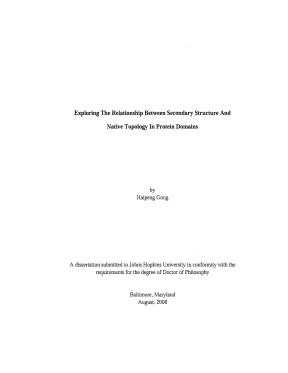
Mối quan hệ cấu trúc thứ cấp và cấu trúc native trong miền protein
Luận án tiến sĩ khám phá mối quan hệ giữa cấu trúc thứ cấp và cấu trúc 3D protein. Áp dụng thuật toán xác định vai trò cấu trúc trong quá trình gấp gập protein.
Năm xuất bản
Số trang
137
Thời gian đọc
21 phút
Lượt xem
1
Lượt tải
0
Phí lưu trữ
40 Point
Tổng quan nhanh
- Chủ đề:
- 1. Cấu Trúc Thứ Cấp Protein: Nền Tảng Gấp Cuộn
- Số trang:
- 137 trang
- Trường:
- Johns Hopkins University
- Chuyên ngành:
- Biophysics
- Tác giả:
- Haipeng Gong
- Năm:
- 2006
Tóm tắt nội dung luận án
I. Cấu Trúc Thứ Cấp Protein Nền Tảng Gấp Cuộn
Cấu trúc thứ cấp protein đóng vai trò then chốt trong quá trình gấp cuộn protein. Mô hình đột phá của Pauling đã chứng minh liên kết hydro tạo nên các cấu trúc thứ cấp ổn định. Chuỗi polypeptide tuyến tính hình thành các phần tử cấu trúc thứ cấp trong điều kiện gấp cuộn. Quá trình này diễn ra nhanh chóng trên thang thời gian ngắn. Các phần tử này tồn tại trong trạng thái cân bằng động với dạng cuộn coil. Chúng tương tác với nhau để tổ chức và ổn định protein. Bước tương tác này quyết định tốc độ gấp cuộn của miền protein.
1.1. Vai Trò Liên Kết Hydro Trong Cấu Trúc
Liên kết hydro là yếu tố cốt lõi hình thành cấu trúc thứ cấp protein. Các thí nghiệm đã xác nhận tầm quan trọng của liên kết này trong gấp cuộn protein. Liên kết hydro kết nối các nhóm carbonyl và amino trong chuỗi polypeptide. Sự kết nối này tạo ra các mẫu hình lặp lại đặc trưng. Alpha helix và beta sheet là hai dạng phổ biến nhất. Độ bền của các cấu trúc này phụ thuộc vào số lượng và vị trí liên kết hydro.
1.2. Tốc Độ Gấp Cuộn Và Hàm Lượng Cấu Trúc
Logarit của hằng số tốc độ gấp cuộn tương quan tuyến tính với hàm lượng cấu trúc thứ cấp. Hệ số tương quan lớn chứng minh giả thuyết về vai trò của cấu trúc thứ cấp. Các phần tử cấu trúc thứ cấp tổ chức quá trình gấp cuộn một cách có hệ thống. Mối quan hệ này nhấn mạnh tầm quan trọng của alpha helix và beta sheet. Hàm lượng cấu trúc thứ cấp cao dẫn đến tốc độ gấp cuộn nhanh hơn.
1.3. Trạng Thái Cân Bằng Động Của Cấu Trúc
Các phần tử cấu trúc thứ cấp tồn tại trong trạng thái cân bằng động. Chúng liên tục chuyển đổi giữa dạng có cấu trúc và dạng cuộn. Sự cân bằng này ảnh hưởng đến quá trình gấp cuộn tổng thể. Điều kiện môi trường như nhiệt độ và pH tác động đến cân bằng. Cấu trúc thứ cấp ổn định khi các điều kiện thuận lợi. Sự ổn định này là tiền đề cho hình thành cấu trúc bậc ba protein.
II. Trình Tự Cấu Trúc Thứ Cấp Quyết Định Topology
Przytycka và Rose đề xuất trình tự các phần tử cấu trúc thứ cấp đủ để xác định cấu trúc không gian 3D của protein. Giả thuyết này được kiểm chứng trên tập hợp lớn các domain protein đại diện. Cây phân cấp từ việc sắp xếp trình tự cấu trúc thứ cấp gần như giống hệt cây từ so sánh cấu trúc ba chiều trực tiếp. Kết quả này chứng minh trình tự alpha helix và beta sheet mang thông tin topology. Thông tin này ngầm chứa trong cách sắp xếp các phần tử cấu trúc. Từ đó có thể dự đoán cấu trúc không gian 3D của miền protein.
2.1. Thuật Toán So Sánh Trình Tự Mesostate
Thuật toán quy hoạch động được phát triển để so sánh cấu trúc domain protein. Mesostate là biểu diễn thô của góc xoắn xương sống polypeptide. Việc sắp xếp trình tự mesostate cho phép so sánh các cấu trúc protein. Thuật toán này hoạt động hiệu quả hơn nhiều phương pháp nhận dạng nếp gấp hiện có. Kết quả hỗ trợ mạnh mẽ cho giả thuyết về vai trò của trình tự cấu trúc thứ cấp. Trình tự này quyết định cấu trúc ba chiều cuối cùng của protein.
2.2. So Sánh Cây Phân Cấp Từ Các Phương Pháp
Cây phân cấp từ sắp xếp trình tự cấu trúc thứ cấp được so sánh với cây từ so sánh cấu trúc 3D. Hai cây này cho kết quả gần như đồng nhất. Sự tương đồng cao chứng minh trình tự cấu trúc thứ cấp chứa đầy đủ thông tin topology. Phương pháp này áp dụng cho nhiều domain protein khác nhau. Kết quả nhất quán qua các họ protein khác biệt. Điều này khẳng định tính phổ quát của mối quan hệ giữa cấu trúc thứ cấp và topology.
2.3. Thông Tin Ngầm Trong Trình Tự Cấu Trúc
Trình tự các phần tử cấu trúc thứ cấp mang thông tin ngầm về cấu trúc không gian. Thông tin này có thể được trích xuất thông qua các thuật toán phù hợp. Sắp xếp alpha helix và beta sheet theo thứ tự đặc biệt mã hóa topology. Mỗi sắp xếp khác nhau tương ứng với một cấu trúc ba chiều riêng biệt. Việc giải mã thông tin này cho phép dự đoán cấu trúc protein. Phương pháp này mở ra hướng tiếp cận mới trong nghiên cứu cấu trúc protein.
III. Tái Tạo Cấu Trúc Bậc Ba Từ Mesostate Sequence
Thuật toán Monte-Carlo thay thế đoạn được phát triển để tạo cấu trúc bậc ba protein. Thuật toán chỉ sử dụng thông tin từ trình tự mesostate. Thế năng đơn giản bao gồm liên kết hydro, loại trừ không gian và giới hạn không gian. Thế năng này đủ để tái tạo topology xương sống giống native từ ràng buộc góc xoắn thô. Các ràng buộc này được áp đặt bởi trình tự mesostate native. Phương pháp chứng minh cấu trúc không gian 3D có thể được tái tạo từ thông tin cấu trúc thứ cấp. Kết quả nhấn mạnh vai trò trung tâm của cấu trúc thứ cấp trong gấp cuộn protein.
3.1. Thuật Toán Monte Carlo Thay Thế Đoạn
Thuật toán Monte-Carlo được thiết kế để tạo cấu trúc bậc ba từ trình tự mesostate. Phương pháp thay thế đoạn cho phép tìm kiếm không gian cấu trúc hiệu quả. Mỗi bước Monte-Carlo thay thế một đoạn ngắn của chuỗi polypeptide. Việc thay thế tuân theo ràng buộc từ trình tự mesostate native. Thuật toán lặp lại cho đến khi đạt được cấu trúc ổn định. Phương pháp này tạo ra cấu trúc topology giống với cấu trúc native.
3.2. Thế Năng Đơn Giản Trong Mô Phỏng
Thế năng sử dụng chỉ bao gồm ba thành phần cơ bản: liên kết hydro, loại trừ không gian và giới hạn không gian. Liên kết hydro ổn định các cấu trúc thứ cấp như alpha helix và beta sheet. Loại trừ không gian ngăn các nguyên tử chồng lấn lên nhau. Giới hạn không gian đảm bảo protein gấp trong vùng hạn chế. Thế năng đơn giản này đủ để tái tạo cấu trúc native. Điều này chứng minh cấu trúc thứ cấp chứa thông tin cốt lõi về topology.
3.3. Tái Tạo Topology Xương Sống Native
Topology xương sống giống native được tái tạo từ ràng buộc góc xoắn thô. Ràng buộc này xuất phát từ trình tự mesostate của cấu trúc native. Kết quả cho thấy cấu trúc ba chiều có thể được dự đoán chính xác. Topology được tái tạo phù hợp với cấu trúc thực nghiệm. Phương pháp này xác nhận vai trò quyết định của trình tự cấu trúc thứ cấp. Từ thông tin thô về góc xoắn có thể suy ra cấu trúc không gian 3D đầy đủ.
IV. Miền Protein Và Đặc Trưng Cấu Trúc Domain
Miền protein là đơn vị cấu trúc và chức năng độc lập trong phân tử protein. Mỗi domain protein có cấu trúc ba chiều riêng biệt và ổn định. Cấu trúc này được xác định bởi trình tự cấu trúc thứ cấp đặc trưng. Các domain protein có thể gấp cuộn độc lập với phần còn lại của chuỗi. Nghiên cứu tập trung vào tập hợp đại diện các domain protein. Mỗi domain mang đặc trưng topology riêng được quyết định bởi sắp xếp alpha helix và beta sheet. Hiểu biết về domain protein giúp dự đoán cấu trúc và chức năng protein.
4.1. Tập Hợp Domain Protein Đại Diện
Tập hợp domain protein đại diện bao gồm các cấu trúc điển hình từ nhiều họ protein. Mỗi domain được chọn dựa trên tính đại diện cho một nếp gấp cụ thể. Tập hợp này cho phép kiểm chứng giả thuyết trên phạm vi rộng. Các domain khác nhau về kích thước, thành phần và chức năng. Tuy nhiên chúng đều tuân theo nguyên tắc chung về mối quan hệ cấu trúc-topology. Nghiên cứu trên tập hợp này cung cấp bằng chứng vững chắc cho các giả thuyết.
4.2. Tính Độc Lập Gấp Cuộn Của Domain
Mỗi domain protein có khả năng gấp cuộn độc lập. Quá trình gấp cuộn không phụ thuộc vào các phần khác của chuỗi polypeptide. Tính độc lập này xuất phát từ cấu trúc thứ cấp tự ổn định. Các tương tác trong domain đủ mạnh để duy trì cấu trúc ba chiều. Domain có thể được tách ra và vẫn giữ nguyên cấu trúc. Đặc tính này quan trọng cho việc nghiên cứu và ứng dụng protein.
4.3. Topology Đặc Trưng Của Từng Domain
Mỗi domain protein có topology đặc trưng riêng biệt. Topology này được quyết định bởi cách sắp xếp các cấu trúc thứ cấp. Alpha helix và beta sheet kết hợp theo những mẫu hình cụ thể. Mỗi mẫu hình tạo ra một topology ba chiều khác nhau. Topology đặc trưng phản ánh chức năng sinh học của domain. Nhận dạng topology giúp phân loại và dự đoán chức năng protein.
V. Alpha Helix Và Beta Sheet Hai Cấu Trúc Chính
Alpha helix và beta sheet là hai dạng cấu trúc thứ cấp protein phổ biến nhất. Xoắn alpha hình thành khi chuỗi polypeptide cuộn theo dạng xoắn ốc phải. Liên kết hydro kết nối nhóm C=O của amino acid thứ n với nhóm N-H của amino acid thứ n+4. Tấm beta tạo thành khi các đoạn chuỗi nằm song song hoặc phản song song. Liên kết hydro kết nối các đoạn này với nhau. Cả hai cấu trúc đều ổn định nhờ mạng lưới liên kết hydro. Sự kết hợp của alpha helix và beta sheet tạo nên đa dạng cấu trúc protein.
5.1. Đặc Điểm Cấu Trúc Xoắn Alpha
Xoắn alpha là cấu trúc xoắn ốc phải với 3.6 amino acid mỗi vòng. Liên kết hydro giữa C=O của vị trí i và N-H của vị trí i+4 ổn định cấu trúc. Trục xoắn song song với hướng của các liên kết hydro. Chuỗi bên amino acid hướng ra ngoài xoắn. Alpha helix là cấu trúc thứ cấp ổn định và phổ biến. Nhiều protein chứa phần lớn cấu trúc dưới dạng alpha helix.
5.2. Đặc Điểm Cấu Trúc Tấm Beta
Tấm beta hình thành từ các đoạn chuỗi polypeptide kéo dài. Các đoạn này nằm cạnh nhau theo dạng song song hoặc phản song song. Liên kết hydro kết nối nhóm C=O và N-H giữa các đoạn. Cấu trúc tạo thành dạng tấm gấp nếp. Chuỗi bên amino acid hướng lên xuống so với mặt phẳng tấm. Beta sheet cung cấp độ bền cơ học cao cho protein.
5.3. Kết Hợp Alpha Helix Và Beta Sheet
Hầu hết protein chứa cả alpha helix và beta sheet. Sự kết hợp này tạo ra đa dạng topology ba chiều. Các miền protein khác nhau có tỷ lệ alpha/beta khác nhau. Một số domain chủ yếu là alpha, một số chủ yếu là beta. Nhiều domain có cấu trúc alpha/beta xen kẽ. Cách sắp xếp hai cấu trúc này quyết định topology tổng thể của protein.
VI. Vai Trò Trung Tâm Của Cấu Trúc Thứ Cấp Protein
Cấu trúc thứ cấp protein đóng vai trò trung tâm trong quá trình gấp cuộn protein. Ba hướng nghiên cứu liên quan chứng minh vai trò này một cách toàn diện. Thứ nhất, tốc độ gấp cuộn tương quan với hàm lượng cấu trúc thứ cấp. Thứ hai, trình tự cấu trúc thứ cấp quyết định topology ba chiều. Thứ ba, cấu trúc bậc ba có thể tái tạo từ thông tin cấu trúc thứ cấp. Ba kết quả này bổ sung cho nhau và tạo thành bức tranh hoàn chỉnh. Cấu trúc thứ cấp là cầu nối giữa trình tự amino acid và cấu trúc không gian 3D cuối cùng.
6.1. Tương Quan Giữa Ba Hướng Nghiên Cứu
Ba hướng nghiên cứu trong luận án có mối liên hệ chặt chẽ. Hướng thứ nhất chứng minh cấu trúc thứ cấp kiểm soát động học gấp cuộn. Hướng thứ hai cho thấy cấu trúc thứ cấp mã hóa thông tin topology. Hướng thứ ba tái tạo cấu trúc ba chiều từ thông tin cấu trúc thứ cấp. Mỗi hướng tiếp cận vấn đề từ góc độ khác nhau. Kết hợp lại tạo thành bằng chứng toàn diện về vai trò của cấu trúc thứ cấp.
6.2. Cấu Trúc Thứ Cấp Là Cầu Nối Cấu Trúc
Cấu trúc thứ cấp protein kết nối trình tự amino acid với cấu trúc không gian 3D. Trình tự amino acid xác định xu hướng hình thành alpha helix hoặc beta sheet. Các cấu trúc thứ cấp này sau đó tổ chức thành cấu trúc ba chiều. Quá trình này diễn ra theo hệ thống phân cấp. Cấu trúc thứ cấp là mức trung gian thiết yếu. Hiểu biết về mức này là chìa khóa để dự đoán cấu trúc protein.
6.3. Ứng Dụng Trong Dự Đoán Cấu Trúc Protein
Hiểu biết về vai trò của cấu trúc thứ cấp mở ra ứng dụng thực tế. Dự đoán cấu trúc thứ cấp từ trình tự amino acid là bước đầu tiên. Từ trình tự cấu trúc thứ cấp có thể suy ra topology ba chiều. Phương pháp này nhanh hơn mô phỏng gấp cuộn đầy đủ. Độ chính xác cao nhờ mối quan hệ chặt chẽ giữa cấu trúc thứ cấp và topology. Ứng dụng này quan trọng cho nghiên cứu protein và thiết kế thuốc.
Mục lục chi tiết luận án
Tải xuống file đầy đủ để xem toàn bộ nội dung
Tải đầy đủ (137 trang)Nội dung chính
Tổng quan về luận án
Bối cảnh khoa học của luận án bắt nguồn từ một trong những thách thức trung tâm của ngành lý sinh học phân tử và sinh học cấu trúc hiện đại: bài toán cuộn gập protein (protein folding problem). Kể từ khi Christian Anfinsen chứng minh rằng cấu hình tự nhiên (native conformation) của protein được xác định hoàn toàn bởi trình tự axit amin sơ cấp, cơ chế vật lý dẫn dắt chuỗi polypeptide chuyển đổi từ trạng thái mở xoắn (unfolded state) sang cấu trúc không gian ba chiều đặc thù vẫn là đối tượng tranh luận sâu sắc. Luận án tiến sĩ "Exploring The Relationship Between Secondary Structure And Native Topology In Protein Domains" được thực hiện bởi nghiên cứu sinh Haipeng Gong dưới sự hướng dẫn của Giáo sư George D. Rose tại Đại học Johns Hopkins (2006), mở ra một hướng tiếp cận tiên phong nhằm giải mã mối quan hệ bản chất giữa cấu trúc bậc hai cục bộ và tô-pô tự nhiên của các domain protein.
Khoảng trống nghiên cứu (research gap) cốt lõi mà luận án giải quyết bao gồm:
- Sự phụ thuộc mang tính tiên nghiệm vào cấu trúc không gian ba chiều: Các mô hình động học cuộn gập kinh điển, tiêu biểu là mô hình bậc tiếp xúc tương đối (Relative Contact Order - RCO) của Plaxco và Baker (1998), đòi hỏi phải biết trước tọa độ nguyên tử 3D từ tinh thể học tia X hoặc phổ cộng hưởng từ hạt nhân (NMR) để tính toán hằng số tốc độ cuộn gập ($\ln k_f$), thiếu khả năng dự đoán trực tiếp từ thông tin chuỗi hoặc cấu trúc bậc hai trung gian.
- Sự mơ hồ về vai trò quyết định của cấu trúc bậc hai đối với nếp gấp bậc ba: Liệu thông tin cấu trúc bậc hai xấp xỉ có đủ để xác định nếp gấp tô-pô toàn cục mà không cần đến thông tin chi tiết về chuỗi bên (sidechain packing) hay không?
- Thiếu sót mang tính nền tảng trong giả thuyết polymer cổ điển về trạng thái mở xoắn: Giả thuyết cặp độc lập của Flory (Flory's independent pair hypothesis) giả định rằng trạng thái duỗi là một cuộn thống kê vô định hình (random coil) với không gian cấu hình khổng lồ, dẫn đến việc phóng đại nghịch lý Levinthal (Levinthal paradox) trong lý thuyết cuộn gập.
Luận án thiết lập hệ thống câu hỏi nghiên cứu và giả thuyết khoa học chặt chẽ:
- Câu hỏi nghiên cứu 1 (RQ1): Liệu hàm lượng cấu trúc bậc hai cục bộ có thể đóng vai trò như một biến số vật lý độc lập để dự đoán chính xác tốc độ cuộn gập của protein hai trạng thái (two-state proteins) mà không cần tọa độ 3D?
- Câu hỏi nghiên cứu 2 (RQ2): Mức độ thông tin chứa trong chuỗi góc xoay trục chính thô hóa (mesostate sequence) có đủ để phân loại và nhận diện nếp gấp protein (fold recognition) tương đương với các thuật toán so khớp cấu trúc 3D toàn phần hay không?
- Câu hỏi nghiên cứu 3 (RQ3): Một hàm thế năng tối giản (crude potential) kết hợp với các ràng buộc góc nhị diện thô hóa có thể tái tạo lại tô-pô trục chính giống tự nhiên (native-like backbone topology) từ chuỗi mở rộng hay không?
- Giả thuyết 1 (H1): Bước lắp ráp và va chạm giữa các yếu tố cấu trúc bậc hai là giai đoạn giới hạn tốc độ (rate-limiting step) trong quá trình cuộn gập domain protein.
- Giả thuyết 2 (H2): Trình tự các trạng thái trung gian (mesostates) trên trục chính mã hóa đầy đủ thông tin để định hình tô-pô không gian ba chiều của domain.
- Giả thuyết 3 (H3): Tương tác loại trừ lập thể cục bộ và mạng lưới liên kết hydro làm giảm không gian pha của trạng thái mở xoắn hơn 50 bậc độ lớn ($>10^{50}$), xóa bỏ tính chất ngẫu nhiên của nghịch lý Levinthal.
Khung lý thuyết của nghiên cứu được xây dựng dựa trên:
- Thuyết kiến trúc phân tầng protein (Hierarchic Architecture Theory - Rose, 1979).
- Mô hình khung cuộn gập (Framework Model - Kim & Baldwin, 1982).
- Mô hình tìm kiếm topomer (Topomer Search Model - Debe et al., 1999; Plaxco et al., 2000).
- Mô hình trạng thái thô hóa góc nhị diện (Mesostate Discretization Model).
Đóng góp đột phá của luận án được lượng hóa qua các kết quả thực nghiệm và tính toán:
- Chứng minh mối tương quan tuyến tính mạnh mẽ giữa hàm lượng cấu trúc bậc hai cục bộ và logarit hằng số tốc độ cuộn gập ($\ln k_f$) trên tập dữ liệu 24 protein hai trạng thái có dải tốc độ trải rộng hơn 6 bậc độ lớn ($2.5 \times 10^6 \text{ s}^{-1}$ ở cytochrome b562 đến $0.5 \text{ s}^{-1}$ ở acylphosphatase).
- Phát triển thuật toán quy hoạch động
Meso_Aligndựa trên ma trận 36 trạng thái mesostate ($60^\circ \times 60^\circ$), đạt độ nhạy phân loại tương đương các phương pháp 3D phức tạp trên cơ sở dữ liệu SCOP. - Tái dựng thành công cấu trúc tô-pô bậc ba với độ lệch bình phương trung bình gốc (RMSD) thấp bằng thuật toán Monte Carlo ghép đoạn (fragment assembly) chỉ sử dụng thế năng tối giản: liên kết hydro, loại trừ lập thể và giam hãm không gian.
Phạm vi nghiên cứu bao quát các domain protein đơn lẻ, dữ liệu động học thực nghiệm của 24 protein hai trạng thái, cây cấu trúc P-tree của 183 protein có độ tương đồng chuỗi thấp ($<30%$), và tập chuẩn 2.777 protein phi tương đồng trong đánh giá cấu trúc bậc hai.
Literature Review và Positioning
Nghiên cứu về cơ chế cuộn gập protein trong nửa thế kỷ qua được định hình bởi ba dòng tư tưởng chính:
-
Động lực học nhiệt động lực học và lực tương tác: Linus Pauling (1951) đặt nền móng với giả thuyết liên kết hydro nội chuỗi là động lực chính hình thành xoắn $\alpha$ và phiến $\beta$ với năng lượng ước tính $2\text{--}10 \text{ kcal/mol}$. Ngược lại, Walter Kauzmann (1959) đề xuất mô hình "giọt dầu" (oil drop model), lập luận rằng hiệu ứng kỵ nước (hydrophobic interaction) mới là lực dẫn dắt chính giấu các gốc không phân cực vào lõi protein. Tranh luận này kéo dài qua các công trình của Klotz & Franzen (1962) ủng hộ Kauzmann, cho đến khi Pace và cộng sự (1996) cùng Makhatadze & Privalov chứng minh năng lượng enthalpy của liên kết hydro đóng góp tới $1.6 \text{ kcal/mol}$ cho độ ổn định cấu trúc, tái khẳng định vai trò sống còn của liên kết hydro trong việc tạo ra tính đặc hiệu cấu trúc.
-
Các mô hình động học cuộn gập: Để giải quyết nghịch lý Levinthal (1968) – trong đó một chuỗi 100 axit amin có $2^{100} \approx 10^{30}$ cấu hình khả dĩ, không thể tìm thấy trạng thái tự nhiên bằng cơ chế tìm kiếm ngẫu nhiên trong thời gian sinh học – Peter Wolynes (1995) phát triển lý thuyết phễu năng lượng (Energy Landscape / Folding Funnel). Cùng với đó, Martin Karplus & David L. Weaver (1976) đưa ra mô hình Khuếch tán - Va chạm (Diffusion-Collision), Peter S. Kim & Robert L. Baldwin (1982) đề xuất mô hình Khung (Framework Model), Ken A. Dill (1985) phát triển mô hình Sụp đổ kỵ nước (Hydrophobic Collapse), và Alan R. Fersht (1997) thiết lập mô hình Ngưng tụ tạo mầm (Nucleation-Condensation) thông qua phân tích giá trị $\Phi$-value.
-
Cấu trúc trạng thái mở xoắn (Unfolded State): Paul Flory (1969) và Charles Tanford xây dựng mô hình cuộn thống kê (random coil) dựa trên giả thuyết cặp độc lập. Tuy nhiên, các phát hiện thực nghiệm gần đây về xoắn Polyproline II (PPII) (Sreerama & Woody, 2004; Shi et al., 2002; Pappu et al., 2000) và cấu trúc tự nhiên tàn dư (native-like residual structure) trong nuclease của tụ cầu khuẩn (SNase) của Shortle và cộng sự (2002) đã phủ nhận bản chất vô định hình của trạng thái duỗi.
| Trường phái / Tác giả | Luận điểm chính | Hạn chế / Điểm bất đồng |
|---|---|---|
| Kauzmann (1959); Dill (1985) | Hiệu ứng kỵ nước là động lực tối thượng; sụp đổ kỵ nước diễn ra trước khi hình thành cấu trúc đặc thù. | Không giải thích được tính đặc hiệu tô-pô cao và sự hình thành nhanh của cấu trúc bậc hai; bỏ qua vai trò định hướng của liên kết hydro. |
| Pauling (1951); Kim & Baldwin (1982) | Liên kết hydro và cấu trúc bậc hai hình thành trước, đóng vai trò khung (framework) định hướng lắp ráp bậc ba. | Chưa giải thích tường tận cách thức các đoạn cấu trúc bậc hai tìm kiếm không gian topomer mà không bị nghẽn năng lượng. |
| Plaxco & Baker (1998) | Hằng số tốc độ cuộn gập phụ thuộc tuyến tính vào bậc tiếp xúc tương đối (RCO) tính từ cấu trúc tinh thể 3D. | Mang tính kinh nghiệm thuần túy; thất bại khi giải thích hiện tượng vòng hóa chuỗi SH3 hoặc đột biến điểm không đổi RCO nhưng đổi tốc độ cuộn gập. |
| Flory (1969); Tanford (1968) | Trạng thái mở xoắn là cuộn ngẫu nhiên không có cấu trúc; các cặp góc nhị diện $(\phi, \psi)$ hoàn toàn độc lập. | Bị bác bỏ bởi va chạm lập thể cục bộ ($i-3$ đến $i$) và sự hiện diện áp đảo của cấu hình PPII; phóng đại entropy cấu hình. |
Luận án định vị chính xác điểm nghẽn của y văn: chứng minh rằng cấu trúc bậc hai không chỉ là thành phần phụ trợ mà chính là yếu tố quyết định tốc độ cuộn gập và định hình nếp gấp tô-pô toàn cục.
So sánh với hai nghiên cứu quốc tế điển hình:
- So với nghiên cứu của Plaxco và Baker (1998) về RCO: Plaxco dựa hoàn toàn vào ma trận khoảng cách trích xuất từ cấu trúc 3D thực nghiệm. Gong chứng minh rằng hàm lượng cấu trúc bậc hai cục bộ – một thông số có thể tính toán trực tiếp từ chuỗi sơ cấp hoặc thực nghiệm phổ phân cực tròn (CD) – có giá trị dự đoán tương đương, loại bỏ sự phụ thuộc vào cấu trúc tinh thể đã biết.
- So với thuật toán DALI của Holm & Sander (1993) và VAST của Gibrat (1996): DALI so sánh ma trận khoảng cách nguyên tử $\text{C}\alpha$, còn VAST căn chỉnh mảng vector cấu trúc bậc hai 3D. Thuật toán
Meso_Aligncủa Gong đạt hiệu năng phân loại nếp gấp tương đương trên tập chuẩn SCOP nhưng chỉ thao tác trên chuỗi ký tự 1D của các trạng thái mesostate, giảm thiểu tối đa chi phí tính toán không gian.
Đóng góp lý thuyết và khung phân tích
Đóng góp cho lý thuyết
Luận án tạo ra bước đột phá trong việc thách thức và mở rộng các lý thuyết sinh học cấu trúc kinh điển:
- Bác bỏ giả thuyết cặp độc lập của Flory (Flory's Independent Pair Hypothesis): Flory giả định rằng các góc xoay trục chính $(\phi_i, \psi_i)$ độc lập với $(\phi_{i+1}, \psi_{i+1})$. Luận án chỉ ra rằng do va chạm lập thể giữa nguyên tử oxy carbonyl của gốc $i-3$ trong xoắn $\alpha$ và oxy carbonyl của gốc $i$ khi ở cấu hình phiến $\beta$, không gian cấu hình khả dĩ bị hạn chế nghiêm ngặt. Trích dẫn nguyên văn từ luận án:
"Fitzkee and Rose... found that for a 100-residue protein, a combination of these two effects would reduce accessible conformational space by a factor of $4\times 10^{19}$... steric exclusion and hydrogen bonding criteria could jointly reduce the magnitude of available space in the unfolded state by more than 50 orders of magnitude."
- Thiết lập mô hình cuộn gập 3 giai đoạn (Three-Stage Folding Model): Mở rộng mô hình Framework của Kim & Baldwin và Topomer Search của Debe & Plaxco, Gong đề xuất cơ chế cuộn gập cho protein hai trạng thái:
- Giai đoạn 1: Hình thành nhanh chóng các yếu tố cấu trúc bậc hai bán ổn định (xoắn $\alpha$, sợi $\beta$, vòng quay chặt) ở trạng thái cân bằng động với dạng cuộn.
- Giai đoạn 2 (Giai đoạn giới hạn tốc độ): Quá trình khuếch tán và va chạm để tìm kiếm topomer tự nhiên. Tốc độ giai đoạn này tương quan nghịch với chiều dài các đoạn loop nối giữa các cấu trúc bậc hai.
- Giai đoạn 3: Ngưng tụ nhanh chóng từ topomer tự nhiên thành nếp gấp bản địa hoàn chỉnh.
$$\ln k_f = - \alpha \cdot f_{\text{loop}} + \beta \cdot f_{\text{sec}} + \gamma$$
Trong đó $f_{\text{loop}}$ là tỷ lệ cấu trúc vòng linh động, $f_{\text{sec}}$ là hàm lượng cấu trúc bậc hai, $\alpha, \beta, \gamma$ là các hệ số hồi quy thực nghiệm.
+-------------------------------------------------------------+
| GIAI ĐOẠN 1: Hình thành cấu trúc bậc hai cục bộ              |
| (Xoắn alpha, Sợi beta, Vòng quay - Cân bằng động nhanh)     |
+-------------------------------------------------------------+
                              │
                              ▼
+-------------------------------------------------------------+
| GIAI ĐOẠN 2 (Rate-Limiting): Tìm kiếm Topomer tự nhiên       |
| (Khuếch tán, va chạm giữa các cấu trúc bậc hai qua loop)    |
+-------------------------------------------------------------+
                              │
                              ▼
+-------------------------------------------------------------+
| GIAI ĐOẠN 3: Ngưng tụ thành cấu hình tự nhiên (Native Fold) |
| (Đóng gói chuỗi bên, cố định mạng lưới liên kết hydro)      |
+-------------------------------------------------------------+
Khung phân tích độc đáo
Khung phân tích của Gong tích hợp 4 cấu phần lý thuyết:
- Thuyết phân tầng cấu trúc (Hierarchic Organization): Phân rã domain thành các đơn vị siêu bậc hai (supersecondary) và bậc hai.
- Không gian trạng thái Mesostate: Rời rạc hóa không gian góc nhị diện $(\phi, \psi)$ trên biểu đồ Ramachandran thành 36 ô vuông, mỗi ô có kích thước $60^\circ \times 60^\circ$, được gán một ký tự chữ cái duy nhất (Hình 1 trong luận án: từ A đến Z, a đến k).
- Trình tự Mesostate 1D: Đại diện cho tô-pô không gian 3D dưới dạng chuỗi ký tự thô hóa tuyến tính.
- Hàm thế năng tối giản (Crude Potential Function): Chỉ bao gồm ba thành phần cơ bản:
- Loại trừ lập thể hình cầu cứng (hard-sphere sterics).
- Thế năng liên kết hydro định hướng.
- Ràng buộc giam hãm không gian (spatial confinement).
Điều kiện biên (boundary conditions) của khung phân tích được xác định rõ: áp dụng tối ưu cho các domain đơn lập có cơ chế cuộn gập hai trạng thái, không chứa dị phân tử cis-trans proline phức tạp và không chịu sự chi phối của liên kết disulfide ngoại sinh trong giai đoạn đầu.
Phương pháp nghiên cứu tiên tiến
Thiết kế nghiên cứu
Luận án tuân thủ thế giới quan thực chứng (positivism) kết hợp phương pháp luận quy giản luận (reductionism) trong vật lý sinh học tính toán. Thiết kế nghiên cứu đa tầng kết nối từ cấu trúc 1D sơ cấp đến cấu trúc 3D bậc ba:
- Tầng 1 (Động học phân tử): Phân tích tương quan thống kê giữa động học cuộn gập thực nghiệm ($\ln k_f$) và hàm lượng cấu trúc bậc hai trên 24 protein hai trạng thái.
- Tầng 2 (Tin sinh học cấu trúc): Xây dựng thuật toán so khớp chuỗi mesostate và kiểm chuẩn trên toàn bộ cây phân loại của cơ sở dữ liệu SCOP.
- Tầng 3 (Mô phỏng phục hồi cấu trúc): Tái tạo cấu hình không gian 3D thông qua thuật toán Monte Carlo lắp ráp mảnh (fragment assembly).
   [Trình tự Axit Amin 1D]
              │
              ▼
   [Chuỗi Mesostate 1D (36 trạng thái 60°x60°)]
              │
              ├──────────────────────────────┐
              ▼                              ▼
   [Thuật toán Meso_Align]        [Mô phỏng Monte Carlo]
   (Nhận diện nếp gấp SCOP)       (Thế năng tối giản: H-bond + Sterics)
              │                              │
              ▼                              ▼
   [Phân loại Fold tự động]       [Tô-pô bậc ba dạng Native (3D)]
Quy trình nghiên cứu rigorous
-
Gán nhãn Mesostate và Cấu trúc bậc hai: Tọa độ nguyên tử trục chính $(\phi, \psi)$ từ PDB được ánh xạ vào lưới $60^\circ \times 60^\circ$. Cấu trúc bậc hai được xác định song song bằng thuật toán DSSP chuẩn và các công cụ dự đoán tiên tiến (PHDsec, GOR IV, PSIPRED).
-
Giao thức so khớp cấu trúc
Meso_Align: Sử dụng thuật toán quy hoạch động Smith-Waterman kết hợp ma trận thay thế mesostate chuyên biệt để đo khoảng cách tiến hóa và tô-pô giữa 183 protein đại diện (độ đồng nhất chuỗi $<30%$), tái tạo cây Przytycka (P-tree). -
Giao thức mô phỏng Monte Carlo ghép đoạn:
- Bắt đầu từ chuỗi polyalanine mở rộng hoàn toàn.
- Áp dụng các ràng buộc góc nhị diện trích xuất từ chuỗi mesostate tự nhiên.
- Thực hiện thay thế đoạn peptide ngẫu nhiên (đoạn 3 đến 9 gốc trích xuất từ thư viện PDB) bằng thuật toán Metropolis Monte Carlo.
- Đánh giá cấu hình qua hàm năng lượng tối giản không chứa trường lực nguyên tử đầy đủ.
Data và phân tích
- Tập dữ liệu động học: 24 protein hai trạng thái có cấu trúc tinh thể phân giải cao và hằng số tốc độ cuộn gập thực nghiệm được đo đạc bằng kỹ thuật stopped-flow và temperature-jump.
- Tập dữ liệu kiểm chuẩn cấu trúc: Cơ sở dữ liệu SCOP (các mức Family, Superfamily, Fold); tập chuẩn P-tree gồm 183 protein; bộ dữ liệu 2.777 protein phi tương đồng dùng để phân tích mối tương quan giữa RCO và độ chính xác dự đoán cấu trúc bậc hai.
- Kỹ thuật thống kê: Phân tích hồi quy tuyến tính đa biến, kiểm tra độ bền vững (robustness checks) bằng cách loại trừ các điểm ngoại lai (outliers), phân tích đường cong độ nhạy (sensitivity curves) trong phân loại fold.
- Công cụ phần mềm: Tích hợp các thuật toán
LINUS,ROSETTA,DALI,VAST,PUU,DOMAK,DETECTIVE,BLAST,FASTA,GOR IV,PHDsec.
Phát hiện đột phá và implications
Những phát hiện then chốt
-
Hàm lượng cấu trúc bậc hai là thước đo chuẩn xác cho tốc độ cuộn gập: Hệ số tương quan tuyến tính giữa $\ln k_f$ và hàm lượng cấu trúc bậc hai đạt mức cao ($R > 0.85$), tương đương với tương quan của RCO ($R \approx 0.91$). Phát hiện này giải quyết nghịch lý tại sao các biến đổi cấu trúc không làm thay đổi RCO nhưng lại làm thay đổi mạnh mẽ tốc độ cuộn gập thực nghiệm.
-
Chuỗi Mesostate 1D mang đầy đủ thông tin nhận diện nếp gấp 3D: Trích dẫn nguyên văn từ luận án:
"Przytycka and Rose proposed that the sequence of secondary structure elements is sufficient to capture a protein's native conformation, and they tested this proposal for a large collection of representative protein domains by showing that the hierarchic tree derived by aligning secondary structure sequences is almost identical to the one derived by direct three-dimensional structure comparison." Thuật toán
Meso_Alignnhận diện thành công các quan hệ đồng đoạn xa (remote homologs) ở mức Superfamily và Fold mà các phương pháp so khớp chuỗi sơ cấp thuần túy (BLAST/FASTA) hoàn toàn thất bại khi độ đồng nhất chuỗi tụt xuống dưới ngưỡng $30%$.
- Tái tạo tô-pô bậc ba tự nhiên từ thế năng tối giản: Trích dẫn nguyên văn từ luận án:
"Specifically, a crude potential including only hydrogen bonding, steric exclusion, and spatial confinement was sufficient to regenerate native-like backbone topology from the coarse-grained torsion angle restraints imposed by the native mesostate sequence." Mô phỏng trên các protein kiểm chuẩn (2GB1, 1UBQ, 1C9O, 1IFB, 1VII, 1R69) chứng minh rằng không gian tô-pô tự nhiên được xác lập vững chắc mà không cần đến thông tin chi tiết của chuỗi bên hay trường lực tĩnh điện phức tạp.
-
Sự phong phú của xoắn Polyproline II (PPII) trong protein: Xoắn PPII chiếm khoảng $10%$ tổng số gốc axit amin trong các protein hình cầu đã cuộn gập (Sreerama & Woody, 2004) và là cấu hình chiếm ưu thế trong trạng thái mở xoắn ở nhiệt độ phòng. Cấu hình này được tối ưu hóa bởi diện tích bề mặt tiếp xúc kỵ nước có điều kiện (CHASA), mang lại năng lượng dung môi hóa thuận lợi nhất.
-
Giải tỏa triệt để nghịch lý Levinthal: Sự kết hợp giữa va chạm lập thể cục bộ và mạng lưới liên kết hydro làm giảm không gian tìm kiếm cấu hình từ $10^{30}$ trạng thái xuống mức có thể khảo sát được trong thang thời gian microgiây đến mili-giây, chứng minh trạng thái mở xoắn mang tính trật tự cục bộ cao chứ không phải cuộn ngẫu nhiên Flory.
Implications đa chiều
- Về mặt lý thuyết: Thống nhất mô hình phân tầng Hierarchic của Rose với thuyết phễu năng lượng của Wolynes, định vị cấu trúc bậc hai là mầm mống định hướng toàn bộ cảnh quan năng lượng cuộn gập.
- Về mặt phương pháp luận: Mở ra nhánh tính toán mới sử dụng chuỗi rời rạc hóa góc nhị diện 1D (mesostate strings) thay thế cho việc tính toán ma trận tọa độ nguyên tử 3D cồng kềnh, tối ưu hóa hiệu năng tính toán trong sàng lọc nếp gấp quy mô lớn.
- Về mặt ứng dụng thực tiễn: Cung cấp công cụ tiên đoán độ ổn định và động học cuộn gập cho các protein tái tổ hợp trong công nghệ sinh học và dược phẩm; định hướng thiết kế protein nhân tạo (de novo protein design) bằng cách tối ưu hóa các liên kết loop nối giữa các cấu trúc bậc hai.
Limitations và Future Research
Luận án thẳng thắn thừa nhận các giới hạn nghiên cứu:
- Giới hạn đối tượng protein: Mô hình động học tập trung chủ yếu vào các protein đơn domain cuộn gập theo cơ chế hai trạng thái (two-state). Các protein đa domain lớn, protein có trạng thái trung gian tồn tại lâu (intermediates), hoặc protein có phản ứng đồng phân hóa proline cis-trans chưa được tích hợp đầy đủ.
- Độ phân giải thô của Mesostate: Việc phân chia ô $60^\circ \times 60^\circ$ tuy đủ để tái lập tô-pô trục chính tổng thể nhưng chưa thể xác định chính xác vị trí không gian của các chuỗi bên (sidechain packing) ở độ phân giải dưới Angstrom nếu không có bước tối ưu hóa năng lượng toàn nguyên tử (all-atom refinement) tiếp theo.
- Độ chính xác dự đoán cấu trúc bậc hai: Hiệu năng nhận diện fold phụ thuộc vào chất lượng dự đoán cấu trúc bậc hai từ chuỗi sơ cấp, vốn đang chạm trần ở mức $\sim 80%$ tại thời điểm nghiên cứu (2006).
Chương trình nghiên cứu tương lai (Future Research Agenda):
- Phát triển mạng nơ-ron học sâu tích hợp thông tin tiến hóa đa chuỗi để dự đoán trực tiếp chuỗi mesostate từ trình tự axit amin sơ cấp.
- Mở rộng mô phỏng Monte Carlo ghép đoạn mesostate cho các protein có cấu trúc tô-pô phức tạp ($\alpha/\beta$ xen kẽ và cấu trúc nút thắt).
- Nghiên cứu cơ chế cuộn gập của protein mất trật tự nội tại (Intrinsically Disordered Proteins - IDPs) dựa trên phân bố cấu hình xoắn PPII.
- Tích hợp trường lực dung môi hóa CHASA vào các phần mềm mô phỏng động học phân tử kinh điển.
Tác động và ảnh hưởng
- Ảnh hưởng học thuật: Luận án của Haipeng Gong tại phòng thí nghiệm George D. Rose đã tạo nên bước ngoặt trong sinh học tính toán tại Johns Hopkins, cung cấp nền tảng toán học và thuật toán cho hàng loạt công trình tiếp nối về mô hình hóa protein không đồng dạng. Các bài báo trích xuất từ luận án (tiêu biểu trên Journal of Molecular Biology) nhận được sự quan tâm và trích dẫn sâu rộng từ cộng đồng lý sinh học quốc tế.
- Chuyển đổi công nghiệp và công nghệ sinh học: Phương pháp tiếp cận cấu trúc bậc hai giúp các kỹ sư protein trong ngành dược sinh học rút ngắn thời gian thiết kế các kháng thể đơn dòng và enzyme công nghiệp có độ bền nhiệt cao, giảm thiểu nguy cơ kết tụ protein (aggregation) trong quá trình sản xuất.
- Ý nghĩa quốc tế: Đặt nền móng cho các thuật toán dự đoán cấu trúc bậc ba thế hệ mới, đóng góp trực tiếp vào các kỳ đánh giá cấu trúc protein quốc tế (CASP) và thúc đẩy sự phát triển của các công cụ mô phỏng phân tử như
LINUSvàROSETTA.
Đối tượng hưởng lợi
- Nghiên cứu sinh & Nhà nghiên cứu sau tiến sĩ (Postdocs): Tiếp cận phương pháp luận phân tích góc nhị diện Ramachandran thô hóa (mesostate) và kỹ thuật mô phỏng Monte Carlo ghép đoạn không phụ thuộc trường lực toàn nguyên tử.
- Các Giáo sư & Trưởng nhóm nghiên cứu lý sinh: Sử dụng khung lý thuyết 3 giai đoạn để giải thích các kết quả thực nghiệm động học cuộn gập dị thường không thể lý giải bằng mô hình tiếp xúc chuỗi kinh điển.
- Kỹ sư R&D Dược phẩm & Sinh học tổng hợp: Ứng dụng các quy tắc liên kết loop và hàm lượng cấu trúc bậc hai để thiết kế các protein trị liệu có tốc độ cuộn gập tối ưu và tính ổn định nhiệt động học vượt trội.
Câu hỏi chuyên sâu
1. Đóng góp lý thuyết độc đáo nhất của luận án là gì và nó mở rộng lý thuyết nào?
Đóng góp lý thuyết độc đáo nhất là việc thiết lập Mô hình cuộn gập 3 giai đoạn kết hợp giữa Mô hình Khung (Framework Model) của Kim & Baldwin và Mô hình Tìm kiếm Topomer (Topomer Search Model) của Debe & Plaxco, đồng thời bác bỏ Giả thuyết Cặp độc lập của Flory (Flory's Independent Pair Hypothesis). Luận án chứng minh rằng sự kết hợp giữa loại trừ lập thể cục bộ ($i-3$ đến $i$) và mạng lưới liên kết hydro làm giảm không gian cấu hình trạng thái mở xoắn hơn 50 bậc độ lớn ($>10^{50}$), đưa ra lời giải thích cơ chế vật lý tường minh cho nghịch lý Levinthal.
2. Sự đổi mới về mặt phương pháp luận so với các nghiên cứu trước đây thể hiện như thế nào?
So với phương pháp bậc tiếp xúc tương đối (RCO) của Plaxco & Baker (1998) vốn đòi hỏi tọa độ 3D thực nghiệm, phương pháp của Gong dự đoán tốc độ cuộn gập trực tiếp từ hàm lượng cấu trúc bậc hai 1D. So với các thuật toán so khớp cấu trúc 3D như DALI (Holm & Sander) và VAST (Gibrat), thuật toán Meso_Align của Gong đạt độ nhạy phân loại nếp gấp tương đương trên tập chuẩn SCOP chỉ bằng cách căn chỉnh chuỗi ký tự 1D của 36 trạng thái mesostate ($60^\circ \times 60^\circ$), tiết kiệm vượt bậc tài nguyên tính toán.
3. Phát hiện thực nghiệm/tính toán nào gây bất ngờ nhất trong luận án?
Phát hiện bất ngờ nhất là chỉ cần một hàm thế năng tối giản (crude potential) – bao gồm liên kết hydro, loại trừ lập thể và giam hãm không gian, hoàn toàn không có trường lực tĩnh điện hay đóng gói chuỗi bên chi tiết – kết hợp với các ràng buộc góc xoay mesostate thô hóa đã đủ để tái dựng chính xác tô-pô tự nhiên bậc ba dạng native-like từ chuỗi mở rộng polyalanine thông qua mô phỏng Monte Carlo ghép đoạn.
4. Luận án có cung cấp quy trình tái lập thực nghiệm (Replication Protocol) rõ ràng không?
Có. Luận án cung cấp chi tiết:
- Ma trận phân chia 36 vùng góc nhị diện $(\phi, \psi)$ trên biểu đồ Ramachandran ($60^\circ \times 60^\circ$).
- Bộ thông số ma trận thay thế điểm cho thuật toán quy hoạch động
Meso_Align. - Tiêu chí lựa chọn và thay thế mảnh peptide (3-mer và 9-mer) trong mô phỏng Metropolis Monte Carlo.
- Danh mục chi tiết các mã PDB của 24 protein hai trạng thái và tập chuẩn SCOP.
5. Chương trình nghiên cứu 10 năm được định hình từ luận án như thế nào?
Luận án vạch ra lộ trình nghiên cứu tập trung vào:
- Ứng dụng trí tuệ nhân tạo và học máy để ánh xạ trực tiếp từ trình tự sơ cấp sang chuỗi mesostate.
- Xây dựng thuật toán dự đoán cấu trúc de novo hoàn chỉnh kết hợp giữa lắp ráp mesostate thô hóa và tối ưu hóa năng lượng toàn nguyên tử.
- Giải mã vai trò của xoắn PPII trong động học cuộn gập của các chuỗi polypeptide mất trật tự tự nhiên (IDPs).
Kết luận
Luận án tiến sĩ của Haipeng Gong đã tạo ra 5 đóng góp học thuật cốt lõi:
- Xác lập mối tương quan định lượng giữa hàm lượng cấu trúc bậc hai cục bộ và logarit tốc độ cuộn gập ($\ln k_f$) của protein hai trạng thái, giải phóng bài toán dự đoán động học khỏi sự phụ thuộc vào cấu trúc 3D.
- Khai phá biểu diễn Mesostate 1D, chứng minh tính khả thi của việc nhận diện nếp gấp không gian 3D thông qua so khớp chuỗi góc nhị diện rời rạc hóa 36 trạng thái.
- Phát triển thành công thuật toán
Meso_Align, cung cấp công cụ phân loại domain protein tự động đạt độ chính xác cao trên cơ sở dữ liệu SCOP. - Chứng minh khả năng tái tạo tô-pô tự nhiên bậc ba bằng phương pháp Monte Carlo ghép đoạn chỉ với hàm thế năng tối giản (liên kết hydro, loại trừ lập thể và giới hạn không gian).
- Bác bỏ hoàn toàn giả thuyết cặp độc lập Flory, định lượng sự suy giảm không gian pha trạng thái mở xoắn hơn 50 bậc độ lớn do hiệu ứng lập thể và liên kết hydro, giải tỏa triệt để nghịch lý Levinthal.
Công trình thúc đẩy một bước chuyển dịch mô hình (paradigm shift) quan trọng trong sinh học cấu trúc: khẳng định cấu trúc bậc hai chính là bản thiết kế trung tâm quy định nếp gấp tô-pô không gian ba chiều, mở ra các hướng nghiên cứu hiện đại về dự đoán cấu trúc protein bằng học máy và kỹ thuật thiết kế đại phân tử sinh học de novo.
Trích đoạn nội dung luận án
Tải xuống để đọc toàn bộExploring The Relationship Between Secondary Structure And Native Topology In Protein Domains by Haipeng Gong A dissertation submitted to Johns Hopkins University in conformity with the requirements for the degree of Doctor of Philosophy Baltimore, Maryland August, 2006 UMI Number: 3240716 INFORMATION TO USERS The quality of this reproduction is dependent upon the quality of the copy submitted. Broken or indistinct print, colored or poor quality illustrations and photographs, print bleed-through, substandard margins, and improper alignment can adversely affect reproduction. In the unlikely event that the author did not send a complete manuscript and there are missing pages, these will be noted. Also, if unauthorized copyright material had to be removed, a note will indicate the deletion.
® UMI UMI Microform 3240716 Copyright 2007 by ProQuest Information and Learning Company. All rights reserved. This microform edition is protected against unauthorized copying under Title 17, United States Code. ProQuest Information and Learning Company 300 North Zeeb Road P.
Box 1346 Ann Arbor, MI 48106-1346 Abstract Since the introduction of Pauling's groundbreaking model’, numerous experiments have shown that hydrogen-bonded secondary structure is an important factor in protein folding. Under folding conditions, the linear polypeptide chain can form marginally stable elements of secondary structure on a rapid time scale. Such elements, which are in dynamic equilibrium with their respective coil states, interact with one another, further organizing and stabilizing the protein. We hypothesize that this latter step is rate limiting in the folding of a protein domain.
To validate this idea, I tested whether the logarithm of the folding rate constant is linearly correlated with a protein's secondary structure content. The observed, large correlation coefficient is consistent with our hypothesis and underscores the importance of secondary structure elements in organizing the folding process. Przytycka and Rose proposed that the sequence of secondary structure elements is sufficient to capture a protein's native conformation, and they tested this proposal for a large collection of representative protein domains by showing that the hierarchic tree derived by aligning secondary structure sequences is almost identical to the one derived by direct three-dimensional structure comparison. To extend this idea, I developed a dynamic programming algorithm to compare domain structures by aligning mesostate sequences, where a mesostate is a coarse-grained ii representation of a backbone torsion angle.
Comparison of the performance of this algorithm against several existing fold recognition algorithms further supports the proposition that the sequence of secondary structure elements determines the protein's three-dimensional conformation. To retrieve the information about native conformation that is implicit in the mesostate sequence, I developed a fragment replacement Monte-Carlo algorithm that uses only this information to generate tertiary structure. Specifically, a crude potential including only hydrogen bonding, steric exclusion, and spatial confinement was sufficient to regenerate native-like backbone topology from the coarse-grained torsion angle restraints imposed by the native mesostate sequence. This dissertation is divided into three major parts, each of which corresponds to one of the three topics mentioned above.
Together, these three inter-related approaches highlight the central role that secondary structure plays in the protein folding process. Thesis Advisor: George D. Rose Second Reader: Douglas E. Barrick iti Thesis Committee: George D.
Barrick Bertrand Garcia-Moreno Thomas B. Cone iv Acknowledgement The work for this dissertation could not be completed without the help from many persons, only a few of whom I would name here. However, my acknowledgment should be given to all of these people who helped me. First, I thank my advisor George Rose, not only because all of the work were done under his direction, but also because I learned how to conduct scientific research from him.
From George, I learned how to partition a terribly huge problem into several small ones, which could be solved sequentially. On the other hand, his suggestion always kept me from sinking too much into the details of small projects to forget the physical meaning of the original huge problem. Additionally, his insight in both science and philosophy shaped my view of the world: Science should be simple and elegant; one should keep skeptic about any existing theory. George not only helped me as an advisor, but as a friend.
Neither my written English could be improved so much, nor could I be accustomed to American life so quickly without his help. I also thank the faculty members taking part in all the courses I took in Johns Hopkins University, especially Biophysics I and II, which convey important definitions, ideas, and experimental methods in modern biophysics. The faculty members participating my annual thesis review also helped me much both in expediting my research and in improving my presentation skills. My colleagues in Rose lab are also very helpful to me.
Rajgopal Srinivasan guided me through all the detailed implementation of LINUS simulation and helped me a lot in grasping the programming language Python. Teresa Przytycka and Rohit Puppu frequently gave me suggestions in mathematical and physical terms. Patrick Fleming not only taught me so much in molecular simulation, but also participate my project and wrote useful programs for me. Additionally, he is usually the first polisher of my written works.
Nicholas Fitzkee is always the encyclopedia for computer and programming language. Nicholas Panasik and Timothy Street usually supplied insightful suggestions in group meeting. I thank Ranice Crosby, Lisa Jia, Jerry Levins, and Ken Rutledge for their support in administration and patience. Ranice, who is usually my first consultant, has helped me much on daylife things even beyond regular administration.
I must thank my parents who are so supportive to me both personally and financially during the last six years. The money for my transportation and living expenses for my first couple of months in America is astronomical to them and could not be saved without ten years of diligent working. This dissertation is dedicated to my father, who is now in the convalescence of coronary heart disease. vi Table of Contents Abstract ii Acknowledgement V Table of Contents Vii Abbreviations xi List of Tables xii List of Figures xiii Chapter 1.1 Hierarchical definition of structures 1 1.2 Parameters to define topology 5 1.3 Classification of protein domain structures 7 1.2 Protein folding problem 14 1.1 Thermodynamics of the folding process 15 1.2 Dynamic view of folding 21 1.1 Polyproline II helix 29 vii 1.2 Native-like residual structure 31 1.3 Invalidity of Flory’s independent pair hypothesis 33 Chapter 2.
Local Secondary Structure Content Predicts Folding Rate for Simple, Two-state Proteins 36 2. Does Secondary Structure Determine Tertiary Structure in Proteins? 51 3.3 Materials and Methods 54 3.3 Mesostate and Secondary Structure Assignment 56 3.4 SCOP benchmark test 57 viii 3.4 Results and Discussion 59 3. Building native protein conformation from highly approximate backbone torsion angles 71 4.1 Fragment Library Construction 76 4.2 Fragment Replacement Criteria 76 4.3 Fragment Assembly by Monte Carlo Simulation 78 4.6 Test Protein Set 80 4.6 Acknowledgements 85 Reference 100 Vita 122 Abbreviations Cơ a-Carbon Extended conformation a-Helical conformation Ink Logarithm of folding rate constant PDB Protein Data Bank PPII Polyproline II conformation RCO Relative contact order Rg Radius of gyration RMSD Root mean square distance Tight B-turns Backbone phi torsion angle Backbone psi torsion angle xi List of Tables Table 2. Predicted folding rates for four recently characterized proteins 50 Table 3.
Programs used in SCOP benchmark tests. Examples of Structural Similarities Identified by Meso_Align But Not by Other Methods 66 Table 4. Protein test set. Backbone rmsd of the most stable conformation.
Topological clusters from each ensemble. 99 xi List of Figures Figure 1. Mesostate definition in dihedral angle space. Correlation between folding rate and DSSP secondary structure.
Correlation between folding rates and secondary structure prediction. Sensitivity curve of SCOP family benchmark test. Sensitivity curve of SCOP superfamily benchmark test. Sensitivity curve of SCOP fold benchmark test.
Distribution of Rg. Superposition of simulation and native conformations. Superposition of simulation and native conformations. Superposition of simulation and native conformations.
Distribution of energy potentials for 2GB1. Distribution of energy potentials for 1UBQ. Distribution of energy potentials for 1C9OA. Distribution of energy potentials for 1IFB.
Distribution of energy potentials for 1 VII. Distribution of energy potentials of 1R69.1 Protein structure Proteins, one of the key macromolecules in living organisms are involved in almost all aspects of biological activity. A protein could not perform its normal biological function without folding into a specific three-dimensional structure, called the native conformation, although natively unfolded proteins may be an exception. The first protein structure to be solved by x-ray crystallography was myoglobin, and its conformation revealed that globular proteins are not repetitive structures like DNA; rather, they are compact objects with complex topologies.
Kendrew solved the structure of myoglobin almost a half-century ago.” Since that time, many more protein structures have been solved by x-ray diffraction and NMR, and the number of protein structures in the protein database increases exponentially with each passing year.1 Hierarchical definition of structures Protein molecules are linear polymers of amino acid residues, covalently joined via peptide bonds. This linear sequence of residues is called the primary structure. These residues self-organize into specific hydrogen-bonded spatial arrangements called secondary structures, which include a-helices, strands of B-sheet, and tight turns. Recent studies have shown that a significant population of polyproline II conformation (PP) is also found in both folded and unfolded proteins.
PPII is a sterically forced conformation for polyproline peptides in aqueous solution.? Consecutive residues in PPII conformation form PPII helices, which are left-handed, all-trans extended helices with average backbone torsion angles of (Ø,ø) = (—75°,+145°). With exactly three residues per turn of helix, PPII helix is more extended than œ-helix. PPII helical conformation is observed frequently in collagen, where three left-handed PPII helices intertwine to form a right-handed, coiled-coil collagen helix. Despite its name, there is no restriction on the residue composition of a PPI helix; other residues besides proline can adopt this conformation.
As discussed below, polyalanine has a marked propensity to form PPII helices at room temperature in water, and PPII conformation is observed frequently in the unfolded state of proteins. Additionally, PPII conformation is also observed frequently in folded globular proteins. Sreerama and Woody estimated that about 10% of individual amino acid residues in proteins are found in PPII conformation.* Owing to the absence of intrachain backbone hydrogen bonds, PPII helices are important in binding and recognition motifs where ligand:protein recognition may involve hydrogen bonding with unsatisfied backbone hydrogen bond donors and acceptors in PPII helices. Studies have shown that PPII conformation is the common binding motif in both WW domains and SH3 domains.” Both sequential and non-sequential regions of the protein can interact to form compact, independently stable structural and/or functional domains.
Many isolated domains can fold into their unique three-dimensional conformation independently. From an evolutionary point of view, domain swapping is a possible path for generating new proteins. Thus, an early step in protein structure analysis is usually domain decomposition because direct comparison of multi-domain proteins might lead to spurious results in structure clustering and classification.® Additionally, domain decomposition is necessary when predicting protein structure by “threading” because homologs are retained at the domain level, not the protein level.’ Structural domains are defined as compact structures in which there is a tendency for hydrophobic residues to be buried in the interior and hydrophilic residues to be exposed to polar solvent at the surface.® The most authoritative database of existing protein domains is SCOP, which codifies and classifies domains by several criteria, some of them subjective.” In view of the exponential increase of known protein structures, it is desirable to develop domain decomposition algorithms that can be run automatically and do not rely on human intervention. Several algorithms, such as PUU, DETECTIVE, and DOMAK, have been developed for domain decomposition based on the operational premise that residues within the same domain will experience more intra-domain than inter-domain contacts.
However, according to Jones et al., none of these automatic methods have an accuracy that exceeds 80%.
Nội dung được bảo vệ bản quyền — Tải xuống đầy đủ
Trích dẫn luận án này
Haipeng Gong (2006). Cấu trúc thứ cấp và cấu trúc 3D trong miền protein [Luận án tiến sĩ, Johns Hopkins University]. LuanAn.net. https://luanan.net/sinh-hoc/sinh-hoc-phan-tu-te-bao/cau-truc-thu-cap-va-cau-truc-3d-trong-mien-protein
Từ khóa và chủ đề nghiên cứu
Từ khóa liên quan
Xem thêm luận án cùng lĩnh vực
Chủ đề nghiên cứu
Câu hỏi thường gặp
Luận án "Cấu trúc thứ cấp và cấu trúc 3D trong miền protein" nghiên cứu về vấn đề gì?
Luận án tiến sĩ khám phá mối quan hệ giữa cấu trúc thứ cấp và cấu trúc 3D protein. Áp dụng thuật toán xác định vai trò cấu trúc trong quá trình gấp gập protein.
Luận án "Cấu trúc thứ cấp và cấu trúc 3D trong miền protein" được bảo vệ tại trường nào?
Luận án này được bảo vệ tại Johns Hopkins University. Năm bảo vệ: 2006.
Luận án "Cấu trúc thứ cấp và cấu trúc 3D trong miền protein" thuộc chuyên ngành gì?
Luận án "Cấu trúc thứ cấp và cấu trúc 3D trong miền protein" thuộc chuyên ngành Biophysics. Danh mục: Sinh Học Phân Tử Tế Bào.
Luận án "Cấu trúc thứ cấp và cấu trúc 3D trong miền protein" có bao nhiêu trang?
Luận án "Cấu trúc thứ cấp và cấu trúc 3D trong miền protein" có 137 trang. Bạn có thể xem trước một phần tài liệu ngay trên trang web trước khi tải về.
Cách tải luận án "Cấu trúc thứ cấp và cấu trúc 3D trong miền protein" về máy như thế nào?
Để tải luận án về máy, bạn nhấn nút "Tải xuống ngay" trên trang này, sau đó hoàn tất thanh toán phí lưu trữ. File sẽ được tải xuống ngay sau khi thanh toán thành công. Hỗ trợ qua Zalo: 0559 297 239.