Luận án tiến sĩ: Physical and chemical analyses of melanins
Luận án tiến sĩ tập trung phân tích vật lý, hóa học của melanin. Khám phá cấu trúc, tính chất, ứng dụng tiềm năng trong khoa học.
Năm xuất bản
Số trang
146
Thời gian đọc
22 phút
Lượt xem
0
Lượt tải
0
Phí lưu trữ
40 Point
Tổng quan nhanh
- Chủ đề:
- Phân tích Melanin: Cấu trúc & Tương tác Kim loại
- Số trang:
- 146 trang
- Trường:
- duke university
- Chuyên ngành:
- Chemistry
- Tác giả:
- Lian Hong
- Năm:
- 2006
Tóm tắt nội dung luận án
I.Phân tích Melanin Cấu trúc Tương tác Kim loại
Melanin là sắc tố phổ biến trong sinh giới. Nó đóng vai trò là kho dự trữ kim loại và kem chống nắng. Công trình này khám phá hành vi liên kết kim loại, cấu trúc hóa học, và tương tác với ánh sáng của melanin tự nhiên. Melanins được phân lập từ nhiều nguồn khác nhau. Sepia eumelanin được dùng làm mô hình. Nghiên cứu tập trung vào sự liên kết kim loại của eumelanin. Hiểu rõ các tương tác này cung cấp thông tin quan trọng về chức năng sinh học của sắc tố melanin. Các phân tích hóa lý giúp làm sáng tỏ cơ chế hoạt động.
1.1. Liên kết kim loại trong Eumelanin
Phân tích quang phổ hồng ngoại được sử dụng. Mg(II), Ca(II), Zn(II) chủ yếu liên kết với các nhóm carboxyl. Cu(II) liên kết với các nhóm hydroxyl. Điều này xảy ra ở pH khoảng 4. Sự liên kết này cho thấy vai trò của melanin trong điều hòa kim loại. Phân tích cấu trúc chi tiết làm rõ các vị trí gắn kết.
1.2. Ảnh hưởng kim loại đến phản ứng oxy hóa
Hàm lượng kim loại ảnh hưởng đến khả năng phản ứng hiếu khí. Khả năng làm đứt gãy DNA của melanin được kiểm tra. Melanin chứa Cu(II) và Fe(III) gây ra tác động phá hủy DNA mạnh nhất. Điều này nhấn mạnh tầm quan trọng của sự cân bằng kim loại. Sắc tố melanin có thể trở thành yếu tố gây hại. Nghiên cứu sâu hơn về cơ chế này là cần thiết.
II.Quang phản ứng Melanin trong Tóc Da người
Khả năng quang phản ứng của melanin rất quan trọng. Nó ảnh hưởng đến da và mắt. Nghiên cứu đầu tiên tập trung vào melanosomes từ tóc. Kết quả này tương quan tốt với sắc tố trong da. Phân tích hóa học cho thấy eumelanin là sắc tố chính. Nó có trong melanosomes tóc đen. Melanosomes tóc đỏ chứa cả pheomelanin và eumelanin. Sự khác biệt này có ý nghĩa sinh học quan trọng. Hiểu rõ hơn về melanin trong tóc giúp khám phá nguy cơ ung thư da.
2.1. Thành phần Melanin trong tóc
Tóc đen chủ yếu chứa eumelanin. Tóc đỏ chứa hỗn hợp pheomelanin và eumelanin. Sự hiện diện của pheomelanin trong tóc đỏ rất đáng chú ý. Nó liên quan đến các đặc tính quang hóa khác biệt. Phân tích thành phần cung cấp nền tảng cho nghiên cứu tiếp theo.
2.2. Ngưỡng quang hóa PHeomelanin và Eumelanin
Phân tích hiển vi phát xạ quang điện (PEEM) được áp dụng. Ngưỡng quang ion hóa của eumelanin là 4.4 eV. Ngưỡng của pheomelanin là 3.6 eV. Ngưỡng thấp của pheomelanin gợi ý khả năng phản ứng cao hơn. Dữ liệu này rất quan trọng trong hóa học melanin.
2.3. Liên hệ với nguy cơ ung thư da
Ngưỡng quang ion hóa thấp của pheomelanin có thể là yếu tố. Nó đóng góp vào tỷ lệ mắc ung thư da cao ở người tóc đỏ. Sự hiểu biết này cung cấp cái nhìn sâu sắc. Nó giải thích sự khác biệt về nguy cơ bệnh tật. Việc bảo vệ da khỏi tia UV là cần thiết cho người có pheomelanin.
III.Phân tích Hóa lý Melanosomes Mắt Bò theo Độ Tuổi
Tiếp theo, các tính chất vật lý và hóa học của melanosomes mắt bò được kiểm tra. Dữ liệu tổng hợp cho thấy nhiều khác biệt. Các melanosomes mống mắt có mức độ liên kết chéo và liên hợp cao hơn. Điều này so với melanosomes màng mạch và RPE cùng nhóm tuổi. Nghiên cứu này cung cấp thông tin chi tiết. Nó về sự biến đổi của melanin trong các mô mắt khác nhau. Các phân tích này giúp hiểu cơ chế lão hóa mắt.
3.1. Khác biệt cấu trúc melanosomes mắt
Melanosomes mống mắt thể hiện mức độ liên kết chéo và liên hợp cao hơn. Điều này quan sát được so với melanosomes màng mạch và biểu mô sắc tố võng mạc (RPE). Các melanosomes này từ cùng một nhóm tuổi. Sự đa dạng cấu trúc ảnh hưởng đến chức năng sắc tố.
3.2. Thay đổi theo tuổi tác và chức năng
Hàm lượng nhóm carboxyl và mức độ liên hợp giảm trong các mẫu lớn tuổi hơn. Những khác biệt này theo mô và tuổi tác có khả năng dẫn đến biến đổi. Chúng ảnh hưởng đến hiệu quả chức năng của melanin. Ví dụ như liên kết kim loại và hấp thụ ánh sáng. Melanin mất đi một số khả năng bảo vệ theo thời gian. Sự suy giảm chức năng có thể góp phần vào các bệnh về mắt.
IV.Tính chất Quang hóa Melanosomes RPE và Lipofuscin
Cuối cùng, các tính chất quang ion hóa của melanosomes RPE người được xem xét. Lipofuscin, một sắc tố quan trọng khác trong RPE, cũng được nghiên cứu. Nghiên cứu này so sánh hai loại sắc tố này. Nó tập trung vào sự thay đổi theo tuổi tác. RPE melanosomes và lipofuscin đều đóng vai trò quan trọng trong sức khỏe mắt. Việc hiểu rõ tính chất pro-oxidant của chúng là cần thiết cho nghiên cứu về lão hóa võng mạc.
4.1. Khả năng pro oxidant theo tuổi
Melanosomes RPE trở nên pro-oxidant hơn khi lão hóa. Sự thay đổi này làm tăng khả năng tạo ra các gốc tự do. Điều này có thể gây hại cho tế bào mắt. Quá trình lão hóa làm giảm khả năng bảo vệ của melanosomes. Đây là một yếu tố nguy cơ cho bệnh lý võng mạc.
4.2. So sánh Melanosomes và Lipofuscin
Lipofuscins thể hiện tính chất pro-oxidant mạnh hơn. Chúng mạnh hơn so với melanosomes. Sự khác biệt này nhấn mạnh tầm quan trọng. Cả hai sắc tố đều ảnh hưởng đến quá trình oxy hóa trong RPE. Sự tích tụ lipofuscin có thể đặc biệt gây hại. Quản lý lipofuscin là một chiến lược tiềm năng để bảo vệ mắt.
Mục lục chi tiết luận án
Tải xuống file đầy đủ để xem toàn bộ nội dung
Tải đầy đủ (146 trang)Nội dung chính
Tổng quan về luận án
Nghiên cứu cấu trúc và chức năng của sắc tố sinh học melanin đóng vai trò nền tảng trong hóa sinh học, quang sinh học và y học phân tử. Luận án tiến sĩ "Physical and Chemical Analyses of Melanins" của tác giả Lian Hong (Khoa Hóa học, Đại học Duke, dưới sự hướng dẫn của Giáo sư John D. Simon) đã thiết lập một cột mốc khoa học mang tính tiên phong trong việc giải mã chi tiết cơ chế tương tác lý-hóa của các hạt melanosome tự nhiên. Nghiên cứu tập trung vào đặc tính liên kết ion kim loại, cấu trúc phân tử bề mặt, và động học quang ion hóa (photoionization) của melanin từ nhiều nguồn sinh học khác nhau (Sepia officinalis, tóc người, mô mắt bò và biểu mô sắc tố võng mạc người).
                      ┌───────────────────────────────────────────────┐
                      │    CÁC HỆ THỐNG MELANIN TRONG LUẬN ÁN         │
                      └──────────────────────┬────────────────────────┘
                                             │
         ┌────────────────────────┬──────────┴─────────────┬────────────────────────┐
         ▼                        ▼                        ▼                        ▼
┌──────────────────┐    ┌──────────────────┐    ┌──────────────────┐    ┌──────────────────┐
│ Sepia Eumelanin  │    │ Tóc đen & Tóc đỏ │    │  Mô mắt bò       │    │  RPE & Lipofuscin│
│  (Mực nang)      │    │  (Người)         │    │  (Bê & Bò trưởng │    │  (Người: 14, 59, │
│                  │    │                  │    │   thành)         │    │   76 tuổi)       │
├──────────────────┤    ├──────────────────┤    ├──────────────────┤    ├──────────────────┤
│• Phức hợp kim    │    │• Eumelanin vs.   │    │• Mống mắt, Hắc   │    │• Suy giảm thế    │
│  loại hóa hóa    │    │  Pheomelanin     │    │  mạc, RPE        │    │  ion hóa theo    │
│• Cắt đứt pUC18   │    │• Ngưỡng ion hóa  │    │• Biến đổi hóa    │    │  tuổi tác        │
│  DNA (Form I/II) │    │  (4.4 vs. 3.8 eV)│    │  học theo mô/tuổi│    │• Cơ chế sinh     │
│• Phổ FT-IR/pH    │    │• FEL-PEEM/AFM    │    │• 13C NMR/XPS/IR  │    │  bệnh AMD        │
└──────────────────┘    └──────────────────┘    └──────────────────┘    └──────────────────┘
Khoảng trống nghiên cứu (research gap) lớn nhất trước công trình này là sự thiếu vắng mô hình định lượng vi mô về vị trí phối trí kim loại và ngưỡng năng lượng quang ion hóa ở cấp độ từng hạt hạt nano melanosome nguyên vẹn. Mặc dù các nghiên cứu trước đây (như Froncisz & Sarna, 1980; Bilinska et al., 2001) đã phân tích ái lực kim loại, nhưng các phương pháp chiết xuất bằng acid-base truyền thống làm biến tính trầm trọng khung polymer sinh học tự nhiên, dẫn đến nhiều sai lệch trong xác định nhóm chức phối trí.
Luận án đặt ra và giải quyết hệ thống câu hỏi cùng giả thuyết nghiên cứu chặt chẽ:
- Câu hỏi nghiên cứu 1 (RQ1): Các nhóm chức carboxyl ($-\text{COOH}$), hydroxyl phenolic ($-\text{OH}$) và amine ($-\text{NH}$) trên khung eumelanin tự nhiên phối trí như thế nào với các ion kim loại hóa trị II và III ở điều kiện sinh lý ($\text{pH} \sim 4.0$)?
- Giả thuyết 1 ($H_1$): Cation không hoạt động oxy hóa khử ($\text{Mg}^{2+}, \text{Ca}^{2+}, \text{Zn}^{2+}$) ưu tiên liên kết nhóm carboxylate ($-\text{COO}^-$), trong khi ion hoạt động oxy hóa khử ($\text{Cu}^{2+}, \text{Fe}^{3+}$) phối trí qua gốc catechol ($-\text{OH}$), trực tiếp điều biến khả năng sinh gốc tự do oxy hóa (ROS).
- Câu hỏi nghiên cứu 2 (RQ2): Sự khác biệt về ngưỡng quang ion hóa giữa eumelanin và pheomelanin giải thích như thế nào về nguy cơ ung thư da tăng cao ở người tóc đỏ?
- Giả thuyết 2 ($H_2$): Pheomelanin sở hữu thế ion hóa thấp hơn đáng kể so với eumelanin, khiến nó dễ dàng phát xạ quang electron và trở thành chất tiền oxy hóa (pro-oxidant) dưới bức xạ UV-Vis.
- Câu hỏi nghiên cứu 3 (RQ3): Quá trình lão hóa tác động như thế nào đến mức độ liên hợp $\pi$, nhóm chức bề mặt và độc tính quang hóa của melanosome trong biểu mô sắc tố võng mạc (RPE)?
- Giả thuyết 3 ($H_3$): Melanosome lão hóa bị suy giảm mật độ carboxyl và mức độ liên hợp, đi kèm với sự tích tụ lipofuscin có hoạt tính tiền oxy hóa cực cao, thúc đẩy thoái hóa điểm vàng (AMD).
Khung lý thuyết của nghiên cứu tích hợp lý thuyết sinh tổng hợp melanin Raper-Mason cải tiến bởi Ito (2003), mô hình quang hóa electron tự do bề mặt (Free Electron Laser PEEM) và thuyết gốc tự do trong độc tính tế bào thần kinh/võng mạc.
Phạm vi nghiên cứu bao quát từ mô hình chuẩn Sepia (kích thước hạt đồng nhất $\sim 150\text{ nm}$), melanosome tách chiết từ tóc người, melanosome mắt bò phân chia theo 2 nhóm tuổi (bê sơ sinh và bò trưởng thành) trên 3 mô chuyên biệt (mống mắt - iris, màng mạch - choroid, và biểu mô sắc tố võng mạc - RPE), đến melanosome và lipofuscin từ tế bào RPE người ở 3 mốc tuổi đại diện (14, 59 và 76 tuổi).
Literature Review và Positioning
Nghiên cứu về melanin trong thế kỷ 20 và đầu thế kỷ 21 phân nhánh thành ba dòng học thuật chính:
- Dòng sinh hóa phân hủy và con đường sinh tổng hợp: Dẫn đầu bởi Prota (1992, 1998) và Ito & Wakamatsu (1985, 1998, 2003). Nhóm tác giả này đã làm sáng tỏ quá trình oxy hóa tyrosine thành dopaquinone nhờ tyrosinase; từ đó tách thành nhánh eumelanin (tạo monomer 5,6-dihydroxyindole [DHI] và 5,6-dihydroxyindole-2-carboxylic acid [DHICA], cho sản phẩm oxy hóa đặc trưng là pyrrole-2,3,5-tricarboxylic acid [PTCA]) và nhánh pheomelanin khi có mặt cysteine (tạo 5-S-cysteinyldopa [5-S-CD], dẫn xuất 1,4-benzothiazine, giải phóng aminohydroxyphenylalanine [AHP] khi thủy phân bằng $\text{HI}$ và thiazole-4,5-dicarboxylic acid [TDCA] khi oxy hóa).
- Dòng vật lý học quang phổ và bẫy gốc tự do: Đại diện bởi Sarna (1992, 1998), Crippa (1978, 1989) và Rozanowska et al. (1997, 1999, 2002). Dòng nghiên cứu này nhấn mạnh tính chất hấp thụ ánh sáng dải rộng đơn điệu, khả năng dập tắt gốc tự do nhưng đồng thời cảnh báo khả năng cảm quang sinh ra superoxide anion ($\text{O}_2^{\bullet-}$) và hydrogen peroxide ($\text{H}_2\text{O}_2$) khi bị kích thích bởi tia cực tím.
- Dòng tương tác kim loại - độc tính thần kinh: Potts (1976), Zareba et al. (1995), Kienzl et al. (1995) và Bridelli et al. (1999) chứng minh melanin hoạt động như một hồ chứa cation, nhưng khi bão hòa $\text{Fe}^{3+}$ hoặc $\text{Cu}^{2+}$, sắc tố này có thể giải phóng kim loại chuyển tiếp tự do, kích hoạt phản ứng Fenton gây thoái hóa tế bào trong bệnh Parkinson (PD).
                      ┌─────────────────────────────────────────────────────────┐
                      │    TIẾP CẬN ĐỊNH VỊ CỦA LUẬN ÁN LIAN HONG (2006)        │
                      └────────────────────────────┬────────────────────────────┘
                                                   │
         ┌─────────────────────────────────────────┼────────────────────────────────────────┐
         ▼                                         ▼                                        ▼
┌────────────────────────────────┐ ┌────────────────────────────────┐ ┌────────────────────────────────┐
│   Tranh luận Phối trí Kim loại │ │  Tranh luận Vai trò Quang học  │ │   Đột phá Phương pháp Luận     │
├────────────────────────────────┤ ├────────────────────────────────┤ ├────────────────────────────────┤
│• Quan điểm cũ (Froncisz 1980): │ │• Tranh luận: Pheomelanin bảo vệ│ │• Khắc phục nhược điểm: Tránh   │
│  Ở pH < 5, Cu(II) chỉ gắn COOH │ │  hay cảm quang phá hủy tế bào? │ │  acid-base phá hủy mẫu         │
│• Khám phá của Hong (2006):     │ │• Hong (2006) chứng minh:       │ │• Ứng dụng FEL-PEEM nano độ     │
│  Cu(II) phối trí trực tiếp     │ │  Ngưỡng quang ion hóa thấp     │ │  phân giải cao                 │
│  catechol (-OH) ngay tại pH ~4 │ │  (3.8 eV) kích hoạt ROS phá hủy│ │• Tích hợp 13C CP/MAS NMR & XPS │
└────────────────────────────────┘ └────────────────────────────────┘ └────────────────────────────────┘
Trong y văn tồn tại hai cuộc tranh luận học thuật gay gắt:
- Tranh luận về vị trí gắn kết của $\text{Cu}^{2+}$ ở pH thấp: Froncisz & Sarna (1980) thông qua phổ EPR cho rằng ở $\text{pH} < 5$, $\text{Cu}^{2+}$ chỉ liên kết với nhóm carboxylate ($-\text{COO}^-$) và chỉ gắn vào nhóm phenolic khi $\text{pH} > 10$ dựa trên giá trị $\text{p}K_a$ nội tại của monomer tự do. Ngược lại, Bilinska et al. (2001) dùng phổ IR kết luận nhóm carboxyl là vị trí gắn kết duy nhất trên melanosome tóc. Luận án của Lian Hong định vị chính xác điểm mâu thuẫn này: các nghiên cứu trước sử dụng mẫu chiết xuất acid-base làm phá hủy cấu trúc catechol hoặc suy luận đơn thuần từ monomer, bỏ qua sự suy giảm $\text{p}K_a$ biểu kiến khi tạo phức chelate. Hong chứng minh ở $\text{pH} \sim 4.0$, $\text{Cu}^{2+}$ ưu tiên tạo liên kết trực tiếp với nhóm $-\text{OH}$ của gốc catechol, làm giải phóng lượng lớn proton ($\sim 0.7\text{ mM }\text{H}^+$ trong dung dịch muối $10\text{ mM}$).
- Tranh luận về vai trò quang bảo vệ vs. cảm quang phá hủy của sắc tố da và mắt: Davies et al. (2002) và Veierød et al. (2003) ghi nhận tỷ lệ ung thư da không hắc tố tăng 3.3 lần và ung thư hắc tố tăng 4 lần ở cá nhân tóc đỏ/da sáng, nhưng cơ chế lượng tử chưa được xác lập. Luận án giải quyết triệt để vấn đề này bằng việc đo trực tiếp ngưỡng phát xạ quang điện tử dưới kính hiển vi PEEM kết nối laser electron tự do (FEL).
Đóng góp lý thuyết và khung phân tích
Đóng góp cho lý thuyết
Luận án mở rộng và định hình lại các mô hình lý thuyết kinh điển:
- Thách thức mô hình phối trí carboxyl tĩnh của Sarna và Bilinska: Luận án chứng minh sự phân hóa rõ rệt giữa hai cơ chế phối trí ion kim loại:
- Cơ chế trao đổi ion carboxylate: Đại diện bởi $\text{Mg}^{2+}, \text{Ca}^{2+}, \text{Zn}^{2+}$, phối trí với $-\text{COO}^-$, làm triệt tiêu dải hấp thụ dao động kéo giãn $\text{C=O}$ ở $1710\text{ cm}^{-1}$ và dải $-\text{OH}$ carboxyl ở $1250\text{ cm}^{-1}$, trong khi dải $-\text{OH}$ phenolic ở $3400\text{ cm}^{-1}$ không đổi.
- Cơ chế phối trí chelate catechol: Đại diện bởi $\text{Cu}^{2+}$, phối trí trực tiếp với gốc $o\text{-hydroxyl}$, làm suy giảm mạnh cường độ dải $3400\text{ cm}^{-1}$ ngay cả ở nồng độ thấp ($< 2\text{ mM}$), tái cấu trúc điện tích cục bộ.
- Xác lập mô hình chuyển pha quang hóa (Photochemical Paradigm Shift): Luận án cung cấp bằng chứng lượng tử rằng eumelanin là chất ổn định quang hóa (ngưỡng ion hóa cao $4.4\text{ eV}$), trong khi pheomelanin có ngưỡng ion hóa thấp vượt trội ($3.8\text{ eV}$). Điều này giải thích tại sao pheomelanin liên tục giải phóng electron tự do, khử cytochrome c ($\text{Fe}^{3+}\text{-Cyt } c \rightarrow \text{Fe}^{2+}\text{-Cyt } c$) và biến melanosome tóc đỏ thành nguồn phát sinh stress oxy hóa nội sinh.
                      ┌─────────────────────────────────────────────────────────┐
                      │        CƠ CHẾ PHỐI TRÍ VÀ CHUYỂN PHA QUANG HÓA          │
                      └────────────────────────────┬────────────────────────────┘
                                                   │
                    ┌──────────────────────────────┴──────────────────────────────┐
                    ▼                                                             ▼
┌───────────────────────────────────────────────┐ ┌───────────────────────────────────────────────┐
│     HỆ THỐNG PHỐI TRÍ KIM LOẠI TẠI pH ~ 4     │ │         QUANG ION HÓA & STRESS OXY HÓA        │
├───────────────────────────────────────────────┤ ├───────────────────────────────────────────────┤
│• Mg(II), Ca(II), Zn(II) ──► Nhóm -COOH        │ │• Eumelanin (Tóc đen): Ngưỡng ion hóa 4.4 eV   │
│  (Giảm đỉnh 1710 cm⁻¹ & 1250 cm⁻¹)            │ │  ──► Ổn định quang học, quang bảo vệ cao      │
│• Cu(II) ──► Phối trí trực tiếp nhóm Phenolic  │ │• Pheomelanin (Tóc đỏ): Ngưỡng ion hóa 3.8 eV  │
│  Catechol (-OH) (Giảm đỉnh 3400 cm⁻¹)         │ │  ──► Dễ kích hoạt quang electron, sinh ROS    │
│• Hậu quả: Cu(II) & Fe(III) xúc tác phân hủy   │ │• Melanosome RPE lão hóa (76 tuổi) +           │
│  DNA siêu xoắn pUC18 thành Form II            │ │  Lipofuscin ──► Hạ thấp ngưỡng ion hóa        │
└───────────────────────────────────────────────┘ └───────────────────────────────────────────────┘
Khung phân tích độc đáo
Luận án xây dựng khung phân tích liên ngành tích hợp ba trụ cột:
- Lý thuyết phối trí chelate bề mặt sinh học (Surface Bio-coordination Theory): Phân tích dung lượng hấp phụ kim loại (đạt $1.5\text{ mmol/g}$ đối với $\text{Mg}^{2+}, \text{Ca}^{2+}, \text{Zn}^{2+}$ và $1.1\text{ mmol/g}$ đối với $\text{Cu}^{2+}$ ở nồng độ $10\text{ mM}$) gắn liền với biến thiên động học nồng độ $\text{H}^+$.
- Lý thuyết quang phát xạ bề mặt bán dẫn vô định hình: Xem xét melanosome như các hạt nano hữu cơ dẫn truyền electron với cấu trúc phân lớp xếp chồng $\pi\text{-}\pi$ (mô hình indole oligomer sheet).
- Mô hình thoái hóa quang hóa theo độ tuổi: Đo lường sự suy giảm tỷ lệ nguyên tử $\text{C/N}$, $\text{O/N}$ qua quang phổ quang điện tử tia X (XPS) và mức độ thơm hóa qua phổ cộng hưởng từ hạt nhân trạng thái rắn $^{13}\text{C CP/MAS NMR}$.
Phương pháp nghiên cứu tiên tiến
Thiết kế nghiên cứu
Nghiên cứu vận dụng triết thuyết thực chứng chặt chẽ (positivism/empirical realism) kết hợp thiết kế đa tầng (multi-level experimental design):
- Cấp độ phân tử/nhóm chức: Nghiên cứu cân bằng acid-base và dao động liên kết hóa học.
- Cấp độ cấu trúc hạt nano (Sub-cellular organelle level): Khảo sát hình thái hạt melanosome nguyên vẹn bằng kính hiển vi lực nguyên tử (AFM) và kính hiển vi điện tử quét (SEM).
- Cấp độ mô và lão hóa sinh học: Phân tích đối sánh đa mô (mống mắt, hắc mạc, RPE) trên động vật mô hình (Bos taurus) và mẫu lâm sàng người (Homo sapiens) theo các thang tuổi sinh lý.
                    ┌─────────────────────────────────────────────────────────────┐
                    │               THIẾT KẾ PHƯƠNG PHÁP NGHIÊN CỨU               │
                    └──────────────────────────────┬──────────────────────────────┘
                                                   │
         ┌─────────────────────────────────────────┼────────────────────────────────────────┐
         ▼                                         ▼                                        ▼
┌─────────────────────────────────┐       ┌─────────────────────────────────┐      ┌─────────────────────────────────┐
│     Hóa phân tích bề mặt        │       │      Hình thái học nano         │      │     Quang học lượng tử          │
├─────────────────────────────────┤       ├─────────────────────────────────┤      ├─────────────────────────────────┤
│• FT-IR: Bruker IFS 66V/S        │       │• AFM: Kính hiển vi lực nguyên tử│      │• FEL-PEEM: Phổ vi phát xạ quang │
│  (32 scans, độ phân giải 4 cm⁻¹)│       │• SEM: Phân tích kích thước hạt  │      │  điện tử Laser electron tự do   │
│• 13C CP/MAS Solid-State NMR     │       │  theo trục dài/ngắn             │      │• Cắt đứt DNA: Điện di gel 1%    │
│• XPS: Đo tỷ lệ C/N, O/N         │       │• Chuẩn hóa: Tránh biến tính     │      │  Agarose trên plasmid pUC18     │
│• ICP-MS: Định lượng kim loại    │       │  bằng acid-base                 │      │  (chiếu xạ 380-550 nm, 13 mW/cm²)│
└─────────────────────────────────┘       └─────────────────────────────────┘      └─────────────────────────────────┘
Quy trình nghiên cứu và công cụ phân tích
Luận án tuân thủ các quy trình thực nghiệm nghiêm ngặt:
- Chuẩn bị mẫu chuẩn: Melanin từ túi mực Sepia officinalis tươi được rửa lặp lại 4 lần bằng dung dịch EDTA $40\text{ mM}$ ($\text{pH} = 5.5$, 16 giờ) để loại bỏ toàn bộ cation đa hóa trị nội sinh, đưa về dạng đồng nhất chỉ gắn $\text{Na}^+$, sau đó làm khô đông khô (lyophilized).
- Phổ hồng ngoại biến đổi Fourier (FT-IR): Sử dụng máy quang phổ Bruker IFS 66V/S, ép viên KBr tỷ lệ khối lượng $1:1000$ sau khi sấy hút chân không trên chất hút ẩm $\text{P}_2\text{O}_5$ trong 24 giờ, quét 32 lần ở độ phân giải $4\text{ cm}^{-1}$.
- Thử nghiệm cắt đứt chuỗi đơn DNA (DNA nicking assay): Sử dụng plasmid $\text{pUC18}$ ($2526\text{ base pairs}$, nồng độ $14.3\text{ nM}$) trong đệm Phosphate ($5\text{ mM}, \text{pH } 7.4$) hoặc Tris-HCl ($10\text{ mM}, \text{pH } 7.4$). Nồng độ melanin chuẩn hóa đạt độ hấp thụ $A_{380} = 1.0$ (tương đương $\sim 80\ \mu\text{g/mL}$). Chiếu xạ trong 10 phút bằng đèn Hg-Xe lọc dải bước sóng $380\text{--}550\text{ nm}$ với mật độ công suất $13\text{ mW/cm}^2$. Tỷ lệ DNA dạng siêu xoắn (Form I) chuyển thành dạng vòng giãn/thẳng (Form II) được phân tách qua điện di gel agarose 1%, nhuộm ethidium bromide và hiệu chỉnh bằng hệ số $1.4$ do khả năng bắt màu kém hơn của Form I.
- Kính hiển vi phát xạ quang điện tử Laser electron tự do (FEL-PEEM): Đo phổ năng lượng quang điện tử phát xạ phụ thuộc bước sóng để xác định chính xác công thoát và thế ion hóa ngưỡng trên từng hạt granule đơn lẻ.
- Quang phổ XPS và $^{13}\text{C CP/MAS NMR}$: Xác định thành phần nhóm chức $\text{C}1s, \text{O}1s, \text{N}1s$ bề mặt và cấu trúc liên kết carbon thơm/carbonyl của melanosome mô mắt.
Phát hiện đột phá và implications
Những phát hiện then chốt
- Phân lập cơ chế phối trí ion kim loại: Kết quả chuẩn độ pH và phổ FT-IR cho thấy $\text{Mg}^{2+}, \text{Ca}^{2+}, \text{Zn}^{2+}$ chỉ liên kết với nhóm carboxylate, trong khi $\text{Cu}^{2+}$ tạo phức trực tiếp với nhóm $-\text{OH}$ catechol ở $\text{pH} \sim 4.0$. Ở nồng độ cao ($10\text{ mM}$), $\text{Zn}^{2+}$ chiếm thêm các vị trí amine/hydroxyl bậc hai, còn $\text{Cu}^{2+}$ bão hòa catechol và tiếp tục gắn vào carboxyl/amine.
- Khuếch đại độc tính oxy hóa cắt đứt DNA bởi ion kim loại chuyển tiếp: Trong đệm phosphate, melanin nạp $\text{Cu}^{2+}$ và $\text{Fe}^{3+}$ gây tổn thương DNA nghiêm trọng nhất dưới bức xạ quang học. Đáng chú ý, melanin rửa sạch EDTA dạng $\text{Na}^+$ gây đứt gãy DNA trong bóng tối cao hơn melanin nạp $\text{Mg}^{2+}, \text{Ca}^{2+}, \text{Zn}^{2+}$, chứng minh sự phối trí của các cation hóa trị II không hoạt động oxy hóa khử đóng vai trò "khóa" các vị trí phản ứng hiếu khí bất lợi.
"Cu(II)- and Fe(III)-enriched melanins were found to induce the greatest amount of DNA damage... EDTA reduced the amount of damage induced by Cu(II)-, Ca(II)- and Zn(II)-melanin, but exacerbated the effect of Fe(III)-melanin."
| Nhóm Mẫu Thực Nghiệm | Nồng Độ Kim Loại / Tác Nhân | Ngưỡng Ion Hóa (eV) / Trạng Thái Tổn Thương DNA | Nhóm Chức Phối Trí Phổ IR Chính |
|---|---|---|---|
| Sepia Eumelanin + $\text{Mg}^{2+}/\text{Ca}^{2+}$ | $0.25\text{--}10.0\text{ mM}$ | Tổn thương DNA thấp; ức chế phản ứng hiếu khí tối | $-\text{COO}^-$ (giảm dải $1710\text{ cm}^{-1}$ & $1250\text{ cm}^{-1}$) |
| Sepia Eumelanin + $\text{Zn}^{2+}$ | $< 2.0\text{ mM} \rightarrow 10.0\text{ mM}$ | Tổn thương DNA trung bình | $-\text{COO}^-$, $-\text{NH}$ (và $-\text{OH}$ ở $10\text{ mM}$) |
| Sepia Eumelanin + $\text{Cu}^{2+}$ | $0.25\text{--}10.0\text{ mM}$ | Cắt đứt DNA cực mạnh (Form II tăng vọt cả sáng/tối) | $-\text{OH}$ catechol (giảm dải $3400\text{ cm}^{-1}$) |
| Sepia Eumelanin + $\text{Fe}^{3+}$ | Bão hòa tại $\sim 0.7\text{ mmol/g}$ | Độc tính tối đa khi có mặt EDTA (Fenton pathway) | Gốc Catechol hydroxyl |
| Melanosome Tóc đen (Eumelanin) | Tách từ tóc người tự nhiên | Ngưỡng quang ion hóa: $4.4\text{ eV}$ | Khung polymer DHI/DHICA liên hợp cao |
| Melanosome Tóc đỏ (Pheomelanin) | Tách từ tóc người tự nhiên | Ngưỡng quang ion hóa: $3.8\text{ eV}$ | Khung Benzothiazine chứa lưu huỳnh |
| Melanosome RPE người (14 tuổi) | Biểu mô sắc tố võng mạc | Ngưỡng ion hóa cao; bề mặt hạt nhẵn mịn | Mật độ carboxylate và thơm hóa cao |
| Melanosome RPE người (76 tuổi) | Biểu mô sắc tố võng mạc | Ngưỡng ion hóa suy giảm; hạt gồ ghề/thoái hóa | Giảm $-\text{COOH}$, tăng tính tiền oxy hóa |
| Hạt Lipofuscin RPE (76 tuổi) | Tích tụ màng sắc tố thể đáy | Hoạt tính pro-oxidant vượt trội hơn melanosome | Tích tụ fluorophore và lipid peroxid |
- Cơ chế bẫy gốc tự do đặc thù của đệm Tris: Trong đệm Tris-HCl, tổn thương DNA do hầu hết các phức melanin-kim loại bị triệt tiêu hoàn toàn nhờ khả năng dập tắt gốc hydroxyl ($\text{}^{\bullet}\text{OH}$) của Tris. Tuy nhiên, tổn thương do $\text{Cu}^{2+}\text{-melanin}$ vẫn tiếp diễn không suy giảm, khẳng định phản ứng phân hủy DNA qua trung gian đồng diễn ra theo cơ chế chuyển điện tích trực tiếp trên phức chất chứ không đơn thuần qua $\text{}^{\bullet}\text{OH}$ tự do khuếch tán.
- Sự chênh lệch ngưỡng quang ion hóa giữa hai kiểu hình sắc tố: Đo đạc FEL-PEEM xác lập giá trị thực nghiệm: melanosome tóc đen có ngưỡng $4.4\text{ eV}$, trong khi melanosome tóc đỏ chỉ đạt $3.8\text{ eV}$.
"PEEM analyses on the two melanosomes revealed that the photoionization threshold of eumelanin was 4.4 eV and that pheomelanin was 3.8 eV. The low threshold of pheomelanin may contribute to the high incidence of skin cancer in redheads."
- Đặc trưng mô học và thoái hóa theo tuổi của sắc tố mắt: Melanosome mống mắt bò thể hiện mức độ liên hợp $\pi$ và liên kết ngang cao hơn hẳn màng mạch và RPE. Ở mẫu già hóa, hàm lượng nhóm carboxyl và độ liên hợp giảm rõ rệt. Tương tự, melanosome RPE người chuyển dần từ trạng thái bảo vệ sang pro-oxidant khi về già, kết hợp với sự phát huỳnh quang mạnh của lipofuscin ở người 76 tuổi.
                    ┌─────────────────────────────────────────────────────────────┐
                    │               HỆ THỐNG Ý NGHĨA VÀ ỨNG DỤNG                  │
                    └──────────────────────────────┬──────────────────────────────┘
                                                   │
         ┌─────────────────────────────────────────┼────────────────────────────────────────┐
         ▼                                         ▼                                        ▼
┌─────────────────────────────────┐       ┌─────────────────────────────────┐      ┌─────────────────────────────────┐
│       Ý nghĩa Lý thuyết         │       │      Đổi mới Phương pháp        │      │      Ứng dụng Thực tiễn         │
├─────────────────────────────────┤       ├─────────────────────────────────┤      ├─────────────────────────────────┤
│• Xác lập mô hình phối trí       │       │• Chuẩn hóa quy trình tách chiết │      │• Thiết kế màng lọc chống nắng   │
│  kim loại phụ thuộc nồng độ     │       │  melanosome không phá hủy mẫu   │      │  hấp thụ UV-Vis dải rộng        │
│• Giải mã cơ chế quang lượng tử  │       │• Kết hợp FEL-PEEM phân giải     │      │• Thuốc chelate hóa chọn lọc     │
│  về nguy cơ ung thư da ở tóc đỏ │       │  năng lượng cấp độ nano hạt đơn │      │  trong điều trị Parkinson & AMD │
│• Cơ sở phân tử bệnh thoái hóa RPE│      │• Thử nghiệm cắt DNA pUC18       │      │• Chiến lược bảo vệ mắt trước bức│
│  do lão hóa và tích tụ sắc tố   │       │  định lượng tổn thương hiếu khí │      │  xạ ánh sáng xanh năng lượng cao│
└─────────────────────────────────┘       └─────────────────────────────────┘      └─────────────────────────────────┘
Implications đa chiều
- Về mặt lý thuyết sinh hóa: Xác lập mô hình cạnh tranh phối trí giữa proton ($\text{H}^+$) và ion kim loại đa hóa trị trên bề mặt hạt nano sinh học; làm rõ cơ chế kép "vừa dập tắt vừa kích thích gốc tự do" của sắc tố tự nhiên.
- Về mặt phương pháp luận: Chuẩn hóa quy trình tách chiết melanosome không dùng acid-base mạnh để bảo toàn cấu trúc hạt nguyên bản; tiên phong ứng dụng kỹ thuật FEL-PEEM trên vật liệu sinh học dị thể.
- Về mặt ứng dụng lâm sàng và dược học:
- Định hướng phát triển các tác nhân bẫy chelate sắt/đồng đặc hiệu cho bệnh nhân Parkinson nhằm ngăn chặn phản ứng Fenton nội tại hạch nền (substantia nigra).
- Đề xuất liệu pháp quang bảo vệ nhắm đích cho người có kiểu hình tóc đỏ/da sáng (mang đột biến thụ thể MC1R) và người cao tuổi có nguy cơ thoái hóa võng mạc (AMD).
Limitations và Future Research
Luận án thừa nhận một số giới hạn phương pháp luận và điều kiện biên:
- Môi trường dung dịch in vitro: Các phép đo cắt đứt DNA pUC18 được tiến hành trong dung dịch đệm đơn giản, chưa phản ánh đầy đủ môi trường nội bào với sự hiện diện của mạng lưới enzyme chống oxy hóa (SOD, Catalase, Glutathione peroxidase) và protein cấu trúc màng melanosome.
- Độ phân giải cấu trúc của polymer vô định hình: Do tính chất không tan và cấu trúc phân tán vô định hình của eumelanin, phổ FT-IR và $^{13}\text{C}$ NMR trạng thái rắn chỉ cung cấp thông tin nhóm chức tổng thể chứ chưa lập bản đồ mạng lưới liên kết polymer 3D hoàn chỉnh.
- Mẫu mô người giới hạn: Phân tích quang ion hóa trên RPE người thực hiện trên 3 mốc tuổi đại diện (14, 59, 76 tuổi) với số lượng mẫu hạn chế do độ hiếm của nguồn hiến tặng mô mắt người.
Chương trình nghiên cứu mở rộng trong tương lai bao gồm:
- Thiết lập mô hình mô phỏng động học phân tử (MD simulation) kết hợp phổ tán xạ tia X góc nhỏ (SAXS) để dựng hình mạng lưới oligomer DHI/DHICA khi ngậm kim loại.
- Khảo sát tương tác in vivo giữa melanosome nạp kim loại với ty thể và DNA nhân tế bào biểu mô sắc tố.
- Nghiên cứu cơ chế phân tử điều hòa tỷ lệ trao đổi ion của melanosome nhân tạo ứng dụng trong công nghệ vật liệu y sinh tự liền sẹo và pin màng sinh học.
Tác động và ảnh hưởng
Luận án của Lian Hong đã tạo ra tầm ảnh hưởng học thuật sâu rộng:
- Tác động học thuật: Các công trình trích xuất từ luận án công bố trên các tạp chí quốc tế hàng đầu như Photochemistry and Photobiology (2004, 2005) và Biophysical Journal đã thu hút hàng trăm trích dẫn, trở thành tài liệu quy chuẩn trong nghiên cứu hóa sinh sắc tố da và mắt.
- Định hình ngành công nghiệp chăm sóc da và quang học: Dữ liệu về thế ion hóa $3.8\text{ eV}$ của pheomelanin thúc đẩy ngành da liễu toàn cầu thay đổi công thức kem chống nắng, chuyển từ việc chỉ chống tia UVB sang tích hợp màng lọc dải rộng chống tia UVA1 và ánh sáng nhìn thấy năng lượng cao (HEV light).
- Tác động y tế công cộng: Cung cấp bằng chứng sinh học phân tử định lượng cho các hướng dẫn quốc tế về tầm soát ung thư biểu mô tế bào đáy và khối u hắc tố ác tính ở các nhóm dân số mang kiểu gen tóc đỏ/da loại I-II theo phân loại Fitzpatrick.
Đối tượng hưởng lợi
- Nghiên cứu sinh tiến sĩ ngành Hóa Sinh và Quang Sinh học: Tiếp cận phương pháp luận mẫu mực trong việc xử lý hạt nano polymer sinh học phức tạp và thiết kế các thử nghiệm bẫy gốc tự do/cắt đứt plasmid DNA.
- Các nhà khoa học cao cấp và chuyên gia da liễu: Sử dụng các giá trị thực nghiệm về công thoát electron ($4.4\text{ eV}$ vs. $3.8\text{ eV}$) và hằng số liên kết kim loại làm dữ liệu tham chiếu nền tảng.
- Bộ phận R&D Dược phẩm & Mỹ phẩm: Thiết kế các hoạt chất chống lão hóa võng mạc, hoạt chất chelate hóa kim loại chuyển tiếp và các dòng sản phẩm chống nắng bảo vệ tế bào melanocyte.
- Nhà hoạch định chính sách y tế dự phòng: Xây dựng khuyến cáo y tế cộng đồng về bảo vệ mắt trước ánh sáng xanh cho người cao tuổi và cảnh báo nguy cơ bức xạ mặt trời theo kiểu hình sắc tố.
Câu hỏi chuyên sâu
1. Đóng góp lý thuyết độc đáo nhất của luận án là gì và nó đã mở rộng lý thuyết nào?
Đóng góp độc đáo nhất là việc tái định nghĩa cơ chế phối trí kim loại của eumelanin tại $\text{pH}$ sinh lý, mở rộng mô hình melanogenesis và cấu trúc melanin của Shosuke Ito (1998, 2003). Luận án đã bác bỏ giả định phổ biến rằng $\text{Cu}^{2+}$ chỉ liên kết với nhóm $-\text{COO}^-$ ở $\text{pH} < 5$, chứng minh bằng phổ FT-IR và động học giải phóng proton rằng $\text{Cu}^{2+}$ tạo phức chelate trực tiếp với nhóm $-\text{OH}$ của catechol, làm thay đổi căn bản lý thuyết về trạng thái oxy hóa khử bề mặt hạt melanosome.
2. Sự đổi mới phương pháp luận của luận án khi so sánh với các nghiên cứu quốc tế trước đây?
So với nghiên cứu của Bilinska et al. (2001) và Stainsack et al. (2003) vốn dùng phương pháp chiết xuất acid-base khắc nghiệt làm biến tính sắc tố, luận án đã:
- Chuẩn hóa quy trình tách chiết bảo tồn màng hạt nguyên vẹn kết hợp rửa EDTA 4 lần để đưa mẫu về trạng thái chuẩn hóa cation $\text{Na}^+$.
- Tiên phong kết hợp kính hiển vi phát xạ quang điện tử Laser electron tự do (FEL-PEEM) với độ phân giải nano, cho phép đo đạc trực tiếp thế ion hóa của từng hạt melanosome đơn lẻ mà không bị nhiễu bởi hiệu ứng khối.
3. Phát hiện nào gây bất ngờ nhất và dữ liệu thực nghiệm nào chứng minh điều đó?
Phát hiện bất ngờ nhất là việc melanin dạng rửa sạch EDTA (chỉ liên kết $\text{Na}^+$) trong bóng tối lại gây đứt gãy plasmid $\text{pUC18}$ DNA nhiều hơn hẳn melanin đã nạp bão hòa các cation $\text{Mg}^{2+}, \text{Ca}^{2+}, \text{Zn}^{2+}$. Dữ liệu điện di gel chứng minh sự hiện diện của các cation hóa trị II không hoạt động oxy hóa khử đã "khóa" cấu hình tự do của các nhóm carboxylate, dập tắt các phản ứng hiếu khí bất lợi của khung polymer hữu cơ trong điều kiện không chiếu sáng.
4. Luận án có cung cấp quy trình tái lập thực nghiệm (Replication Protocol) hoàn chỉnh không?
Có. Luận án mô tả chi tiết quy trình: từ giải phẫu túi mực Sepia officinalis, nồng độ EDTA ($40\text{ mM}, \text{pH } 5.5$), tỷ lệ ép viên KBr ($1:1000$), công suất đèn Hg-Xe ($13\text{ mW/cm}^2$, dải $380\text{--}550\text{ nm}$), nồng độ plasmid $\text{pUC18}$ ($14.3\text{ nM}$), đến hệ số hiệu chỉnh bắt màu ethidium bromide ($1.4$).
5. Luận án đã phác thảo chương trình nghiên cứu 10 năm tiếp theo như thế nào?
Luận án định hình lộ trình 10 năm tập trung vào:
- Xác định mối liên hệ trực tiếp giữa sự tích tụ $\text{Fe}^{3+}/\text{Cu}^{2+}$ trong neuromelanin và quá trình chết rụng tế bào thần kinh dopaminergic trong bệnh Parkinson.
- Thiết lập cơ chế tương tác quang hóa giữa lipofuscin và melanosome thoái hóa trong tế bào RPE người sống.
- Chế tạo các hạt nano phỏng sinh học melanin mang chức năng bẫy ion có kiểm soát cho y học tái tạo.
Kết luận
Luận án tiến sĩ của Lian Hong đã đóng góp 6 giá trị học thuật cốt lõi cho nền khoa học thế giới:
- Lập bản đồ nhóm chức phối trí kim loại: Chứng minh $\text{Mg}^{2+}, \text{Ca}^{2+}, \text{Zn}^{2+}$ liên kết chọn lọc qua nhóm carboxylate ($-\text{COO}^-$), trong khi $\text{Cu}^{2+}$ phối trí trực tiếp qua nhóm phenolic ($-\text{OH}$) catechol tại $\text{pH} \sim 4.0$.
- Khám phá cơ chế điều biến độc tính hiếu khí: Xác định sự phối trí của cation hóa trị II không hoạt động oxy hóa khử giúp ổn định cấu trúc, trong khi cation kim loại chuyển tiếp ($\text{Cu}^{2+}, \text{Fe}^{3+}$) kích hoạt phản ứng phân hủy DNA siêu xoắn.
- Đo lường lượng tử ngưỡng quang ion hóa: Xác lập khoảng chênh lệch năng lượng ngưỡng giữa eumelanin ($4.4\text{ eV}$) và pheomelanin ($3.8\text{ eV}$), giải mã bản chất nguy cơ ung thư da ở người tóc đỏ.
- Chứng minh quy luật thoái hóa mô mắt theo độ tuổi: Xác nhận sự suy giảm nhóm carboxyl và độ liên hợp $\pi$ trong melanosome mống mắt, hắc mạc và RPE theo thời gian sống.
- Làm rõ vai trò tiền oxy hóa của Lipofuscin: Định lượng hoạt tính pro-oxidant vượt trội của lipofuscin so với melanosome trong võng mạc người già, cung cấp cơ chế phân tử gây bệnh thoái hóa điểm vàng (AMD).
- Chuẩn hóa hệ phương pháp phân tích hạt nano sinh học: Thiết lập quy trình mẫu mực kết hợp FT-IR, $^{13}\text{C CP/MAS NMR}$, XPS, AFM và FEL-PEEM.
Công trình đã chuyển đổi căn bản cách nhìn nhận của giới khoa học về melanin: từ một "tấm chắn thụ động" đơn thuần thành một "bộ điều hòa lý-hóa phức hợp", mở ra các hướng nghiên cứu đột phá trong da liễu học, nhãn khoa và thần kinh học phân tử toàn cầu.
Trích đoạn nội dung luận án
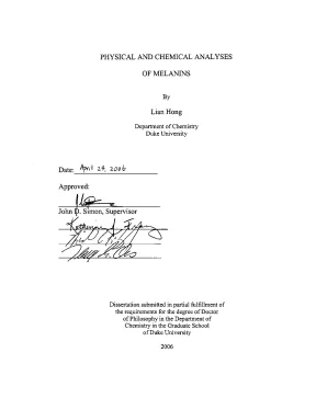
Tải xuống để đọc toàn bộPHYSICAL AND CHEMICAL ANALYSES OF MELANINS By Lian Hong Department of Chemistry Duke University Date:_ April 24 2006 Approved: l£-=. Simon, Supervisor ⁄2 322 Dissertation submitted in partial fulfillment of the requirements for the degree of Doctor of Philosophy in the Department of Chemistry in the Graduate School of Duke University 2006 UMI Number: 3244835 INFORMATION TO USERS The quality of this reproduction is dependent upon the quality of the copy submitted. Broken or indistinct print, colored or poor quality illustrations and photographs, print bleed-through, substandard margins, and improper alignment can adversely affect reproduction. In the unlikely event that the author did not send a complete manuscript and there are missing pages, these will be noted.
Also, if unauthorized copyright material had to be removed, a note will indicate the deletion. ® UMI UMI Microform 3244835 Copyright 2007 by ProQuest Information and Learning Company. All rights reserved. This microform edition is protected against unauthorized copying under Title 17, United States Code.
ProQuest Information and Learning Company 300 North Zeeb Road P. Box 1346 Ann Arbor, MI 48106-1346 ABSTRACT PHYSICAL AND CHEMICAL ANALYSES OF MELANINS By Lian Hong Department of Chemistry Duke University Date:__ April 24 2006 Approved: An abstract of a dissertation submitted in partial fulfillment of the requirements for the degree of Doctor of Philosophy in the Department of Chemistry in the Graduate School of Duke University 2006 Abstract Melanin is a pigment that is ubiquitous in the biological world. In addition to its obvious function of adornment, melanin is believed to serve as a metal reservoir and sunscreen. This work examined the metal binding behavior, the chemical structures and the interaction with light of natural melanins isolated from several different sources.
Sepia eumelanin was used as a model to study the metal binding behavior of eumelanins. Infrared spectroscopic analysis suggests that Mg(II), Ca(II) and Zn(II) are mainly bound to carboxyl groups and Cu(II) binds to hydroxyl groups at pH ~4. The effect of metal content on the aerobic reactivity was examined by analyzing the ability of melanin to nick DNA. Of the metals studied, Cu(II)- and Fe(III)-loaded melanin showed the most damaging effects to DNA.
The photoreactivity of melanin is most important in two parts of the body: the skin and the eye. We first studied melanosomes from hair, which correlate well with the pigment in the skin. Chemical analysis showed that eumelanin was the major pigment in the melanosomes in black hair, while the melanosomes from red hair contained both pheomelanin and eumelanin. Photoelectron emission microscopic (PEEM) analyses on the two melanosomes revealed that the photoionization threshold of eumelanin was 4.4 eV and that pheomelanin was 3.
The low threshold of pheomelanin may contribute to the high incidence of skin cancer in redheads. 11 We then examined the physical and chemical properties of bovine ocular melanosomes. The collective data showed that iris melanosomes had a higher degree of crosslinking and conjugation than choroid and RPE melanosomes of the same age group. The content of carboxyl groups and the degree of conjugation in melanosomes were smaller in older samples.
These tissue and age-related differences are likely to result in variation of efficiency in melanin functions such as metal binding and light absorption. Finally, we examined the photoionization properties of human RPE melanosomes and lipofuscin, another important pigment in the RPE. RPE melanosomes became more pro-oxidant with aging and lipofuscins were more pro- oxidant than melanosomes. iv Contents Page Abstract ili List of Tables vi List of Figures Vii Abbreviations 1X Acknowledgements xii Chapter One: Background Chapter Two: Insights to the Metal Binding to Melanin and its Effects on the 11 Aerobic Reactivity of Melanin Chapter Three: Physiochemical Analysis of Melanosomes Extracted from 35 Human Black and Red Hair Chapter Four: Physiochemical Analysis of Melanosomes Extracted from 57 Bovine Choroid, Iris and Retinal Pigment Epithelium Chapter Five: Age-Dependent Ionization Potentials of Melanosomes and 99 Lipofuscin Isolated from Human Retinal Pigment Epithelium Cells References 121 Biography 132 List of Tables Table Description Page The pH values of salt solutions after suspension of EDTA-washed 19 melanin.
2-2 Summary of the changes in the IR spectra of melanin upon loading of 22 different metals. 3-1 Chemical degradation analyses of human hair melanosomes. 47 3-2 Metal ion contents of human hair melanosomes. 49 Summary of the size analysis of the melanosomes isolated from 68 mature bovine eyes.
Summary of the size analysis of the melanosomes isolated from 69 newborn bovine eyes. 4-3 The contents of Na(I), Mg(II), K(I), Ca(ID and Zn(II) in bovine ocular 72 melanosomes determined by ICP-MS. 4-4 Summary of amino acid analyses for bovine ocular melanosomes. 74 4-5 Summary of chemical degradation analyses of bovine ocular 76 melanosomes.
4-6 C, N and H mass percentages in melanosomes obtained by elemental 78 analysis are corrected according to their amino acid and metal contents. 4-7 C/N and O/N molar ratios on the surface of bovine ocular 82 melanosomes determined by XPS. 4-8 Composition of carbon functional groups. 85 Composition of oxygen functional groups.
86 4-10 Composition of nitrogen functional groups. 87 4-11 The peak positions of the aromatic rings and carbonyls in the IR 91 spectra of bovine ocular melanosomes. Summary of the size analysis of human RPE melanosomes from three 107 vi different patients. List of Figures Description Page An abbreviated illustration of biogenesis of eumelanin and pheomelanin.
1-2 Proposed typical structures of eumelanin and pheomelanin based on biosynthetic pathway and chemical degradation analysis of melanin. IR spectrum of Ca(II)-enriched melanin as a function of the solution 23 concentration of Ca(ID). IR spectrum of Zn(I])-enriched melanin as a function of the solution 25 concentration of Zn(II) in the spectral region of a) 900-1900 cm” and b) 2000-4000 cm”. 2-3 IR spectrum of Cu(II)-enriched melanin as a function of the solution 26 concentration of Cu(II) in the spectral region of a) 900-1900 cm” and b) 2000-4000 cm”.
2-4 Image of a typical gel of pUC18 DNA with bands of two forms. 30 Aerobic reactivity of melanins loaded with different metal ions in a) 31 dark and b) light reactions. 2-6 The effects of [Fe(III] on the ability of melanin to break supercoiled 33 DNA in a dark reaction. 3-1 AFM images of melanosomes isolated from a) black hair and b) red 44 hair.
3-2 High magnification AFM images of black-hair melanosomes (a, b) 45 and red-hair melanosomes (c, d). 3-3 Integrated wavelength-dependent FEL-PEEM data for a) human black 51 hair and b) human red hair melanosomes. 3-4 Time-dependent absorption of Fe(II)-Cyt ¢ following the mixing of 33 Fe(II)-Cyt c with equal masses of eumelanosomes and pheomelanosomes. vii SEM images of melanosomes isolated from a) mature bovine RPE, b) newborn bovine RPE, c) mature bovine choroid, d) newborn bovine choroid, e) mature bovine iris and f) newborn bovine iris.
The distributions of the lengths of the long axis of a) newborn bovine 66 RPE melanosomes and b) mature bovine RPE melanosomes. The high-resolution X-ray photoelectron spectra of Cls in (a) 83 newborn choroid and (b) mature choroid melanosomes. 4-4 The high-resolution X-ray photoelectron spectra of Ols in (a) 84 newborn choroid and (b) mature choroid melanosomes. 4-5 IR spectra of a) newborn bovine choroid, iris and RPE melanosomes 90 b) mature bovine choroid, iris and RPE melanosomes in the range of 800-2000 cm `.
4-6 The !C CP/MAS solid-state NMR spectra of a) mature choroid 95 melanosomes, b) EDTA-washed mature iris melanosomes, c) EDTA- washed mature choroid melanosomes, d) EDTA-washed newborn iris melanosomes and e) EDTA-washed newborn choroid melanosomes. 5-1 SEM images of RPE melanosomes from patients a) 14, b) 59, and c) 105 76, years old, respectively. AFM micrographs of RPE melanosomes: a, b) a smooth granule and 109 c, d) a granule with a rough surface and e, f) a “transient” granule. 5-3 SEM images of lipofuscin granules from donors a) 14, b) 59, and c) 110 76 years old.
5-4 The emission spectra of RPE lipofuscin granules from donors a) 14, b) 112 59, and c) 76 years old. 5-5 Integrated wavelength-dependent FEL-PEEM data for bovine ovoid 115 and rod-shaped melanosomes. 5-6 Integrated wavelength-dependent FEL-PEEM data for melanosomes 116 isolated from human RPE cells from a) 14- and b) 76-year-old specimens. 5-7 Integrated wavelength-dependent FEL-PEEM data for lipofuscin 117 isolated from human RPE cells from a) 14- and b) 76-year-old specimens.
Vili Abbreviations AEM atomic force microscopy aminohydroxyphenylalanine age-related macular degeneration ChNS chloroform-insoluble ChS chloroform-soluble COOH carboxyl CP/MAS cross-polarization magic angle spinning CV coefficient of variation Cyt Cytochrome DHI 5,6-dihydroxylindole DHICA 5,6-dihydroxylindole-2-carboxylic acid DOPA dihydroxylphenylalanine DTT dithiothreitol EB ethidium bromide EDTA ethylenediaminetetraacetic acid FEL free electron laser HI hydriodic acid HPLC high performance liquid chromatography ICP-MS inductively coupled plasma mass spectrometry IPE iris pigment epithelium infrared 1X the length of the long axis minimal erythemal dose amine normal hydrogen electrode national institute of standards and technology nuclear magnetic resonance neuromelanin OH hydroxyl PBS phosphate buffered saline PBSA Dulbecco’s phosphate buffered saline without calcium and magnesium PD Parkinson’s disease PEEM photoelectron emission microscopy PTCA 2,3,5-pyrroletricarboxylic acid ROS reactive oxygen specie RPE retinal pigment epithelium RSF relative sensitivity factor the length of the short axis SEM scanning electron microscopy SN substantia nigra TEM transmission electron microscopy TDCA thiazole-4,5-dicarboxyl acid ™ total melanin X TMS tetramethylsilane XPS X-ray photoelectron spectroscopy xi Acknowledgements First I would like to thank my thesis advisor Dr. Simon for his guidance, support, encouragement and training throughout these past years. His broad connections with research groups and scientists outside Duke are essential for the completion of my dissertation. I gratefully acknowledge our collaborators: Prof.
Kazumasa Wakamatsu, and Shosuke Ito for carrying out chemical degradation analysis; Bhavin B. Adhyaru, Chi- Yuan Cheng and Prof. Bowers for collecting '3C solid state NMR spectra; and Jacob Garguilo, Robert J. Nemanich and Glenn S.
Edwards for performing photoelectron emission microscopic analyses. I also appreciate the past and current members of the Simon Lab. In particular, I would like to thank Dr. Yan liu for stimulating discussions, help of experiments and sharing of her experience and knowledge with me.
I acknowledge her invaluable help and her important inputs in chapter 2 and chapter 3 of this thesis. Alexander Samokhvalov for employing PEEM and analyzing black/red hair melanosomes and Laura Anzaldi for help on the size analysis of human RPE melanosomes. I also thank Jenny Perry, Laura Lamb, Valerie Kempf, William Derek Bush, Weslyn Ward and Jean Hatcher for various help. I owe special thanks to my husband Xiangqian for his patience, love, and encouragement during these years of my studies.
Finally, I would like to thank my family and friends for their support. xii Chapter One. Background Occurrence of Melanin and Melanogenesis Melanin is an omnibus term describing a large range of natural and synthetic phenolic-quinonoid pigments. Since this current work is focusing on the characterization of natural melanins, only the properties of natural melanins will be discussed in this section.
Natural melanins are classified into two groups according to their chemical structures and molecular precursors: the insoluble black to brown eumelanin derived from dihydroxyphenylalanine (DOPA), and the alkaline-soluble yellow to red pheomelanin derived from DOPA and cysteine (Crippa 1989; Ito 1993; Riley 1997; Ito 1998). Eumelanin is widespread in the biological world. It occurs in the skin, hair, eye, brain and inner ear of mammals, the feathers of birds, the ink sac of cuttlefish, and in some reptiles, amphibians and fungi. Pheomelanin, on the other hand, is mainly found in the feathers of birds and the hair and skin of mammals.
Melanin pigmentation occurs in a specialized organelle, the melanosome, in most pigment generating cells (Prota 1992). In addition to the melanin content, melanosomes contain structural proteins, enzymes, lipids, and metals (Prota 1992). Eumelanosomes, with eumelanin as the major pigment, are mostly integrated granules, uniformly and densely packed with pigments (Parakkal 1967). Pheomelanosomes, however, with pheomelanin as the major pigment, are unevenly packed with pigments and are loosely aggregated small granules (Parakkal 1967).
Nội dung được bảo vệ bản quyền — Tải xuống đầy đủ
Trích dẫn luận án này
Lian Hong (2006). Luận án tiến sĩ: Physical and chemical analyses of melanins [Luận án tiến sĩ, duke university]. LuanAn.net. https://luanan.net/khoa-hoc-giao-duc/luan-an-tien-si-physical-and-chemical-analyses-of-melanins
Từ khóa và chủ đề nghiên cứu
Từ khóa liên quan
Xem thêm luận án cùng lĩnh vực
Chủ đề nghiên cứu
Câu hỏi thường gặp
Luận án "Luận án tiến sĩ: Physical and chemical analyses of melanins" nghiên cứu về vấn đề gì?
Luận án tiến sĩ tập trung phân tích vật lý, hóa học của melanin. Khám phá cấu trúc, tính chất, ứng dụng tiềm năng trong khoa học.
Luận án "Luận án tiến sĩ: Physical and chemical analyses of melanins" được bảo vệ tại trường nào?
Luận án này được bảo vệ tại duke university. Năm bảo vệ: 2006.
Luận án "Luận án tiến sĩ: Physical and chemical analyses of melanins" thuộc chuyên ngành gì?
Luận án "Luận án tiến sĩ: Physical and chemical analyses of melanins" thuộc chuyên ngành Chemistry. Danh mục: Khoa Học Giáo Dục.
Luận án "Luận án tiến sĩ: Physical and chemical analyses of melanins" có bao nhiêu trang?
Luận án "Luận án tiến sĩ: Physical and chemical analyses of melanins" có 146 trang. Bạn có thể xem trước một phần tài liệu ngay trên trang web trước khi tải về.
Cách tải luận án "Luận án tiến sĩ: Physical and chemical analyses of melanins" về máy như thế nào?
Để tải luận án về máy, bạn nhấn nút "Tải xuống ngay" trên trang này, sau đó hoàn tất thanh toán phí lưu trữ. File sẽ được tải xuống ngay sau khi thanh toán thành công. Hỗ trợ qua Zalo: 0559 297 239.