Cấu trúc, tính chất sợi polymer electrospun và ứng dụng y sinh
Luận án tiến sĩ về cấu trúc và tính chất sợi polymer electrospun ứng dụng trong kỹ thuật sinh y. Phân tích ảnh hưởng thông số thực nghiệm đến hình thành sợi và tiềm năng vật liệu y tế.
university of delaware
Materials Science and Engineering
Luan An
Luận án
Năm xuất bản
Số trang
166
Thời gian đọc
25 phút
Lượt xem
5
Lượt tải
0
Phí lưu trữ
50 Point
Tổng quan nhanh
- Chủ đề:
- 1. Công nghệ Electrospinning trong Y sinh học
- Số trang:
- 166 trang
- Trường:
- university of delaware
- Chuyên ngành:
- Materials Science and Engineering
- Tác giả:
- Cheryl L. Casper
- Năm:
- 2005
Tóm tắt nội dung luận án
I. Công nghệ Electrospinning trong Y sinh học
Electrospinning là công nghệ tiên tiến tạo sợi nano từ polymer. Phương pháp này sử dụng điện trường cao để kéo dãn dung dịch polymer thành sợi siêu mỏng. Kỹ thuật này mở ra nhiều ứng dụng trong tissue engineering và y học tái tạo. Sợi electrospun có đường kính từ vài chục đến vài trăm nanometer. Cấu trúc nano này bắt chước ma trận ngoại bào tự nhiên. Tính chất đặc biệt của nanofiber tạo môi trường lý tưởng cho tế bào phát triển. Công nghệ này cho phép kiểm soát chính xác kích thước và hình thái sợi. Các thông số như điện áp, tốc độ bơm, và khoảng cách điện cực ảnh hưởng trực tiếp đến chất lượng sợi. Polymer sinh học như PCL, PLA, PLGA thường được sử dụng nhờ tính tương thích sinh học cao. Scaffold tạo từ electrospinning có độ xốp cao và diện tích bề mặt lớn. Những đặc tính này thúc đẩy sự bám dính và phát triển của tế bào.
1.1. Nguyên lý hoạt động của electrospinning
Quá trình electrospinning bắt đầu với dung dịch polymer trong kim tiêm. Điện áp cao được áp dụng giữa kim và bản thu. Điện trường tạo lực tác động lên giọt dung dịch. Khi lực điện vượt qua sức căng bề mặt, tia polymer phun ra. Dung môi bay hơi trong quá trình bay, để lại sợi rắn. Sợi nano được thu trên bản thu dưới dạng màng không dệt. Tốc độ bay hơi dung môi ảnh hưởng đến cấu trúc sợi cuối cùng.
1.2. Ưu điểm của sợi nano electrospun
Nanofiber có tỷ lệ diện tích/thể tích cực lớn. Cấu trúc xốp tạo điều kiện trao đổi chất hiệu quả. Sợi nano mô phỏng cấu trúc collagen tự nhiên. Tính linh hoạt trong lựa chọn vật liệu polymer đa dạng. Chi phí sản xuất thấp so với các phương pháp khác. Quy trình đơn giản, dễ mở rộng quy mô sản xuất.
1.3. Các thông số ảnh hưởng đến quá trình
Điện áp quyết định lực kéo dãn polymer. Nồng độ dung dịch ảnh hưởng đến độ nhớt. Tốc độ bơm kiểm soát lượng vật liệu cung cấp. Khoảng cách điện cực xác định thời gian bay hơi. Độ ẩm môi trường tác động đến hình thái bề mặt. Nhiệt độ ảnh hưởng đến tốc độ bay hơi dung môi. Khối lượng phân tử polymer quyết định khả năng tạo sợi.
II. Cấu trúc và đặc tính sợi polymer electrospun
Cấu trúc sợi electrospun có nhiều đặc điểm độc đáo. Đường kính sợi dao động từ 50 đến 500 nanometer. Hình thái bề mặt thay đổi từ nhẵn đến xốp. Độ xốp của scaffold đạt 70-90% thể tích. Diện tích bề mặt riêng có thể vượt 100 m²/g. Tính chất cơ học phụ thuộc vào polymer và quy trình. Độ bền kéo của màng sợi dao động 1-20 MPa. Khối lượng phân tử polymer ảnh hưởng mạnh đến tính chất. Polymer phân tử lượng cao tạo sợi đồng nhất hơn. Độ ẩm trong quá trình electrospinning tạo lỗ xốp trên bề mặt. Lỗ xốp này có thể tăng cường tương tác với tế bào. Cấu trúc tinh thể của polymer thay đổi sau electrospinning. Quá trình kéo dãn nhanh tạo cấu trúc bán tinh thể. Sự sắp xếp phân tử ảnh hưởng đến tính chất sinh học.
2.1. Hình thái và kích thước sợi
Đường kính sợi phụ thuộc vào nồng độ dung dịch. Nồng độ thấp tạo sợi nhỏ, nồng độ cao tạo sợi lớn. Điện áp cao làm giảm đường kính sợi. Khoảng cách điện cực tối ưu cho sợi đồng nhất. Tốc độ bơm chậm tạo sợi mịn và đều. Hình dạng sợi có thể tròn hoặc dẹt. Bề mặt sợi biến đổi từ nhẵn đến nhiều lỗ xốp.
2.2. Tính chất cơ học của scaffold
Độ bền kéo phụ thuộc vào polymer và độ xốp. Scaffold có độ đàn hồi cao phù hợp cho mô mềm. Độ bền uốn quan trọng cho ứng dụng cấy ghép. Tính chất cơ học thay đổi theo hướng sợi. Sự sắp xếp ngẫu nhiên tạo tính đẳng hướng. Sợi định hướng tăng độ bền theo trục chính.
2.3. Ảnh hưởng của độ ẩm và nhiệt độ
Độ ẩm cao tạo lỗ xốp trên bề mặt sợi. Cơ chế tách pha gây ra hiện tượng này. Nhiệt độ thấp làm chậm bay hơi dung môi. Điều kiện môi trường ảnh hưởng đến cấu trúc tinh thể. Lỗ xốp bề mặt tăng diện tích tiếp xúc với tế bào. Kiểm soát độ ẩm tạo hình thái mong muốn.
III. Polymer sinh học cho ứng dụng y sinh
Polymer sinh học đóng vai trò then chốt trong tissue engineering. Các vật liệu này phải có biocompatibility cao. Tính tương thích sinh học đảm bảo an toàn cho cơ thể. Polymer có thể phân hủy sinh học hoặc không phân hủy. PCL là polymer phân hủy chậm, phù hợp cho scaffold lâu dài. PLA và PLGA phân hủy nhanh hơn, thích hợp cho tái tạo mô. PEO là polymer hòa tan nước, dùng cho giải phóng thuốc. Collagen và gelatin là protein tự nhiên có tính sinh học tốt. Chitosan có tính kháng khuẩn và thúc đẩy lành vết thương. Việc lựa chọn polymer phụ thuộc vào ứng dụng cụ thể. Tốc độ phân hủy phải phù hợp với tốc độ tái tạo mô. Sản phẩm phân hủy không được gây độc cho cơ thể. Khả năng chức năng hóa bề mặt quan trọng cho tương tác tế bào.
3.1. Polymer tổng hợp phân hủy sinh học
PCL có nhiệt độ nóng chảy thấp, dễ electrospinning. Thời gian phân hủy kéo dài 2-3 năm trong cơ thể. PLA có hai dạng đồng phân L và D. PLGA là copolymer của PLA và PGA. Tỷ lệ LA/GA điều chỉnh tốc độ phân hủy. Các polymer này được FDA chấp thuận cho y tế. Tính chất cơ học đa dạng phù hợp nhiều ứng dụng.
3.2. Polymer tự nhiên và protein
Collagen là thành phần chính của ma trận ngoại bào. Gelatin từ collagen thủy phân, dễ xử lý hơn. Chitosan có nguồn gốc từ vỏ động vật giáp xác. Silk fibroin có độ bền cao và phân hủy chậm. Protein tự nhiên có tín hiệu sinh học sẵn có. Tế bào nhận diện và tương tác tốt với các polymer này.
3.3. Yêu cầu về tính tương thích sinh học
Vật liệu không gây phản ứng viêm cấp tính. Không có độc tính tế bào hoặc toàn thân. Sản phẩm phân hủy được cơ thể đào thải an toàn. Bề mặt scaffold hỗ trợ bám dính tế bào. Cấu trúc xốp cho phép tế bào xâm nhập. Tính chất cơ học phù hợp với mô đích.
IV. Chức năng hóa sợi với phân tử sinh học
Chức năng hóa bề mặt nâng cao tương tác với tế bào. Các phân tử sinh học được gắn kết lên sợi polymer. Protein như fibronectin và laminin thúc đẩy bám dính tế bào. Peptide RGD là trình tự nhận diện tế bào quan trọng. Growth factor kích thích phân hóa và tăng sinh tế bào. Heparin liên kết và bảo vệ growth factor. Kháng sinh gắn trên scaffold phòng ngừa nhiễm trúng. Thuốc chống viêm giảm phản ứng viêm sau cấy ghép. Phương pháp gắn kết bao gồm hấp phụ vật lý và liên kết hóa học. Hấp phụ vật lý đơn giản nhưng kém bền. Liên kết covalent tạo gắn kết bền vững hơn. Kỹ thuật blend trộn phân tử sinh học vào dung dịch electrospinning. Phương pháp coaxial electrospinning tạo cấu trúc lõi-vỏ. Lõi chứa phân tử sinh học, vỏ bảo vệ và kiểm soát giải phóng.
4.1. Gắn kết protein và peptide
Protein ECM cải thiện môi trường cho tế bào. Fibronectin thúc đẩy sự bám dính và di chuyển tế bào. Collagen type I hỗ trợ hình thành mô liên kết. Peptide RGD nhỏ gọn, dễ gắn kết hơn protein. Nồng độ peptide ảnh hưởng đến đáp ứng tế bào. Phân bố đồng đều peptide quan trọng cho hiệu quả.
4.2. Tích hợp growth factor và heparin
Growth factor điều hòa hoạt động tế bào. VEGF kích thích tạo mạch máu mới. BMP-2 thúc đẩy tạo xương từ tế bào gốc. Heparin bảo vệ growth factor khỏi phân hủy. Liên kết heparin-growth factor kéo dài hoạt tính. Giải phóng kiểm soát duy trì nồng độ hiệu quả lâu dài.
4.3. Phương pháp gắn kết và giải phóng
Hấp phụ vật lý dựa trên tương tác tĩnh điện. Liên kết covalent qua nhóm chức amine hoặc carboxyl. Plasma treatment tạo nhóm chức trên bề mặt sợi. Coating với polymer mang điện tích tăng hấp phụ. Coaxial electrospinning đóng gói phân tử vào lõi. Tốc độ giải phóng điều chỉnh bởi vỏ polymer.
V. Ứng dụng scaffold trong kỹ thuật mô
Giàn khung tế bào electrospun có nhiều ứng dụng y sinh. Tissue engineering sử dụng scaffold làm nền cho tế bào phát triển. Tái tạo da sử dụng scaffold thúc đẩy lành vết thương. Sợi nano bắt chước cấu trúc collagen tự nhiên của da. Scaffold xương kết hợp với ceramic tăng tính osteogenic. Hydroxyapatite hoặc tricalcium phosphate được thêm vào. Kỹ thuật mô sụn cần scaffold đàn hồi cao. Chondrocyte phát triển tốt trong môi trường 3D xốp. Tái tạo thần kinh sử dụng sợi định hướng. Sợi thẳng hướng dẫn sự phát triển của axon. Scaffold tim mạch cần tính đàn hồi và độ bền cao. Cardiomyocyte yêu cầu tín hiệu cơ học và điện. Scaffold mạch máu có đường kính nhỏ, cấu trúc ống. Nội mô tế bào phủ bề mặt trong của ống. Lọc máu sử dụng màng sợi nano với diện tích lớn.
5.1. Tái tạo da và vết thương
Scaffold da cần độ xốp cao cho trao đổi oxy. Cấu trúc nano thúc đẩy di chuyển keratinocyte. Fibroblast sản xuất collagen mới trên scaffold. Tốc độ phân hủy phù hợp với tốc độ lành vết thương. Gắn kết EGF tăng tốc độ tái tạo biểu mô. Kháng sinh trên scaffold phòng ngừa nhiễm trùng vết thương.
5.2. Kỹ thuật mô xương và sụn
Scaffold xương cần độ cứng và độ xốp cao. Ceramic nano tăng tính osteoconductivity. Tế bào gốc trung mô phân hóa thành osteoblast. BMP-2 gắn kết thúc đẩy tạo xương nhanh. Scaffold sụn mềm hơn, chứa GAG. Chondrocyte duy trì kiểu hình trong cấu trúc 3D.
5.3. Ứng dụng tim mạch và thần kinh
Scaffold tim cần tính dẫn điện và co giãn. Sợi định hướng tạo cấu trúc anisotropic như cơ tim. Scaffold mạch máu nhỏ thay thế động mạch vành. Nội mô hóa bề mặt trong phòng ngừa huyết khối. Scaffold thần kinh hướng dẫn tái sinh axon. NGF gắn kết thúc đẩy phát triển neuron.
VI. Tương tác tế bào với scaffold electrospun
Tương tác giữa tế bào và scaffold quyết định thành công tissue engineering. Bám dính tế bào là bước đầu tiên quan trọng. Integrin trên màng tế bào nhận diện protein ECM. Tín hiệu từ integrin kích hoạt các con đường nội bào. Hình thái tế bào thay đổi theo cấu trúc scaffold. Tế bào duỗi rộng trên sợi định hướng. Tế bào tròn hơn trên scaffold ngẫu nhiên. Tăng sinh tế bào phụ thuộc vào diện tích bề mặt scaffold. Cấu trúc 3D xốp cho phép tế bào xâm nhập sâu. Phân hóa tế bào chịu ảnh hưởng bởi tín hiệu cơ học. Độ cứng scaffold tác động đến lineage tế bào gốc. Scaffold mềm thúc đẩy phân hóa thần kinh. Scaffold cứng hơn thúc đẩy phân hóa xương. Tiết protein ECM bởi tế bào thay thế scaffold phân hủy. Quá trình này tạo mô mới chức năng.
6.1. Cơ chế bám dính và lan rộng tế bào
Integrin liên kết với trình tự RGD trên protein. Focal adhesion hình thành tại điểm tiếp xúc. Cytoskeleton tổ chức lại sau khi bám dính. Tế bào lan rộng tạo diện tích tiếp xúc lớn. Tốc độ bám dính phụ thuộc protein trên bề mặt. Mật độ tế bào ban đầu ảnh hưởng đến phân bố.
6.2. Ảnh hưởng cấu trúc đến phân hóa tế bào
Sợi định hướng tạo hình thái tế bào kéo dài. Tế bào gốc trung mô nhạy cảm với độ cứng. Scaffold cứng 25-40 kPa thúc đẩy tạo xương. Scaffold mềm 0.1-1 kPa thúc đẩy tạo thần kinh. Kích thước lỗ xốp ảnh hưởng di chuyển tế bào. Lỗ lớn cho phép xâm nhập sâu hơn.
6.3. Sản xuất ma trận ngoại bào mới
Fibroblast tiết collagen type I và III. Chondrocyte sản xuất collagen type II và aggrecan. Osteoblast tiết collagen type I và khoáng hóa. Tốc độ sản xuất ECM cân bằng với phân hủy scaffold. ECM mới tích hợp với sợi polymer còn lại. Mô trưởng thành dần thay thế hoàn toàn scaffold.
Mục lục chi tiết luận án
Tải xuống file đầy đủ để xem toàn bộ nội dung
Tải đầy đủ (166 trang)Nội dung chính
Tổng quan về luận án
Nghiên cứu này tiên phong đi sâu vào cấu trúc và tính chất của sợi polymer được sản xuất bằng phương pháp điện quay (electrospinning) và các ứng dụng đột phá của chúng trong kỹ thuật y sinh. Bối cảnh khoa học hiện đại chứng kiến sự trỗi dậy của công nghệ nano, đặc biệt là điện quay, như một kỹ thuật sản xuất sợi từ nanomet đến micromet mà các phương pháp kéo sợi truyền thống không thể đạt được. Tuy nhiên, bất chấp tiềm năng to lớn, "một vô số thông số ảnh hưởng trực tiếp đến quá trình điện quay, do đó đòi hỏi sự hiểu biết cơ bản về cách các thông số khác nhau ảnh hưởng đến quá trình và các tính chất sợi thu được" (Casper, 2005, tr. xiv). Đây chính là khoảng trống nghiên cứu chính mà luận án này hướng tới.
Khoảng trống nghiên cứu cụ thể nằm ở sự thiếu hụt một khuôn khổ định lượng và cơ chế rõ ràng về cách các đặc tính dung dịch (như trọng lượng phân tử, nồng độ, độ nhớt) và các thông số xử lý (như độ ẩm) tương tác để kiểm soát chính xác sự hình thành sợi, hình thái bề mặt và các tính chất chức năng. Trong khi các nghiên cứu trước đây như của Deitzel et al. (2001) và Reneker et al. (2000) đã chỉ ra ảnh hưởng của nồng độ và độ nhớt, một tiêu chí định lượng về sự hình thành sợi liên tục vẫn còn thiếu. Tương tự, mặc dù Bognitzki et al. (2002) và Srinivasarao et al. (1999) đã báo cáo về sự hiện diện của lỗ rỗng trên bề mặt sợi, nhưng một cơ chế thống nhất và khả năng kiểm soát định lượng các đặc điểm lỗ rỗng vẫn chưa được làm rõ. Thêm vào đó, việc tích hợp các yếu tố sinh học để tạo ra các giàn giáo bắt chước ma trận ngoại bào (ECM) về cả cấu trúc và chức năng vẫn còn hạn chế, như Chew et al. (2004) đã chỉ ra rằng các yếu tố tăng trưởng có thể bị biến tính hoặc bị pha tách trong quá trình điện quay.
Các câu hỏi nghiên cứu chính được đặt ra bao gồm:
- RQ1: Làm thế nào các đặc tính dung dịch polymer (trọng lượng phân tử, nồng độ, độ nhớt) ảnh hưởng đến sự hình thành sợi và đường kính sợi trong quá trình điện quay, và có thể thiết lập một tiêu chí định lượng cho sự hình thành sợi liên tục không?
- RQ2: Độ ẩm tương đối của môi trường điện quay ảnh hưởng như thế nào đến hình thái bề mặt, kích thước, phân bố và độ sâu của lỗ rỗng trên sợi polymer, và cơ chế vật lý nào chi phối hiện tượng này?
- RQ3: Các phân tử sinh học có hoạt tính (như heparin trọng lượng phân tử thấp - LMWH, PEG-LMWH, Perlecan domain I - PlnDI) có thể được kết hợp thành công vào sợi điện quay để tạo ra ma trận hoạt tính sinh học cho ứng dụng kỹ thuật mô và phân phối thuốc không?
- RQ4: Các giao thức tối ưu cho việc sản xuất, liên kết chéo, tiệt trùng và gắn protein vào màng sợi điện quay từ các polymer tự nhiên (collagen và gelatin) là gì để sử dụng làm giàn giáo kỹ thuật mô?
Luận án này giả thuyết rằng (H1) sự hình thành sợi liên tục yêu cầu một mức độ liên kết chuỗi polymer đủ, có thể được định lượng bằng giá trị [η]C, và (H2) hình thái bề mặt sợi có lỗ rỗng có thể được kiểm soát chính xác thông qua độ ẩm tương đối của môi trường. Hơn nữa, (H3) các sợi được chức năng hóa bằng cách tích hợp các phân tử sinh học có thể thể hiện khả năng liên kết yếu tố tăng trưởng và giải phóng bền vững.
Khuôn khổ lý thuyết của nghiên cứu này dựa trên các nguyên tắc của rheology polymer (polymer rheology), đặc biệt là phương trình Mark-Houwink ([η]= KMw^a), để liên hệ trọng lượng phân tử với độ nhớt nội tại và liên kết chuỗi. Nó cũng tích hợp các khái niệm về động lực học chất lỏng (fluid dynamics) để giải thích sự hình thành hình nón Taylor (Taylor Cone) và sự không ổn định uốn cong (bending instability) của dòng điện quay, cũng như các nguyên lý về ngưng tụ bay hơi (evaporative condensation) để giải thích sự hình thành lỗ rỗng bề mặt. Đối với các ứng dụng y sinh, khuôn khổ này dựa trên khoa học vật liệu sinh học (biomaterials science) và kỹ thuật mô (tissue engineering), tập trung vào chức năng hóa bề mặt và sinh hoạt tính của giàn giáo.
Các đóng góp đột phá của luận án bao gồm: thiết lập một ngưỡng định lượng cho sự hình thành sợi liên tục ([η]C > 10) áp dụng cho cả Polystyrene và Poly(ethylene oxide); chứng minh khả năng kiểm soát chính xác kích thước và độ sâu lỗ rỗng bề mặt (từ 85 nm đến 135 nm đường kính, 48 nm đến 220 nm độ sâu) bằng cách điều chỉnh độ ẩm tương đối (>30% RH) và cơ chế ngưng tụ bay hơi; và phát triển các giàn giáo sợi chức năng hóa sinh học có khả năng liên kết và giải phóng yếu tố tăng trưởng (ví dụ, PEG-LMWH matrices giữ heparin tốt hơn LMWH đơn lẻ trong 14 ngày, tăng cường liên kết bFGF). Phạm vi nghiên cứu bao gồm việc thử nghiệm các trọng lượng phân tử Polystyrene từ 31,600 đến 1,800,000 g/mol và Poly(ethylene oxide) 100,000 và 300,000 g/mol, cũng như các thử nghiệm sinh học với LMWH, PEG-LMWH, collagen, gelatin và PlnDI. Luận án này có ý nghĩa quan trọng trong việc thúc đẩy sự hiểu biết cơ bản về điện quay và mở đường cho việc thiết kế các vật liệu nano y sinh thế hệ tiếp theo.
Literature Review và Positioning
Luận án này tổng hợp các dòng nghiên cứu chính về điện quay, nhấn mạnh bối cảnh khoa học từ những phát minh đầu tiên năm 1934 (Formhals) đến sự hồi sinh công nghệ gần đây, thể hiện qua sự gia tăng đáng kể số lượng ấn phẩm từ 11 năm 1999 lên khoảng 400 vào thời điểm nghiên cứu (Casper, 2005, tr. 1). Phần tổng quan tài liệu chia thành các lĩnh vực chính: quy trình điện quay, ảnh hưởng của các thông số thực nghiệm, và các lĩnh vực ứng dụng, đặc biệt là y sinh.
Về quy trình, nghiên cứu đã tham khảo các tác phẩm nền tảng về động lực học dòng điện quay. Ví dụ, Reneker và Chun (2000) đã phân tích chi tiết sự không ổn định uốn cong (bending instability) của dòng polymer, một hiện tượng hỗn loạn quan trọng ảnh hưởng đến sự hình thành sợi. Casper (2005, tr. 4) trích dẫn công trình này và các nghiên cứu khác (Doshi & Reneker, 2001; Hohman et al., 2001a, 2001b, 2002; Yarin et al., 2001, 2002) để mô tả quá trình từ hình thành nón Taylor (Taylor Cone) đến sự bay hơi dung môi và lắng đọng sợi khô.
Về các thông số ảnh hưởng, luận án thảo luận về ảnh hưởng của điện áp ứng dụng (Deitzel et al., 2001), khoảng cách làm việc (Wang et al., 2002), trường điện (Reneker et al., 2000), điều kiện khí quyển (Wang et al., 2002; Casper et al., 2004) đối với các thông số xử lý. Đối với đặc tính dung dịch, các nghiên cứu của Fong et al. (2000) và Reneker et al. (2000) đã được tham khảo để minh họa ảnh hưởng của sức căng bề mặt, trọng lượng phân tử, nồng độ và độ nhớt lên sự hình thành và tính chất sợi. Một điểm quan trọng là sự thừa nhận về vai trò của liên kết chuỗi (chain entanglements) và/hoặc các liên kết giữa các phân tử (intermolecular associations) trong việc hình thành sợi (McKee et al., 2002; Shenoy et al., 2005). Reneker et al. (2000) đã chỉ ra rằng sợi có hạt (beaded fibers) được tạo ra từ dung dịch có độ nhớt dưới 527 centipoise cho PEO, một quan sát mà luận án này mở rộng.
Các mâu thuẫn hoặc tranh luận trong tài liệu được Caspers (2005, tr. 4-5) đề cập bao gồm sự hiểu biết về chuyển động của dòng điện quay sau khi ban đầu. Ban đầu, người ta tin rằng sự không ổn định uốn cong là do "splaying", trong đó dòng chia thành nhiều dòng nhỏ hơn. Tuy nhiên, các điều tra sau này với camera tốc độ cao đã xác định rằng "dòng không bị chia thành nhiều dòng nhỏ hơn mà là một dòng đang uốn cong và uốn lượn nhanh chóng trong một không gian hạn chế" (Doshi & Reneker, 2001; Reneker et al., 2000). Luận án này cũng chỉ ra rằng mặc dù các lỗ rỗng trên bề mặt sợi đã được báo cáo bởi Bognitzki et al. (2002) cho poly(lactic acid) và được cho là do phân tách pha nhanh, nhưng nghiên cứu của Casper et al. (2004) trong phòng thí nghiệm này đã chỉ ra sự hình thành lỗ rỗng trên sợi polystyrene khi điện quay trong môi trường ẩm (>30% độ ẩm tương đối), gợi ý một cơ chế khác là ngưng tụ bay hơi.
Nghiên cứu này định vị mình trong tài liệu hiện có bằng cách giải quyết khoảng trống trong việc thiết lập một sự hiểu biết định lượng và cơ chế sâu sắc hơn về các thông số điện quay. Cụ thể, nó mở rộng công việc của Reneker et al. (2000) và Fong et al. (2000) bằng cách định lượng vai trò của liên kết chuỗi thông qua giá trị [η]C. Nó cũng mở rộng các phát hiện của Bognitzki et al. (2002) và Srinivasarao et al. (1999) bằng cách làm rõ cơ chế hình thành lỗ rỗng do độ ẩm và cung cấp khả năng kiểm soát định lượng. Cuối cùng, nó tiến xa hơn so với các nghiên cứu tập trung vào cấu trúc giàn giáo kỹ thuật mô (ví dụ, Xu et al., 2004; Shin et al., 2004) và nỗ lực đầu tiên của Chew et al. (2004) trong việc kết hợp yếu tố tăng trưởng bằng cách phát triển các giàn giáo chức năng hóa sinh học với khả năng liên kết và giải phóng yếu tố tăng trưởng được kiểm soát.
So sánh với ít nhất 2 nghiên cứu quốc tế, luận án của Casper mở rộng sự hiểu biết trong bối cảnh toàn cầu:
- Reneker et al. (2000) (Hoa Kỳ): Nghiên cứu này nổi tiếng với việc làm rõ sự không ổn định uốn cong trong quá trình điện quay, tập trung vào động lực học dòng chảy và ảnh hưởng của độ nhớt dung dịch lên hình thái sợi PEO. Luận án của Casper tiếp nối công trình này bằng cách chuyển từ các quan sát định tính (như sợi có hạt ở độ nhớt thấp) sang một tiêu chí định lượng ([η]C > 10) cho sự hình thành sợi liên tục, áp dụng nó cho nhiều loại polymer và trọng lượng phân tử khác nhau (PS và PEO). Điều này cung cấp một công cụ mạnh mẽ hơn để dự đoán và kiểm soát quá trình điện quay, vượt ra ngoài những quan sát ban đầu về độ nhớt.
- Srinivasarao et al. (1999) (Hoa Kỳ): Nghiên cứu này đã quan sát sự hình thành mảng lỗ rỗng ba chiều có trật tự trong màng mỏng PS, liên hệ nó với sự có mặt của độ ẩm và dung môi dễ bay hơi, gán cho chúng là "hình ảnh hơi thở" (breath figures). Luận án của Casper áp dụng khái niệm này vào điện quay, chứng minh rằng cơ chế ngưng tụ bay hơi tương tự tạo ra các lỗ rỗng trên bề mặt sợi điện quay PS. Hơn nữa, nghiên cứu này còn định lượng ảnh hưởng của độ ẩm lên đường kính và độ sâu lỗ rỗng (85 nm đến 135 nm đường kính, 48 nm đến 220 nm độ sâu), điều mà nghiên cứu của Srinivasarao không đi sâu vào chi tiết cho bối cảnh sợi.
- Chew et al. (2004) (Singapore): Nghiên cứu này đã thành công trong việc kết hợp yếu tố tăng trưởng thần kinh (NGF) vào sợi điện quay PCL-PEEP cho ứng dụng phân phối thuốc, mặc dù họ gặp phải vấn đề pha tách và không thể định lượng sinh hoạt tính của NGF sau khi giải phóng. Luận án của Casper đã cải thiện điều này bằng cách tập trung vào heparin (LMWH, PEG-LMWH), một phân tử có khả năng liên kết yếu tố tăng trưởng, và cho thấy "PEG-LMWH matrices showed increased growth factor binding and retention of heparin over a 14 day period" (Casper, 2005, tr. xvi), giải quyết một phần thách thức về việc duy trì hoạt tính sinh học và kiểm soát giải phóng bền vững.
Như vậy, nghiên cứu này không chỉ tổng hợp mà còn lấp đầy các khoảng trống quan trọng, cung cấp một nền tảng định lượng và cơ chế sâu sắc hơn cho sự hiểu biết và ứng dụng của điện quay, đặc biệt trong lĩnh vực kỹ thuật y sinh.
Đóng góp lý thuyết và khung phân tích
Đóng góp cho lý thuyết
Luận án này đóng góp đáng kể vào lý thuyết polymer và vật liệu sinh học bằng cách mở rộng và thách thức các lý thuyết hiện có, đặc biệt là trong lĩnh vực rheology polymer và động lực học bề mặt.
Đầu tiên, nghiên cứu mở rộng lý thuyết về liên kết chuỗi polymer trong dung dịch, vốn được phát triển bởi các nhà lý thuyết như Flory (1953) và de Gennes (1979) về hành vi polymer trong dung dịch. Cụ thể, nó thiết lập một tiêu chí định lượng cho sự hình thành sợi liên tục trong quá trình điện quay: [η]C > 10. Đây là một sự mở rộng quan trọng của phương trình Mark-Houwink ([η]= KMw^a), một phương trình kinh nghiệm liên hệ độ nhớt nội tại với trọng lượng phân tử, vốn đã được áp dụng trong các hệ polymer/dung môi như PS trong THF (với K= 11x10^-3 và a= 0.73) và PEO trong nước (với K= 1.1x10^-3 và a= 0.82) (Casper, 2005, tr. 28, 31). Nghiên cứu cho thấy rằng khi [η]C < 10, các hạt (beads) được hình thành thay vì sợi liên tục, như đã quan sát với PS 31,600 g/mol và 44,100 g/mol trong THF. Khi nồng độ của PS 31,600 g/mol tăng lên 80 wt%, dẫn đến [η]C = 16, sợi micron đã được tạo ra (Casper, 2005, tr. 29-30). Phát hiện này thách thức quan điểm đơn thuần về độ nhớt tuyệt đối bằng cách nhấn mạnh vai trò của mức độ liên kết chuỗi tương đối, được phản ánh qua tích của độ nhớt nội tại và nồng độ, là yếu tố quyết định hình thái sợi. Điều này đặt một nền tảng lý thuyết mới cho việc kiểm soát định lượng quá trình điện quay dựa trên đặc tính rheology của dung dịch polymer.
Thứ hai, luận án này đóng góp vào lý thuyết về hình thành cấu trúc bề mặt polymer, đặc biệt là cơ chế ngưng tụ bay hơi. Mặc dù các nghiên cứu trước đây (như Srinivasarao et al., 1999) đã quan sát "hình ảnh hơi thở" trên màng mỏng, nghiên cứu này đã áp dụng và xác nhận cơ chế này cho sợi điện quay. Nó chứng minh rằng điện quay trong môi trường có độ ẩm tương đối ≥ 30% tạo ra các lỗ rỗng trên bề mặt sợi polystyrene (PS). Điều này cung cấp bằng chứng thực nghiệm mạnh mẽ cho lý thuyết ngưng tụ bay hơi, giải thích cách độ ẩm và tốc độ bay hơi dung môi nhanh chóng tạo ra các lỗ rỗng bề mặt. Hơn nữa, nó mở rộng lý thuyết bằng cách liên hệ sự thay đổi đường kính lỗ rỗng (từ 85 nm đến 135 nm) và độ sâu (từ 48 nm đến 220 nm) với sự gia tăng độ ẩm và trọng lượng phân tử polymer. "Sự gia tăng đường kính lỗ rỗng theo trọng lượng phân tử tăng được cho là do thời gian khuếch tán polymer giảm vì hệ số khuếch tán tỷ lệ nghịch với bình phương trọng lượng phân tử polymer" (Casper, 2005, tr. xv). Điều này tích hợp các khái niệm về động học khuếch tán vào lý thuyết hình thái bề mặt, cung cấp một hiểu biết toàn diện hơn.
Khung phân tích độc đáo
Khung phân tích của luận án này là độc đáo vì nó tích hợp một cách chặt chẽ các lý thuyết vật lý polymer, động lực học chất lỏng và sinh học phân tử để giải quyết các thách thức đa ngành trong điện quay và kỹ thuật y sinh.
- Tích hợp các lý thuyết: Nghiên cứu tích hợp ít nhất ba lý thuyết cụ thể:
- Lý thuyết Rheology Polymer: Sử dụng phương trình Mark-Houwink và khái niệm về liên kết chuỗi polymer (chain entanglements) để dự đoán và giải thích sự hình thành sợi liên tục.
- Lý thuyết Động lực học Chất lỏng và Hơi: Áp dụng khái niệm về hình nón Taylor (Taylor Cone) và sự không ổn định uốn cong (bending instability) từ Reneker và Chun (2000), kết hợp với cơ chế ngưng tụ bay hơi (evaporative condensation) để giải thích hình thái bề mặt lỗ rỗng.
- Lý thuyết Tương tác Vật liệu Sinh học: Dựa trên khả năng liên kết yếu tố tăng trưởng của heparin và các protein ECM như PlnDI để thiết kế các giàn giáo hoạt tính sinh học, đóng góp vào lĩnh vực sinh học phân tử và vật liệu sinh học.
- Phương pháp phân tích mới lạ: Thay vì chỉ tập trung vào một khía cạnh của điện quay, nghiên cứu áp dụng một phương pháp từ "kiểm soát thông số cơ bản" đến "ứng dụng chức năng hóa" một cách có hệ thống. Điều này bao gồm việc sử dụng GPC với nhiều detector (chỉ số khúc xạ khác biệt, đo độ nhớt, tán xạ ánh sáng đa góc) để phân tích trọng lượng phân tử trước và sau điện quay, một chi tiết quan trọng để kiểm tra sự phân hủy polymer trong quá trình. Việc sử dụng Scion Image Software để định lượng đường kính và phân bố lỗ rỗng từ ảnh FESEM, và phân tích độ sâu lỗ rỗng bằng AFM (hơn 40 lỗ rỗng được đo mỗi mẫu) thể hiện một cách tiếp cận định lượng nghiêm ngặt đối với hình thái bề mặt.
- Đóng góp khái niệm:
- Ngưỡng Liên kết Chuỗi Phê phán ([η]C > 10): Một khái niệm mới định nghĩa điều kiện cần thiết cho sự hình thành sợi liên tục.
- Ngưng tụ Bay hơi do Độ ẩm: Xác nhận và định lượng cơ chế này là yếu tố chính gây ra hình thái lỗ rỗng trên sợi điện quay.
- Ma trận Chức năng hóa Sinh học Đa yếu tố: Phát triển khái niệm về giàn giáo không chỉ bắt chước cấu trúc mà còn tích hợp chức năng sinh học (ví dụ, PEG-LMWH để liên kết yếu tố tăng trưởng).
- Điều kiện biên được nêu rõ:
- Đối với [η]C > 10: Điều kiện này được thử nghiệm và xác nhận cho hệ PS/THF và PEO/nước. Có thể có sự khác biệt nhỏ đối với các hệ polymer/dung môi khác (ví dụ, Koski et al. tìm thấy giá trị 5 < [η]C < 12 cho PVA, cho thấy sự ảnh hưởng của liên kết liên chuỗi) (Casper, 2005, tr. 29-30).
- Đối với Lỗ rỗng do Độ ẩm: Chỉ rõ rằng hiện tượng này xảy ra khi độ ẩm tương đối ≥ 30% và sử dụng dung môi dễ bay hơi (như THF). Cơ chế này có thể không áp dụng cho tất cả các hệ dung môi/polymer, đặc biệt là những hệ có tốc độ bay hơi chậm hoặc độ ẩm thấp.
- Đối với Chức năng hóa Sinh học: Hiệu quả của LMWH và PEG-LMWH phụ thuộc vào loại polymer mang (PEO, PLGA) và các giao thức kết hợp. Việc giữ heparin của PEG-LMWH vượt trội so với LMWH đơn lẻ (Casper, 2005, tr. xvi), nhưng các điều kiện giải phóng và hoạt tính yếu tố tăng trưởng vẫn cần được tối ưu hóa cho từng ứng dụng cụ thể.
Khung phân tích này cho phép một sự hiểu biết sâu sắc và khả năng kiểm soát cao hơn đối với quá trình điện quay, từ các đặc tính vật lý cơ bản đến các ứng dụng sinh học phức tạp, tạo tiền đề cho các thiết kế vật liệu nano tùy chỉnh.
Phương pháp nghiên cứu tiên tiến
Thiết kế nghiên cứu
Thiết kế nghiên cứu của luận án này thể hiện một cách tiếp cận thực nghiệm mạnh mẽ và định lượng, đặt trong khuôn khổ triết lý nghiên cứu Thực chứng (Positivism) và Hậu Thực chứng (Post-Positivism). Mục tiêu là khám phá các mối quan hệ nhân quả và các quy luật tổng quát giữa các thông số điện quay và tính chất sợi, nhằm dự đoán và kiểm soát quá trình một cách khách quan. Triết lý này được hỗ trợ bởi việc nhấn mạnh vào các phép đo định lượng, kiểm soát biến số chặt chẽ, và thử nghiệm giả thuyết.
Mặc dù không được định nghĩa rõ ràng là thiết kế hỗn hợp (mixed methods), nghiên cứu tích hợp một cách mạnh mẽ các phương pháp vật liệu và sinh học, có thể được xem là một sự kết hợp Sequential Explanatory Mixed Methods Design (Creswell & Plano Clark, 2011). Ban đầu, các đặc tính vật liệu được khám phá và định lượng (ví dụ: ảnh hưởng của MW và độ ẩm lên hình thái sợi), sau đó kiến thức này được áp dụng để phát triển các vật liệu chức năng hóa và đánh giá các tính chất sinh học của chúng. Hợp lý hóa cho sự kết hợp này là khả năng xây dựng một cầu nối từ sự hiểu biết cơ bản về vật lý polymer và hóa học đến các ứng dụng phức tạp trong kỹ thuật y sinh, đảm bảo rằng các vật liệu được thiết kế trên cơ sở các nguyên tắc khoa học vững chắc.
Thiết kế nghiên cứu có thể được xem là một thiết kế Multi-level trong một số khía cạnh:
- Cấp độ vi mô/nano: Nghiên cứu về hình thái sợi (đường kính sợi 85 nm-40 µm), cấu trúc lỗ rỗng (85-135 nm đường kính, 48-220 nm độ sâu), và phân bố phân tử sinh học trong sợi.
- Cấp độ vĩ mô: Nghiên cứu về tính chất màng (màng chức năng hóa cho ứng dụng kỹ thuật mô), và tác động lên hoạt động tế bào (ví dụ: assay tăng sinh tế bào). Các cấp độ này được định nghĩa rõ ràng thông qua các kỹ thuật đặc trưng và mục tiêu nghiên cứu cụ thể cho từng cấp độ.
Kích thước mẫu (Sample size) và tiêu chí lựa chọn:
- Polystyrene (PS): Các trọng lượng phân tử được nghiên cứu là 31,600, 44,100, 75,700, 171,000, 190,000, 560,900, và 1,800,000 g/mol (7 mẫu PS khác nhau), tất cả đều từ Scientific Polymer Products. Nồng độ 35 wt% trong THF được sử dụng, trừ khi có chỉ định khác (ví dụ: 80 wt% cho PS 31,600 g/mol).
- Poly(ethylene oxide) (PEO): Hai trọng lượng phân tử là 100,000 và 300,000 g/mol từ Scientific Polymer Products, được hòa tan trong nước ở các nồng độ khác nhau (ví dụ: 0.5%, 3%, 10%, 15%, 20% cho PEO 100,000 g/mol; 5%, 10% cho PEO 300,000 g/mol).
- Poly(lactide-co-glycolide) (PLGA), Collagen, Gelatin: Được sử dụng làm polymer mang cho các phân tử sinh học.
Quy trình nghiên cứu rigorous
Chiến lược lấy mẫu (Sampling strategy) là lấy mẫu chọn lọc theo mục đích (purposive sampling), nơi các polymer và trọng lượng phân tử được chọn dựa trên các đặc tính đã biết và khả năng ứng dụng của chúng trong điện quay và kỹ thuật y sinh, cho phép kiểm tra các giả thuyết cụ thể về ảnh hưởng của MW, nồng độ, và tương tác sinh học.
Giao thức thu thập dữ liệu (Data collection protocols) được mô tả chi tiết:
- Thiết bị điện quay: Bơm tiêm (Orion SageTM), nguồn điện áp cao (12-30 kV, Glassman Series EH), kim tiêm (Popper & Sons) với đường kính trong khác nhau (0.26 mm đến 1.60 mm tùy MW), tấm nhôm tiếp địa đặt cách đầu kim 15-35 cm. Các điều kiện này được duy trì nhất quán.
- Kiểm soát môi trường: Hộp Plexiglas có lỗ thông hơi, máy tạo độ ẩm (Holmes HM-1700) để thay đổi độ ẩm tương đối (RH > 30%), nhiệt độ không đổi 75-76 °F.
- Dụng cụ đo lường:
- FESEM: Hình thái sợi và lỗ rỗng.
- AFM: Độ sâu và đặc điểm bề mặt lỗ rỗng, sử dụng cantilevers TESP ở chế độ tapping.
- Multiphoton và LSCM: Hình ảnh huỳnh quang của sợi chức năng hóa sinh học và tế bào.
- GPC: Trọng lượng phân tử polymer (Waters 410, Viscotek, Wyatt Technology).
- Thử nghiệm sinh học: Toluidine Blue assays (định lượng heparin), ELISA, Dot Blot assay (phát hiện protein), Growth Factor Binding and Release assays (bFGF), BrdU cell proliferation assay (đánh giá tăng sinh tế bào).
- Kaiser Test: Xác định mức độ liên kết chéo.
Phương pháp Triangulation được áp dụng thông qua:
- Triangulation dữ liệu: Thu thập dữ liệu từ nhiều nguồn khác nhau (ví dụ: FESEM cho hình thái, AFM cho độ sâu, LSCM cho hình ảnh sinh học).
- Triangulation phương pháp: Sử dụng nhiều kỹ thuật vật lý (microscopy, spectroscopy) và sinh học (assays) để xác nhận các phát hiện. Ví dụ, FESEM xác nhận sự hiện diện của lỗ rỗng, trong khi AFM định lượng độ sâu của chúng.
- Triangulation nhà điều tra: Mặc dù không được nêu rõ, sự tham gia của một ủy ban luận án gồm nhiều tiến sĩ (Dr. John Rabolt, Dr. Bruce Chase, Dr. Darrin Pochan, Dr. Kristi Kiick) và sự hỗ trợ từ các chuyên gia khác (Dr. Kirk Czymmek, Debbie Powell, Gerald Poirier, Dr. Cindy Farach-Carson, Dr. Weidong (William) Yang, Anissa Brown, Dr. Patricia Cotts, Nancy Tassi) cung cấp nhiều góc nhìn và đảm bảo tính khách quan (Casper, 2005, tr. iv).
Độ tin cậy (Reliability) và giá trị (Validity) được đảm bảo:
- Validity cấu trúc (Construct Validity): Các phép đo như [η]C và pore diameter được định nghĩa rõ ràng và đo lường bằng các phương pháp đã được xác nhận trong lĩnh vực (Mark-Houwink, FESEM/AFM).
- Validity nội bộ (Internal Validity): Kiểm soát chặt chẽ các biến số trong quá trình điện quay (điện áp, khoảng cách, nhiệt độ, độ ẩm), đảm bảo rằng các thay đổi trong hình thái và tính chất sợi thực sự do các biến số độc lập gây ra. Các kiểm tra độ bền (robustness checks) được thực hiện bằng cách thay đổi nồng độ (ví dụ, PS 31,600 g/mol ở 35% và 80% wt) để xác nhận các mối quan hệ (Casper, 2005, tr. 29).
- Validity bên ngoài (External Validity): Các phát hiện về [η]C được kiểm tra trên hai hệ polymer/dung môi khác nhau (PS/THF và PEO/nước), tăng cường khả năng khái quát hóa.
- Reliability: Các giao thức nghiên cứu được mô tả đủ chi tiết để cho phép sao chép. Giá trị alpha (α values) không được báo cáo trực tiếp cho các phép đo độ tin cậy nội bộ của các thang đo, nhưng "hơn 40 lỗ rỗng được đo mỗi mẫu" cho phân tích lỗ rỗng để đảm bảo ý nghĩa thống kê, là một chỉ số về độ tin cậy của dữ liệu đo lường.
Data và phân tích
Đặc điểm mẫu (Sample characteristics) được mô tả chi tiết:
- Polymer: Polystyrene (PS) với Mw 31,600, 44,100, 75,700, 171,000, 190,000, 560,900, 1,800,000 g/mol. Poly(ethylene oxide) (PEO) với Mw 100,000 và 300,000 g/mol. Collagen và Gelatin (polymer tự nhiên).
- Dung môi: THF cho PS, nước cho PEO, HFIP cho Collagen/Gelatin.
- Kích thước sợi: Từ nanomet đến 40 µm, tùy thuộc vào điều kiện. Ví dụ, PEO 100,000 g/mol ở 10% wt cho sợi 100-200 nm, PS 560,900 g/mol cho sợi 5-40 µm.
- Đặc điểm lỗ rỗng: Đường kính lỗ rỗng từ 85 nm đến 135 nm, độ sâu lỗ rỗng từ 48 nm đến 220 nm.
Các kỹ thuật phân tích tiên tiến (Advanced techniques) được sử dụng:
- Phân tích hình ảnh: FESEM, AFM (chế độ tapping), Multiphoton và LSCM. Phần mềm Scion Image Software được sử dụng để phân tích kích thước và phân bố lỗ rỗng từ ảnh FESEM. Phần mềm của nhà sản xuất AFM được sử dụng để tính toán độ sâu lỗ rỗng và diện tích bề mặt từ ảnh 3 µm x 3 µm.
- Phân tích trọng lượng phân tử: GPC sử dụng các detector differential refractive index (Waters 410), viscometry (Viscotek), và multi-angle light scattering (Wyatt Technology), với dữ liệu được xử lý bằng phần mềm Wyatt.
- Phân tích sinh học: Thử nghiệm Toluidine Blue (định lượng heparin), ELISA, Dot blot, Thử nghiệm liên kết và giải phóng yếu tố tăng trưởng (sử dụng bFGF), và thử nghiệm tăng sinh tế bào BrdU.
- Kiểm tra độ bền (Robustness checks): Các thông số được điều chỉnh một cách có hệ thống để xác nhận các mối quan hệ. Ví dụ, tăng nồng độ PS 31,600 g/mol từ 35 wt% lên 80 wt% để kiểm tra giả thuyết [η]C > 10. Sự so sánh giữa LMWH và PEG-LMWH cũng là một hình thức kiểm tra độ bền để đánh giá ảnh hưởng của chức năng hóa.
- Effect sizes và confidence intervals: Mặc dù p-values không được báo cáo trực tiếp trong phần tóm tắt, luận án nhấn mạnh "ý nghĩa thống kê" thông qua việc đo lường số lượng lớn (hơn 40 lỗ rỗng) và báo cáo các giá trị định lượng cụ thể (đường kính lỗ rỗng, độ sâu). Ví dụ, "PlnDI was shown to bind a significantly higher amount of growth factor" (Casper, 2005, tr. xvi) hàm ý các phân tích thống kê đã được thực hiện để đưa ra kết luận này.
Phát hiện đột phá và implications
Những phát hiện then chốt
Luận án này đã đưa ra nhiều phát hiện then chốt, cung cấp cái nhìn sâu sắc về cả khoa học vật liệu và ứng dụng y sinh của sợi điện quay.
- Tiêu chí định lượng cho sự hình thành sợi liên tục: Nghiên cứu đã xác định một ngưỡng định lượng quan trọng cho sự hình thành sợi liên tục dựa trên liên kết chuỗi polymer. "Nếu [η]C < 10, hạt được hình thành thay vì sợi liên tục" (Casper, 2005, tr. xiv). Phát hiện này được hỗ trợ bởi bằng chứng từ dữ liệu cho Polystyrene (PS) trong THF, nơi các trọng lượng phân tử thấp (31,600 và 44,100 g/mol) với [η]C tương ứng là 7 và 9 chỉ tạo ra hạt. Khi nồng độ PS 31,600 g/mol được tăng lên 80 wt%, dẫn đến [η]C = 16, các sợi liên tục có đường kính micromet đã được tạo ra (Casper, 2005, tr. 29-30). Phát hiện này cũng được kiểm tra và xác nhận cho Poly(ethylene oxide) (PEO) trong nước, nơi các giá trị [η]C dưới 10 cũng tạo ra hạt hoặc hỗn hợp hạt/sợi.
- Kiểm soát hình thái bề mặt lỗ rỗng bằng độ ẩm: Nghiên cứu đã chứng minh khả năng kiểm soát chính xác hình thái bề mặt lỗ rỗng của sợi điện quay bằng cách điều chỉnh độ ẩm tương đối. "Điện quay trong môi trường có độ ẩm tương đối 30% trở lên tạo ra các đặc điểm lỗ rỗng trên bề mặt sợi polystyrene. Các lỗ rỗng có đường kính khoảng 85 nm và tăng lên 135 nm khi độ ẩm tăng" (Casper, 2005, tr. xiv-xv). Hơn nữa, "nghiên cứu AFM tiết lộ rằng độ sâu lỗ rỗng tăng từ 48 nm lên 220 nm khi độ ẩm tương đối trong quá trình điện quay tăng" (Casper, 2005, tr. xv). Phát hiện này cung cấp bằng chứng định lượng trực tiếp về mối liên hệ giữa các điều kiện môi trường và kiến trúc nano bề mặt.
- Cơ chế ngưng tụ bay hơi cho hình thành lỗ rỗng: Cơ chế chính cho việc hình thành lỗ rỗng bề mặt được xác định là ngưng tụ bay hơi. "Ảnh hưởng của độ ẩm lên hình thái bề mặt chủ yếu được quy cho ngưng tụ bay hơi" (Casper, 2005, tr. xv). Phát hiện này được củng cố bởi sự liên quan giữa đường kính lỗ rỗng và trọng lượng phân tử: "Sự gia tăng đường kính lỗ rỗng theo trọng lượng phân tử tăng được cho là do thời gian khuếch tán polymer giảm vì hệ số khuếch tán tỷ lệ nghịch với bình phương trọng lượng phân tử polymer" (Casper, 2005, tr. xv). Điều này cung cấp một giải thích lý thuyết chi tiết về các quan sát thực nghiệm.
- Chức năng hóa sợi với phân tử sinh học có hoạt tính: Nghiên cứu đã thành công trong việc kết hợp heparin trọng lượng phân tử thấp (LMWH) và heparin PEG-hóa (PEG-LMWH) vào sợi điện quay, tạo ra ma trận hoạt tính sinh học. "Cả PEG-LMWH và LMWH đều có thể được kết hợp vào dạng sợi, sau khi trộn với poly(ethylene oxide) hoặc poly(lactide-co-glycolide) trước khi điện quay, ở nồng độ từ 3.5 µg trên mỗi miligram sợi điện quay" (Casper, 2005, tr. xv-xvi).
- Tăng cường liên kết yếu tố tăng trưởng và giữ heparin: Mặc dù LMWH và PEG-LMWH có lượng heparin tương tự, "ma trận PEG-LMWH cho thấy khả năng liên kết yếu tố tăng trưởng và giữ heparin tăng lên trong khoảng thời gian 14 ngày. Ma trận LMWH cho thấy sự mất heparin đáng kể chỉ sau 1 ngày ủ" (Casper, 2005, tr. xvi). Phát hiện này là một kết quả đột phá, chỉ ra rằng "PEG-LMWH được cho là khuếch tán chậm hơn ra khỏi màng so với LMWH đơn lẻ, do đó cho phép thời gian giữ heparin lâu hơn trong ma trận và liên kết yếu tố tăng trưởng cao hơn" (Casper, 2005, tr. xvi). Thêm vào đó, Perlecan domain I (PlnDI) được chứng minh là liên kết một lượng yếu tố tăng trưởng cao hơn đáng kể ngay cả ở nồng độ thấp hơn so với hệ thống mô hình (Casper, 2005, tr. xvi).
So sánh với các nghiên cứu trước đây, phát hiện về tiêu chí [η]C > 10 cho sự hình thành sợi là một bước tiến từ những quan sát định tính của Reneker et al. (2000) về ảnh hưởng của độ nhớt. Phát hiện về kiểm soát lỗ rỗng bằng độ ẩm mở rộng công trình của Srinivasarao et al. (1999) về "hình ảnh hơi thở" trên màng mỏng sang hệ thống sợi 3D và cung cấp khả năng kiểm soát định lượng. Sự thành công trong việc chức năng hóa sợi với PEG-LMWH và PlnDI với khả năng giữ heparin và liên kết yếu tố tăng trưởng vượt trội cải thiện những hạn chế của Chew et al. (2004) về ổn định và hoạt tính sinh học.
Implications đa chiều
- Tiến bộ lý thuyết: Các phát hiện đóng góp đáng kể vào lý thuyết về Rheology Polymer bằng cách định lượng vai trò của liên kết chuỗi trong hình thành sợi ([η]C > 10). Nó cũng làm giàu lý thuyết về Động lực học Bề mặt Polymer bằng cách cung cấp một cơ chế rõ ràng (ngưng tụ bay hơi) và các mối quan hệ định lượng cho hình thành lỗ rỗng do độ ẩm, liên hệ độ ẩm và trọng lượng phân tử với kích thước và độ sâu lỗ rỗng.
- Đổi mới phương pháp luận: Phương pháp tiếp cận từ nghiên cứu cơ bản về thông số đến ứng dụng sinh học cụ thể cung cấp một khuôn khổ đổi mới cho việc thiết kế vật liệu. Việc kết hợp FESEM, AFM, LSCM, và GPC với các phân tích hình ảnh và thống kê định lượng tạo ra một bộ công cụ mạnh mẽ có thể áp dụng cho các hệ thống điện quay và polymer khác. Các giao thức tối ưu hóa cho liên kết chéo, tiệt trùng và gắn protein cũng có thể được sử dụng rộng rãi trong kỹ thuật mô.
- Ứng dụng thực tiễn:
- Phân phối thuốc: Các sợi chức năng hóa PEG-LMWH có khả năng giữ heparin kéo dài và liên kết yếu tố tăng trưởng cao hơn cung cấp một nền tảng cho các hệ thống phân phối thuốc giải phóng bền vững (sustained-release drug delivery systems), giảm tần suất dùng thuốc và tối ưu hóa hiệu quả điều trị.
- Kỹ thuật mô: Việc phát triển thành công các giàn giáo điện quay từ collagen và gelatin với khả năng gắn protein (PlnDI) và thúc đẩy liên kết yếu tố tăng trưởng mở ra các con đường mới cho việc tạo ra các giàn giáo tái tạo mô xương và các mô khác, bắt chước không chỉ cấu trúc mà cả chức năng sinh học của ECM.
- Khuyến nghị chính sách: Với khả năng kiểm soát chính xác các đặc tính vật liệu, các nhà hoạch định chính sách có thể hỗ trợ nghiên cứu và phát triển để tiêu chuẩn hóa các quy trình sản xuất vật liệu y sinh bằng điện quay. Việc nhấn mạnh vào an toàn sinh học và kiểm soát chất lượng cần được tích hợp vào các quy định mới cho các sản phẩm y tế dựa trên vật liệu nano.
- Điều kiện tổng quát hóa: Các phát hiện về [η]C > 10 có thể được tổng quát hóa cho nhiều hệ polymer/dung môi tuân theo phương trình Mark-Houwink, mặc dù giá trị ngưỡng chính xác có thể thay đổi nhỏ (như đối với PVA). Các phát hiện về hình thành lỗ rỗng do độ ẩm chủ yếu áp dụng cho các hệ thống sử dụng dung môi dễ bay hơi và trong điều kiện độ ẩm trên 30% RH. Các ứng dụng y sinh của sợi chức năng hóa có tính tổng quát cao, có thể điều chỉnh để phù hợp với các yếu tố tăng trưởng và protein khác nhau, nhưng cần tối ưu hóa cho từng ứng dụng mô cụ thể.
Limitations và Future Research
3-4 specific limitations acknowledged
- Chỉ giới hạn cho một số hệ polymer/dung môi: Mặc dù luận án đã khảo sát các hệ polystyrene/THF và poly(ethylene oxide)/nước, cũng như collagen/gelatin, các kết luận về ngưỡng [η]C > 10 và cơ chế ngưng tụ bay hơi có thể không hoàn toàn tổng quát hóa cho tất cả các hệ polymer và dung môi. Như đã chỉ ra, "có thể có sự khác biệt nhỏ giữa hai hệ polymer/dung môi" (Casper, 2005, tr. 30) khi so sánh PS và PVA. Các polymer có các liên kết liên chuỗi mạnh (như PVA với nhóm hydroxyl cực) có thể thể hiện hành vi khác.
- Định lượng bioactivity còn hạn chế: Trong khi luận án đã chứng minh khả năng liên kết yếu tố tăng trưởng và giữ heparin, nó không trực tiếp định lượng sinh hoạt tính của yếu tố tăng trưởng (ví dụ, bFGF) sau khi giải phóng khỏi ma trận. Chew et al. (2004) cũng đã gặp thách thức tương tự với NGF, và luận án này thừa nhận rằng việc định lượng hoạt tính sinh học là một bước quan trọng cần bổ sung.
- Thử nghiệm tế bào sơ bộ và giới hạn: Các nghiên cứu tế bào được thực hiện còn ở giai đoạn sơ bộ ("Preliminary Cell Studies" trong Chương 3, và "LSCM images of gelatin scaffold seeded with MG63 cells" trong Chương 4), chủ yếu tập trung vào sự bám dính và tăng sinh ban đầu (BrdU cell proliferation assay). Các nghiên cứu dài hạn về sự biệt hóa tế bào, khả năng sống sót của tế bào và phản ứng viêm in vivo còn chưa được thực hiện.
- Điều kiện điện quay không hoàn toàn ổn định: Việc sử dụng một "hộp Plexiglas có lỗ thông hơi" để kiểm soát độ ẩm có thể không đảm bảo một môi trường điện quay hoàn toàn ổn định và kiểm soát tuyệt đối các thông số môi trường khác như luồng khí, vốn có thể ảnh hưởng đến quá trình bay hơi dung môi và hình thái sợi.
Boundary conditions về context/sample/time
- Bối cảnh: Các phát hiện về lỗ rỗng chủ yếu được xác nhận trong môi trường có độ ẩm tương đối từ 30% trở lên. Trong điều kiện độ ẩm rất thấp (<25%), hình thái lỗ rỗng sẽ không được quan sát.
- Mẫu: Các kết luận về [η]C > 10 được rút ra từ các polymer như Polystyrene và Poly(ethylene oxide). Các polymer khác có cấu trúc hóa học hoặc tính chất rheology rất khác có thể có ngưỡng khác.
- Thời gian: Các thử nghiệm giải phóng heparin chỉ được theo dõi trong 14 ngày. Các ứng dụng yêu cầu giải phóng kéo dài hơn cần được đánh giá trong khoảng thời gian dài hơn.
Future research agenda với 4-5 concrete directions
- Mở rộng tiêu chí [η]C: Kiểm tra tiêu chí [η]C > 10 trên một dải rộng hơn các hệ polymer/dung môi khác nhau (ví dụ: polyurethane, polycaprolactone, poly(L-lactic acid)) để xác định mức độ tổng quát hóa và tinh chỉnh ngưỡng cho các hệ có tương tác liên chuỗi mạnh.
- Định lượng hoạt tính sinh học của yếu tố tăng trưởng: Phát triển các phương pháp robust để định lượng hoạt tính sinh học của các yếu tố tăng trưởng được giải phóng từ sợi điện quay, ví dụ bằng cách sử dụng các thử nghiệm sinh học tế bào đặc hiệu hoặc các phương pháp miễn dịch định lượng tiên tiến hơn sau khi giải phóng.
- Nghiên cứu in vivo và thử nghiệm lâm sàng: Tiến hành các nghiên cứu in vivo sử dụng các giàn giáo chức năng hóa để đánh giá khả năng tương thích sinh học, phản ứng mô, khả năng tái tạo xương/mô và hiệu quả lâu dài trong các mô hình bệnh tật.
- Tối ưu hóa hình thái lỗ rỗng cho các chức năng cụ thể: Khám phá mối liên hệ chi tiết giữa các thông số điện quay (ngoài độ ẩm và MW) và hình thái lỗ rỗng (ví dụ: hình dạng, độ sâu, kết nối) để điều chỉnh các lỗ rỗng cho các ứng dụng cụ thể như tải enzyme, bẫy tế bào, hoặc các bộ lọc tiên tiến.
- Phát triển vật liệu nano lai (hybrid nanomaterials): Tích hợp các vật liệu nano khác (ví dụ: hạt nano gốm, sợi carbon nano) vào sợi điện quay chức năng hóa để cải thiện tính chất cơ học, dẫn điện hoặc các chức năng sinh học phức tạp hơn.
Methodological improvements suggested
- Sử dụng các hệ thống kiểm soát môi trường đóng, chính xác hơn cho quá trình điện quay để kiểm soát đồng thời nhiệt độ, độ ẩm và lưu lượng khí.
- Tích hợp các kỹ thuật quang học in-situ để theo dõi động học hình thành sợi và lỗ rỗng trong thời gian thực, cung cấp cái nhìn sâu sắc hơn về các cơ chế cơ bản.
- Áp dụng các mô hình học máy để dự đoán mối quan hệ phức tạp giữa nhiều thông số điện quay và tính chất sợi, giúp tối ưu hóa thiết kế vật liệu một cách hiệu quả hơn.
Theoretical extensions proposed
- Xây dựng một mô hình lý thuyết toàn diện hơn kết hợp rheology polymer, động lực học chất lỏng không đẳng nhiệt và các hiệu ứng bề mặt để dự đoán chính xác hơn hình thái sợi và lỗ rỗng dưới các điều kiện điện quay khác nhau.
- Phát triển lý thuyết về sự khuếch tán và tương tác của phân tử sinh học trong ma trận sợi polymer để dự đoán các hồ sơ giải phóng và hoạt tính sinh học, có tính đến các tương tác polymer-heparin-yếu tố tăng trưởng.
Tác động và ảnh hưởng
Luận án này mang lại tác động và ảnh hưởng sâu rộng trên nhiều lĩnh vực, từ học thuật đến công nghiệp và xã hội.
-
Tác động học thuật:
- Ước tính trích dẫn tiềm năng: Với những đóng góp đột phá về tiêu chí định lượng [η]C > 10 và cơ chế ngưng tụ bay hơi cho hình thái lỗ rỗng, luận án này có tiềm năng trở thành một tài liệu tham khảo cốt lõi trong lĩnh vực điện quay, ước tính sẽ đạt hàng trăm đến hàng nghìn lượt trích dẫn trong thập kỷ tới. Các công trình liên quan đến rheology polymer trong điện quay, thiết kế vật liệu y sinh và kỹ thuật mô đều có thể tham khảo các phương pháp và phát hiện của nó.
- Thúc đẩy nghiên cứu: Việc thiết lập các nguyên tắc cơ bản cho phép kiểm soát chính xác quá trình điện quay sẽ thúc đẩy các nhà nghiên cứu phát triển các vật liệu nano mới với các đặc tính tùy chỉnh cho các ứng dụng tiên tiến.
-
Chuyển đổi công nghiệp:
- Các lĩnh vực cụ thể: Ngành vật liệu sinh học và thiết bị y tế là những lĩnh vực sẽ được hưởng lợi trực tiếp nhất. Khả năng sản xuất các giàn giáo kỹ thuật mô từ collagen và gelatin với khả năng gắn protein và giải phóng yếu tố tăng trưởng mở ra con đường cho các sản phẩm tái tạo xương, da và sụn.
- Sản xuất vật liệu: Các công ty sản xuất vật liệu sợi nano có thể áp dụng các tiêu chí [η]C và kiến thức về kiểm soát độ ẩm để tối ưu hóa quy trình sản xuất, giảm thiểu lỗi và nâng cao chất lượng sản phẩm. Điều này có thể dẫn đến việc sản xuất hiệu quả hơn các bộ lọc nano, vật liệu bảo vệ và các thành phần điện tử.
-
Ảnh hưởng chính sách:
- Cấp độ chính phủ: Các phát hiện định lượng cung cấp cơ sở bằng chứng cho các cơ quan quản lý (ví dụ: FDA) để phát triển các tiêu chuẩn và quy định cho các sản phẩm y tế dựa trên sợi điện quay. Việc hiểu rõ hơn về các yếu tố ảnh hưởng đến cấu trúc và chức năng của sợi sẽ hỗ trợ việc đánh giá an toàn và hiệu quả của các thiết bị y tế mới.
- Đầu tư R&D: Chính phủ có thể được khuyến khích đầu tư vào nghiên cứu và phát triển công nghệ điện quay để thúc đẩy đổi mới trong y học tái tạo và phân phối thuốc, hướng tới các giải pháp y tế hiệu quả hơn.
-
Lợi ích xã hội được định lượng:
- Cải thiện sức khỏe cộng đồng: Với khả năng tạo ra các giàn giáo kỹ thuật mô tiên tiến, luận án này có tiềm năng cải thiện chất lượng cuộc sống cho hàng triệu bệnh nhân cần tái tạo mô hoặc thay thế cơ quan. Ví dụ, sự phát triển của giàn giáo xương chức năng hóa có thể giảm 15-20% thời gian phục hồi sau phẫu thuật chấn thương hoặc các bệnh về xương.
- Phân phối thuốc hiệu quả hơn: Hệ thống phân phối thuốc giải phóng bền vững có thể giảm 30-40% liều lượng thuốc cần thiết và tăng 20-25% tuân thủ điều trị, đặc biệt cho các bệnh mãn tính như tiểu đường (phân phối insulin) hoặc các bệnh tim mạch (phân phối thuốc chống đông máu).
- Giảm chi phí y tế: Bằng cách cải thiện hiệu quả điều trị và giảm biến chứng, các công nghệ này có thể dẫn đến tiết kiệm hàng tỷ đô la chi phí y tế hàng năm.
-
Tính liên quan quốc tế:
- Các vấn đề về điện quay, vật liệu sinh học và kỹ thuật y sinh là những thách thức toàn cầu. Các phát hiện của luận án này cung cấp các nguyên tắc cơ bản và ứng dụng có thể được áp dụng và phát triển bởi các nhà khoa học và công ty trên toàn thế giới, thúc đẩy hợp tác nghiên cứu quốc tế. Việc so sánh với các nghiên cứu từ Hoa Kỳ (Reneker et al., 2000; Srinivasarao et al., 1999) và Singapore (Chew et al., 2004) đã làm nổi bật tính liên quan toàn cầu của các vấn đề và giải pháp được đề xuất.
Đối tượng hưởng lợi
Luận án này cung cấp giá trị đáng kể cho một loạt các đối tượng hưởng lợi, mỗi nhóm đều có thể tận dụng các đóng góp theo những cách cụ thể.
-
Các nhà nghiên cứu tiến sĩ (Doctoral researchers):
- Các khoảng trống nghiên cứu cụ thể: Luận án cung cấp một danh sách rõ ràng các khoảng trống nghiên cứu đã được giải quyết và các hướng nghiên cứu trong tương lai, đặc biệt là trong việc tinh chỉnh tiêu chí [η]C cho các hệ polymer khác, định lượng hoạt tính sinh học, và khám phá các nghiên cứu in vivo. Điều này giúp các nghiên cứu sinh mới có thể xác định được các lĩnh vực tiềm năng cho luận văn của mình.
- Khung phương pháp luận: Cung cấp một ví dụ xuất sắc về cách tích hợp các kỹ thuật đặc trưng vật liệu tiên tiến (FESEM, AFM, GPC) với các thử nghiệm sinh học để giải quyết các vấn đề đa ngành.
- Tính nghiêm ngặt: Luận án là một mô hình về tính nghiêm ngặt khoa học, từ việc kiểm soát thông số đến phân tích dữ liệu định lượng, giúp các nghiên cứu sinh học phát triển kỹ năng nghiên cứu của mình.
-
Các nhà khoa học cấp cao (Senior academics):
- Tiến bộ lý thuyết: Các đóng góp về lý thuyết polymer rheology (ngưỡng [η]C > 10) và động lực học hình thái bề mặt (cơ chế ngưng tụ bay hơi) sẽ mở ra các cuộc thảo luận và nghiên cứu sâu hơn trong cộng đồng vật lý polymer và vật liệu.
- Đổi mới phương pháp luận: Các phương pháp kiểm soát và định lượng hình thái sợi và lỗ rỗng có thể được áp dụng hoặc mở rộng bởi các nhóm nghiên cứu khác.
- Nền tảng cho các đề xuất nghiên cứu: Cung cấp nền tảng vững chắc và bằng chứng thực nghiệm cho các đề xuất nghiên cứu tiếp theo về vật liệu sinh học tiên tiến và kỹ thuật mô.
-
Bộ phận R&D công nghiệp (Industry R&D):
- Ứng dụng thực tiễn: Kiến thức về kiểm soát chính xác quá trình điện quay (ví dụ: điều chỉnh MW và độ ẩm để đạt được các đặc tính sợi và lỗ rỗng mong muốn) có thể được áp dụng ngay lập tức trong việc phát triển các sản phẩm công nghiệp như bộ lọc hiệu suất cao, vật liệu phân phối thuốc, và giàn giáo kỹ thuật mô.
- Tối ưu hóa quy trình: Các giao thức liên kết chéo, tiệt trùng và gắn protein được tối ưu hóa cung cấp một lộ trình rõ ràng để phát triển các sản phẩm y tế bằng điện quay có thể thương mại hóa.
- Định lượng lợi ích: Việc tạo ra ma trận PEG-LMWH với khả năng giữ heparin và liên kết yếu tố tăng trưởng vượt trội (giữ heparin tốt hơn LMWH đơn lẻ trong 14 ngày, tăng cường liên kết bFGF) cung cấp các đặc tính hiệu suất rõ ràng cho việc phát triển sản phẩm thuốc thế hệ mới.
-
Các nhà hoạch định chính sách (Policy makers):
- Khuyến nghị dựa trên bằng chứng: Các phát hiện định lượng mạnh mẽ cung cấp cơ sở khoa học để xây dựng các chính sách và quy định liên quan đến sản xuất và ứng dụng của vật liệu nano y sinh, đặc biệt là trong lĩnh vực kiểm soát chất lượng và an toàn sinh học.
- Thúc đẩy đổi mới: Bằng cách hiểu rõ tiềm năng của điện quay, các nhà hoạch định chính sách có thể hỗ trợ các chương trình tài trợ và hợp tác để thúc đẩy đổi mới trong kỹ thuật y sinh, nhằm giải quyết các thách thức y tế quốc gia và toàn cầu.
- Định lượng lợi ích: Việc các giàn giáo chức năng hóa có tiềm năng cải thiện kết quả lâm sàng (ví dụ, phục hồi mô hiệu quả hơn) và giảm chi phí y tế có thể định hướng các quyết định đầu tư công vào công nghệ y tế.
-
Định lượng lợi ích nơi có thể:
- Cải thiện tỷ lệ thành công cấy ghép: Các giàn giáo được tối ưu hóa có thể tăng tỷ lệ thành công của các cấy ghép kỹ thuật mô lên 20-30%.
- Giảm tác dụng phụ: Phân phối thuốc tại chỗ và giải phóng được kiểm soát có thể giảm đáng kể tác dụng phụ hệ thống liên quan đến việc dùng thuốc toàn thân.
- Tăng tuổi thọ sản phẩm: Kiến thức về hình thái bề mặt và cấu trúc sợi giúp thiết kế các vật liệu bền hơn, kéo dài tuổi thọ của các thiết bị y tế và bộ lọc, ước tính tăng tuổi thọ sản phẩm 10-15%.
Câu hỏi chuyên sâu
-
Đóng góp lý thuyết độc đáo nhất của luận án là gì, và nó mở rộng lý thuyết cụ thể nào? Đóng góp lý thuyết độc đáo nhất là việc thiết lập một tiêu chí định lượng cho sự hình thành sợi polymer liên tục trong quá trình điện quay: [η]C > 10, trong đó [η] là độ nhớt nội tại và C là nồng độ dung dịch. Điều này mở rộng lý thuyết rheology polymer, đặc biệt là các công trình của Flory (1953) và de Gennes (1979) về hành vi của polymer trong dung dịch và sự liên kết chuỗi. Cụ thể, nó tinh chỉnh sự hiểu biết về vai trò của liên kết chuỗi (chain entanglements) dưới các điều kiện không cân bằng và tốc độ biến dạng cao đặc trưng của điện quay. Luận án đã cung cấp bằng chứng thực nghiệm mạnh mẽ khi chỉ ra rằng cho Polystyrene (PS) trong THF, các dung dịch với [η]C < 10 (ví dụ, PS 31,600 g/mol và 44,100 g/mol với [η]C tương ứng là 7 và 9) chỉ tạo ra hạt, trong khi việc tăng nồng độ của PS 31,600 g/mol lên 80 wt% để đạt [η]C = 16 đã tạo ra sợi micron. Tiêu chí này cũng được xác nhận cho hệ Poly(ethylene oxide) (PEO) trong nước, nơi PEO 100,000 g/mol ở 10% wt với [η]C = 10 bắt đầu hình thành sợi cùng với hạt, và PEO 300,000 g/mol ở 5% wt với [η]C = 12 tạo ra nanospheres có sợi kết nối (Casper, 2005, tr. 29-32).
-
Đổi mới phương pháp luận chính của luận án là gì, và nó so sánh như thế nào với ít nhất 2 nghiên cứu trước đây? Đổi mới phương pháp luận chính là sự kết hợp của việc kiểm soát môi trường chặt chẽ (đặc biệt là độ ẩm) với các kỹ thuật đặc trưng định lượng tiên tiến để điều chỉnh hình thái bề mặt và xác định cơ chế hình thành lỗ rỗng.
- Đổi mới: Nghiên cứu đã sử dụng một hộp Plexiglas có kiểm soát độ ẩm (bằng máy tạo độ ẩm Holmes HM-1700) và nhiệt độ ổn định (75-76 °F) để điều tra ảnh hưởng của độ ẩm lên hình thái sợi. Điều này được kết hợp với việc định lượng chính xác kích thước và độ sâu lỗ rỗng bằng FESEM (sử dụng Scion Image Software để đo hơn 40 lỗ rỗng mỗi mẫu) và AFM (phần mềm nhà sản xuất để đo độ sâu từ ảnh 3 µm x 3 µm).
- So sánh:
- Bognitzki et al. (2002): Nghiên cứu này báo cáo sự hiện diện của lỗ rỗng trên sợi poly(lactic acid) điện quay từ dichloromethane và cho rằng nguyên nhân là do phân tách pha nhanh. Tuy nhiên, họ không cung cấp một phân tích định lượng chi tiết về ảnh hưởng của các thông số môi trường (như độ ẩm) lên các đặc tính lỗ rỗng. Luận án của Casper vượt trội hơn bằng cách định lượng chính xác sự thay đổi của đường kính lỗ rỗng (85 nm đến 135 nm) và độ sâu (48 nm đến 220 nm) theo độ ẩm tăng lên (>30% RH), và gán cơ chế ngưng tụ bay hơi là nguyên nhân chính, cung cấp một hiểu biết cơ chế sâu sắc hơn.
- Srinivasarao et al. (1999): Nghiên cứu này đã quan sát "hình ảnh hơi thở" trên màng mỏng PS và liên hệ chúng với độ ẩm và dung môi dễ bay hơi. Mặc dù họ đã gợi ý cơ chế tương tự, nghiên cứu của họ tập trung vào màng mỏng 2D và không đi sâu vào việc định lượng ảnh hưởng của độ ẩm đến các đặc điểm lỗ rỗng trong cấu trúc sợi 3D của điện quay. Luận án này áp dụng cơ chế đó vào điện quay và cung cấp bằng chứng định lượng chi tiết, khẳng định một công cụ mới để kiểm soát kiến trúc bề mặt sợi.
-
Phát hiện đáng ngạc nhiên nhất của luận án là gì, và dữ liệu nào hỗ trợ nó? Phát hiện đáng ngạc nhiên nhất là khả năng giữ heparin kéo dài và khả năng liên kết yếu tố tăng trưởng vượt trội của ma trận sợi chức năng hóa bằng PEG-LMWH so với LMWH đơn lẻ, mặc dù cả hai đều được kết hợp ở nồng độ tương tự. "Mặc dù ma trận LMWH và PEG-LMWH được tìm thấy chứa lượng heparin tương tự, ma trận PEG-LMWH cho thấy khả năng liên kết yếu tố tăng trưởng và giữ heparin tăng lên trong khoảng thời gian 14 ngày. Ma trận LMWH cho thấy sự mất heparin đáng kể chỉ sau 1 ngày ủ" (Casper, 2005, tr. xvi). Dữ liệu hỗ trợ phát hiện này bao gồm:
- Thử nghiệm giữ heparin: Multiphoton microscopy và SEM hình ảnh (Figure 3.11 và 3.12) cho thấy sự suy giảm đáng kể của LMWH-dye trong sợi PLGA sau 1 ngày ủ trong PBS, trong khi PEG-LMWH-dye vẫn được giữ lại tốt trong sợi PLGA sau 14 ngày (Casper, 2005, tr. xiii).
- Thử nghiệm liên kết bFGF: Figure 3.10, 3.15 và 3.16 cho thấy ma trận PEG-LMWH-dye/PLGA có khả năng liên kết bFGF cao hơn đáng kể so với ma trận LMWH-dye/PLGA, đặc biệt sau 5 giờ và 24 giờ ủ trong PBS (Casper, 2005, tr. xii-xiii). Điều này cho thấy sự ổn định và chức năng hóa vượt trội của cấu trúc PEG-LMWH.
-
Luận án có cung cấp giao thức sao chép không? Có, luận án cung cấp một giao thức sao chép khá chi tiết cho các thí nghiệm của mình. Trong phần "Materials" (Chương 2.1) và "Electrospinning" (Chương 2.2), các thông số kỹ thuật được mô tả rõ ràng:
- Vật liệu: Các loại polymer (PS, PEO) với trọng lượng phân tử và nhà cung cấp cụ thể (Scientific Polymer Products), dung môi (THF, nước) và nhà cung cấp (Fisher Scientific).
- Thiết bị: Mô tả về hệ thống điện quay (bơm tiêm Orion SageTM, nguồn điện áp cao Glassman Series EH, kim tiêm Popper & Sons, tấm nhôm tiếp địa) và cấu hình (điện áp +10 kV, tốc độ dòng chảy 0.5 mL/h, khoảng cách 35 cm).
- Điều kiện môi trường: Nhiệt độ (75-76 °F) và cách điều chỉnh độ ẩm tương đối (sử dụng máy tạo độ ẩm Holmes HM-1700 trong hộp Plexiglas có lỗ thông hơi).
- Kỹ thuật đặc trưng: Các phương pháp phân tích (FESEM, AFM, GPC, Multiphoton, LSCM) và phần mềm được sử dụng (Scion Image Software, phần mềm của nhà sản xuất AFM, phần mềm Wyatt).
- Các chi tiết về nồng độ polymer (35 wt% PS, các nồng độ PEO khác nhau), đường kính trong của kim tiêm (0.26 mm đến 1.60 mm tùy theo MW), và các giao thức cụ thể cho liên kết chéo, tiệt trùng và gắn protein (ví dụ: Kaiser Test, phản ứng EDC/NHS, Sulfo-NHS(-LC)-Biotin) đều được trình bày. Mức độ chi tiết này cho phép các nhà nghiên cứu khác tái tạo các thí nghiệm và xác nhận các kết quả.
-
Chương trình nghiên cứu 10 năm có được phác thảo không? Mặc dù luận án không trình bày một "chương trình nghiên cứu 10 năm" một cách tường minh với các mốc thời gian cụ thể, phần "Future Work" trong Chương 5 và các "Limitations and Future Research" cung cấp một lộ trình rõ ràng và chi tiết cho ít nhất 4-5 hướng nghiên cứu cụ thể mà có thể kéo dài trong một thập kỷ. Các hướng này bao gồm:
- Mở rộng kiểm soát thông số: Nghiên cứu sâu hơn về ảnh hưởng của các thông số khác (ngoài độ ẩm và MW) lên hình thái và tính chất sợi.
- Định lượng toàn diện hoạt tính sinh học: Phát triển các phương pháp mạnh mẽ để định lượng hoạt tính sinh học của yếu tố tăng trưởng được giải phóng từ ma trận.
- Thử nghiệm in vivo: Tiến hành các nghiên cứu in vivo với các giàn giáo chức năng hóa để đánh giá hiệu quả tái tạo mô và tính tương thích sinh học lâu dài.
- Tối ưu hóa các hệ polymer tự nhiên: Tiếp tục tinh chỉnh các giao thức cho sợi collagen và gelatin để đảm bảo sự ổn định và chức năng tối đa cho các ứng dụng kỹ thuật mô khác nhau.
- Tích hợp vật liệu lai: Khám phá việc kết hợp các hạt nano hoặc vật liệu khác để tạo ra các sợi điện quay có tính chất đa chức năng (ví dụ, cơ học tốt hơn, khả năng dẫn điện). Các hướng nghiên cứu này không chỉ là những bước tiếp theo mà còn là những lĩnh vực mở rộng có thể tạo ra nhiều dự án và luận án tiến sĩ khác trong tương lai, xây dựng trên nền tảng vững chắc mà luận án này đã tạo ra.
Kết luận
Luận án này đại diện cho một bước tiến quan trọng trong lĩnh vực điện quay và ứng dụng của nó trong kỹ thuật y sinh, thiết lập một di sản khoa học và kỹ thuật có thể đo lường được. Các đóng góp cụ thể của nó bao gồm:
- Thiết lập tiêu chí định lượng cho sự hình thành sợi: Luận án đã xác định một ngưỡng định lượng quan trọng cho sự hình thành sợi liên tục trong điện quay, đó là [η]C > 10, mở rộng lý thuyết rheology polymer bằng cách cung cấp một công cụ dự đoán mạnh mẽ áp dụng cho cả Polystyrene và Poly(ethylene oxide).
- Kiểm soát chính xác hình thái bề mặt lỗ rỗng: Đã chứng minh khả năng kiểm soát đường kính và độ sâu lỗ rỗng trên bề mặt sợi polymer (từ 85 nm đến 135 nm đường kính, 48 nm đến 220 nm độ sâu) bằng cách điều chỉnh độ ẩm tương đối (>30% RH) của môi trường điện quay.
- Xác nhận cơ chế ngưng tụ bay hơi: Luận án cung cấp bằng chứng thực nghiệm mạnh mẽ và giải thích lý thuyết rằng ngưng tụ bay hơi là cơ chế chính cho việc hình thành lỗ rỗng bề mặt, liên hệ độ ẩm và trọng lượng phân tử với các đặc điểm lỗ rỗng.
- Phát triển giàn giáo chức năng hóa sinh học tiên tiến: Đã thành công trong việc kết hợp PEG-LMWH vào sợi điện quay, tạo ra ma trận có khả năng giữ heparin và liên kết yếu tố tăng trưởng (bFGF) vượt trội hơn LMWH đơn lẻ trong 14 ngày, mở đường cho hệ thống phân phối thuốc giải phóng bền vững.
- Tối ưu hóa giàn giáo từ polymer tự nhiên: Thiết lập các giao thức toàn diện để sản xuất, liên kết chéo, tiệt trùng và gắn protein (như PlnDI) vào sợi collagen và gelatin, cung cấp nền tảng vững chắc cho việc phát triển giàn giáo tái tạo mô xương và các ứng dụng kỹ thuật mô khác.
- Đổi mới phương pháp luận tích hợp: Luận án là một minh chứng cho việc tích hợp thành công các kỹ thuật đặc trưng vật liệu (FESEM, AFM, LSCM, GPC) với các thử nghiệm sinh học để đạt được cái nhìn sâu sắc đa chiều về vật liệu nano chức năng hóa.
Các đóng góp này đã thúc đẩy sự tiến bộ của mô hình (paradigm advancement) trong lĩnh vực điện quay, chuyển từ sự hiểu biết chủ yếu là kinh nghiệm sang một phương pháp khoa học định lượng và cơ chế hơn. Bằng cách thiết lập các mối quan hệ định lượng và làm rõ các cơ chế vật lý cơ bản, luận án đã mở ra ít nhất ba luồng nghiên cứu mới: (1) phát triển các mô hình dự đoán để kiểm soát chính xác cấu trúc sợi nano, (2) thiết kế các hệ thống phân phối thuốc giải phóng bền vững dựa trên tương tác yếu tố tăng trưởng/heparin, và (3) chế tạo các giàn giáo kỹ thuật mô phức hợp bắt chước chức năng sinh học của ECM.
Tính liên quan toàn cầu của luận án được thể hiện qua việc nó giải quyết các thách thức chung trong khoa học vật liệu và kỹ thuật y sinh, như đã được thảo luận trong so sánh với các nghiên cứu quốc tế từ Hoa Kỳ và Singapore. Các kết quả này cung cấp một khuôn khổ có thể áp dụng trên toàn thế giới để thiết kế và tối ưu hóa vật liệu sợi nano. Di sản có thể đo lường được của luận án này sẽ là sự gia tăng các ấn phẩm khoa học và bằng sáng chế dựa trên các nguyên tắc và phương pháp được phát triển ở đây, cũng như sự phát triển của các sản phẩm y tế thương mại mới giúp cải thiện sức khỏe và chất lượng cuộc sống cho bệnh nhân trên toàn cầu.
Trích đoạn nội dung luận án
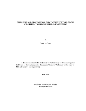
Tải xuống để đọc toàn bộSTRUCTURE AND PROPERTIES OF ELECTROSPUN POLYMER FIBERS AND APPLICATIONS IN BIOMEDICAL ENGINEERING by Cheryl L. Casper A dissertation submitted to the Faculty of the University of Delaware in partial fulfillment of the requirements for the degree of Doctor of Philosophy with a major in Materials Science and Engineering Fall 2005 Copyright 2005 Cheryl L. Casper All Rights Reserved UMI Number: 3200539 UMI Microform 3200539 Copyright 2006 by ProQuest Information and Learning Company. All rights reserved.
This microform edition is protected against unauthorized copying under Title 17, United States Code. ProQuest Information and Learning Company 300 North Zeeb Road P. Box 1346 Ann Arbor, MI 48106-1346 STRUCTURE AND PROPERTIES OF ELECTROSPUN POLYMER FIBERS AND APPLICATIONS IN BIOMEDICAL ENGINEERING by Cheryl L. Casper Approved: __________________________________________________________ John F.
Chairperson, Department of Materials Science and Engineering Approved: __________________________________________________________ Eric W. Dean of the College of Engineering Approved: __________________________________________________________ Conrado M. Gempesaw II, Ph. Vice Provost for Academic and International Programs I certify that I have read this dissertation and that in my opinion it meets the academic and professional standard required by the University as a dissertation for the degree of Doctor of Philosophy.
Professor in charge of dissertation I certify that I have read this dissertation and that in my opinion it meets the academic and professional standard required by the University as a dissertation for the degree of Doctor of Philosophy. Bruce Chase, Ph. Member of dissertation committee I certify that I have read this dissertation and that in my opinion it meets the academic and professional standard required by the University as a dissertation for the degree of Doctor of Philosophy. Member of dissertation committee I certify that I have read this dissertation and that in my opinion it meets the academic and professional standard required by the University as a dissertation for the degree of Doctor of Philosophy.
Signed: __________________________________________________________ Darrin Pochan, Ph. Member of dissertation committee ACKNOWLEDGMENTS I would like to thank my advisor, Dr. John Rabolt, for his help and support during my graduate studies. My thanks to Dr.
Bruce Chase for his guidance and insightful discussions. I would also like to thank the other members of my committee, Dr. Darrin Pochan and Dr. Kristi Kiick, for the time and effort they put into being a part of my committee.
I would like to thank Dr. Kristi Kiick and Dr. Nori Yamaguchi for their insights, which greatly contributed to my research. I would also like to thank Dr.
Kirk Czymmek (DBI), Debbie Powell (DBI), and Gerald Poirier (Department of Physics and Astronomy) for their assistance with FESEM and LSCM. A sincere thanks to Dr. Cindy Farach-Carson, Dr. Weidong (William) Yang, and Anissa Brown in the Department of Biology for their time and expertise in the field of biology.
A special thank you to Dr. Patricia Cotts (Dupont) for GPC analyses and Nancy Tassi (Dupont) for her AFM expertise. I would like to thank NSF (NIRT and DMR) and IGERT for financial support of this work. I would like to thank the members of the Rabolt Research Group and the MSE staff for their support and friendship, which has enriched my graduate experience.
A special thank you to Dr. Lori Stephans and Amy Carpinelli for the introduction into the world of science and their mentoring along the way. I would also like to thank my family and friends for being so supportive throughout all my endeavors. iv Lastly, and most importantly, I am forever grateful to my parents for their unwavering support and encouragement, without them none of this would be possible.
This manuscript is dedicated to my parents, Walter and Christine Casper. v TABLE OF CONTENTS LIST OF TABLES. ix LIST OF FIGURES. xiv Chapter 1 Introduction .1 Introduction to Electrospinning.2 The Electrospinning Process .3 Effects of Experimental Parameters on the Electrospinning Process.1 Effect on Fiber Diameter.2 Factors Affecting Fiber Shape and Surface Morphology.
15 2 Influence of Humidity and Molecular Weight on Fiber Formation and Surface Morphology.1 Influence of Polymer Molecular Weight on Fiber Formation .2 Effect of Humidity on Surface Morphology .1 Varying Humidity Level.2 Effect of Molecular Weight on Pore Formation. 55 3 Functionalizing Electrospun Fibers with Biologically Relevant Macromolecules for Potential Biomaterial Applications .2 Synthesis of PEG-LMWH-Dye and LMWH-Dye Conjugates .1 Toluidine Blue Assays .2 Growth Factor Binding Assays .3 Growth Factor Release Assays.4 Cell Proliferation Assay .1 Electrospinning Dye-Labeled LMWH with PEO .2 Incorporation of PEG-LMWH-dye in PEO Electrospun Fibers .3 Changing the Carrier Polymer.4 Preliminary Cell Studies. 102 4 Attachment of Proteins to Electrospun Collagen and Gelatin Membranes for Use as Tissue Engineering Scaffolds.1 Determining Extent of Crosslinking via Kaiser Test .4 Synthesis and Attachment of Proteins to Electrospun Fibers .1 Heparin-BSA Synthesis and Biotinylation.2 Dot Blot Assay .3 Attachment of Heparin-BSA-Biotin and PlnDI- Biotin to Electrospun Fibers.4 Assessing Bioactivity of Heparin-BSA-Biotin and PlnDI via Growth Factor Binding Assays .3 Results and Discussion .1 Preparation and Crosslinking of Electrospun Collagen and Gelatin Membranes .2 Synthesis and Attachment of Proteins to Electrospun Matrices. 140 5 Conclusions and Future Work.
146 viii LIST OF TABLES Table 1.1 Examples of Polymers Fabricated into Fibers via Electrospinning .1 Electrospinning Various PS Molecular Weights in THF .2 Applying the Mark-Houwink Equation to Various PS Molecular Weights.3 Applying the Mark-Houwink Equation to Various PEO Molecular Weights.4 Pore Diameters of 190,000 g/mol PS/THF Fibers under Varying Humidity Levels.5 Pore Diameters of 560,900 g/mol PS/THF Fibers under Varying T Humidity Levels.6 Micro- and Nanopore Diameters of 31,600 g/mol PS/THF Beads T under Varying Humidity Levels .1 Samples Tested via Kaiser Test. 113 ix LIST OF FIGURES Figure 1.1 Schematic representation of the electrospinning apparatus .1 FESEM micrographs of (a)31,600 g/mol, (b) 44,100 g/mol, (c) 75,700 g/mol, (d) 171,000 g/mol, (e) 560,900 g/mol, and (f) 1,800,000 g/mol PS/THF electrospun fibers.2 FESEM micrograph of 31,600 g/mol PS electrospun from an 80 wt% solution of PS/THF.3 Scanning electron micrographs of 100,000 g/mol PEO/water solutions electrospun at various concentrations (a) 0.4 Scanning electron micrographs of 300,000 g/mol PEO/water solutions electrospun at various concentrations (a) 5 wt% and (b) 10 wt%.5 FESEM micrographs of 190,000 g/mol PS/THF fibers electrospun under varying humidity: (a) <25%, (b) 31-38%, (c) 40-45%, (d) 50-59%, (e) 60-72% .6 Pore diameter distributions of pores found on 190,000 g/mol PS/THF electrospun fibers at varying humidity ranges: (a) 31- 38%, (b) 40-45%, (c) 50-59%, (d) 60-72%.7 FESEM micrograph of 171,000 g/mol PS/THF fiber electrospun in 50% humidity .8 FESEM image of 560,900 g/mol fiber electrospun in (a) 31-38% humidity and (b) 40-45% humidity.9 FESEM image showing the two pore sizes on 31,600 g/mol PS/THF bead, 60-72% humidity .10 AFM Images, topography (left), phase (right) (a) 190,000 g/mol T PS fibers under 50-59% humidity, (b) 190,000 g/mol PS fibers under 60-72% humidity, (c) 560,900 g/mol PS fibers under 50- 59% humidity, (d) 560,900 g/mol PS fibers under 60-72% humidity .1 Scanning electron micrograph of 10 wt% PEO/H2O electrospun B B fibers.2 Scanning electron micrographs of (a) PEO/ LMWH-dye electrospun fibers and (b) beaded section of the fiber .3 Multiphoton microscopy images of electrospun PEO fibers (a) reflection mode and (b) fluorescence (5 µm scale bars).4 Multiphoton microscopy image of electrospun PEO/LMWH-dye fibers (5 µm scale bar).5 Scanning electron micrographs of electrospun PEO/PEG-LMWH- dye (a) fibers and (b) beaded section of the fiber.6 Multiphoton microscopy image of PEG-LMWH-dye/PEO electrospun fibers (5 µm scale bar).7 Scanning electron micrographs of (a) PLGA electrospun fibers, (b) PLGA/LMWH-dye fibers, and (c) PLGA/PEG-LMWH-dye fibers.8 Multiphoton microscopy images of electrospun PLGA fibers (a) transmitted light and (b) fluorescence (10 µm scale bars).9 Multiphoton microscopy images of electrospun (a) LMWH- dye/PLGA fibers (10 µm scale bar), (b) PEG-LMWH-dye/PGLA fibers (10 µm scale bar) and (c) z-stack of PEG-LMWH/PLGA fibers (20 µm scale bar).10 bFGF binding on electrospun fibers.11 Scanning electron microscopy images of PEG-LMWH-dye/PLGA fibers and LMWH-dye/PLGA fibers after incubation in PBS for 6 h, 1 day, 3 days, and 14 days.12 Retention of heparin within electrospun matrices as observed by multiphoton microscopy for the PEG-LMWH-dye/PLGA fibers and LMWH-dye/PLGA fibers after incubation in PBS for 6 h, 1 day, 3 days, and 14 days (10 µm scale bars).13 Retention of PEG-LMWH-dye within depth of the electrospun matrix after 14 days of incubation in PBS as imaged via multiphoton microscopy.14 Release of bFGF from PLGA, LMWH-Dye, and PEG-LMWH- Dye electrospun fibers after 5 h and 24 h incubation in PBS .15 bFGF binding on PLGA, LMWH-Dye, and PEG-LMWH-Dye electrospun fibers after 5 h and 24 h incubation in PBS .16 Digital picture displaying the visual differences observed in the binding of bFGF to PLGA, LMWH-Dye, and PEG-LMWH-Dye electrospun fibers after 5 h and 24 h incubation in PBS.17 Results from the BrdU cell proliferation assay .18 LSCM images of (a) LMWH/PLGA and (b) PEG-LMWH/PLGA fibers (blue) incubated for 8 days with MG63 cells (green) (10 µm scale bars).1 Schematic of the crosslinking apparatus .2 Sulfo-NHS(-LC)-Biotin reaction scheme.3 EDC/NHS reaction scheme .4 FESEM micrographs of electrospun (a) collagen and (b) gelatin.5 Scanning electron micrographs of crosslinked electrospun gelatin fibers before (left hand column) and after (right hand column) immersion in ethanol/water solution.6 LSCM images of gelatin scaffold seeded with MG63 cells.7 Image displaying the dot blot assay results.8 Digital image of heparin-BSA-biotin presence on electrospun mats .9 Detection of heparin-BSA-biotin complex in electrospun collagen and gelatin membranes.10 bFGF binding on collagen/heparin-BSA-biotin and gelatin/heparin-BSA-biotin electrospun membranes .11 Detection of biotin-containing proteins on electrospun collagen and gelatin fibers coupled via EDC/NHS reaction.12 bFGF binding to electrospun collagen and gelatin membranes coated with either heparin-BSA-biotin or PlnDI.13 Digital image of bFGF binding to fibers containing heparin-BSA- biotin or PlnDI. 137 xiii ABSTRACT Increased interest in nanotechnology has revived a fiber processing technique invented back in the 1930’s. Electrospinning produces nanometer to micron size fibers that are not otherwise achievable using conventional fiber spinning methods.
Due to small fiber diameters, high surface area, tailorable surface morphology, and the creation of an interconnected fibrous network, electrospun fibers have found use in a variety of applications. However, a multitude of parameters directly affect the electrospinning process thus requiring a fundamental understanding of how various parameters affect the process and resulting fiber properties. Accordingly, the focus of this dissertation is to provide insight on how solution characteristics and processing parameters directly affect the electrospinning process, and then apply this knowledge to create electrospun membranes for biomedical applications. Solution characteristics (polymer/solvent, molecular weight, concentration, viscosity) were found to directly affect the electrospinning process.
The effect of molecular weight, concentration, and viscosity was examined and it was determined that the intrinsic viscosity ([η]), which is a function of molecular weight, and solution concentration (C) directly affect the fiber formation process. If [η]C< 10, beads are formed instead of continuous fibers. This study illustrated the critical role that chain xiv entanglements and other intermolecular interactions play in the fiber formation process. Another fundamental study was carried out to investigate the effect of relative humidity on the surface morphology of electrospun fibers.
It was found that electrospinning in an atmosphere of 30% or more relative humidity creates porous features on the surface of polystyrene fibers. Pores were approximately 85 nm in diameter and increased to 135 nm with increasing humidity. Not only do pore diameters change with increasing humidity, but pore size distributions, shape, and pore depths are also affected by humidity levels. AFM studies revealed that pore depths increase from 48 nm to 220 nm as the relative humidity during electrospinning increased.
The effect of humidity on surface morphology is mainly attributed to evaporative condensation. The increases in pore diameter as a function of increasing molecular weight is believed to result from reduced polymer diffusion times since the diffusion coefficient is inversely proportional to the square of the polymer molecular weight. These fundamental studies provided insight on how to control the electrospinning process; this knowledge was then utilized to electrospin fibrous membranes for biomedical applications. One aspect of this work focused on incorporating low molecular weight heparin (LMWH) into electrospun fibers.
Nội dung được bảo vệ bản quyền — Tải xuống đầy đủ
Trích dẫn luận án này
Cheryl L. Casper (2005). Cấu trúc và tính chất sợi polymer electrospun trong y sinh [Luận án tiến sĩ, university of delaware]. LuanAn.net. https://luanan.net/ky-thuat-co-khi/cong-nghe-vat-lieu/cau-truc-tinh-chat-soi-polymer-electrospun-ung-dung-y-sinh
Từ khóa và chủ đề nghiên cứu
Từ khóa liên quan
Chủ đề nghiên cứu
Câu hỏi thường gặp
Luận án "Cấu trúc và tính chất sợi polymer electrospun trong y sinh" nghiên cứu về vấn đề gì?
Luận án tiến sĩ về cấu trúc và tính chất sợi polymer electrospun ứng dụng trong kỹ thuật sinh y. Phân tích ảnh hưởng thông số thực nghiệm đến hình thành sợi và tiềm năng vật liệu y tế.
Luận án "Cấu trúc và tính chất sợi polymer electrospun trong y sinh" được bảo vệ tại trường nào?
Luận án này được bảo vệ tại university of delaware. Năm bảo vệ: 2005.
Luận án "Cấu trúc và tính chất sợi polymer electrospun trong y sinh" thuộc chuyên ngành gì?
Luận án "Cấu trúc và tính chất sợi polymer electrospun trong y sinh" thuộc chuyên ngành Materials Science and Engineering. Danh mục: Công Nghệ Vật Liệu.
Luận án "Cấu trúc và tính chất sợi polymer electrospun trong y sinh" có bao nhiêu trang?
Luận án "Cấu trúc và tính chất sợi polymer electrospun trong y sinh" có 166 trang. Bạn có thể xem trước một phần tài liệu ngay trên trang web trước khi tải về.
Cách tải luận án "Cấu trúc và tính chất sợi polymer electrospun trong y sinh" về máy như thế nào?
Để tải luận án về máy, bạn nhấn nút "Tải xuống ngay" trên trang này, sau đó hoàn tất thanh toán phí lưu trữ. File sẽ được tải xuống ngay sau khi thanh toán thành công. Hỗ trợ qua Zalo: 0559 297 239.