Sirtuins Hst3p và Hst4p điều hòa ổn định bộ gen trong Saccharomyces cerevisiae
Luận án tiến sĩ khám phá vai trò sirtuins Hst3p và Hst4p trong điều hòa ổn định bộ gen. Nghiên cứu cơ chế deacetylase histone H3 lysine 56 và ảnh hưởng đến sửa chữa DNA.
Molecular Biology and Genetics
Luan An
Luận án
Năm xuất bản
Số trang
197
Thời gian đọc
30 phút
Lượt xem
0
Lượt tải
0
Phí lưu trữ
50 Point
Tổng quan nhanh
- Chủ đề:
- 1. Sirtuins Hst3p và Hst4p: Vai trò điều hòa bộ gen
- Số trang:
- 197 trang
- Trường:
- The Johns Hopkins University
- Chuyên ngành:
- Molecular Biology and Genetics
- Tác giả:
- Ivana Celic
- Năm:
- 2006
Tóm tắt nội dung luận án
I. Sirtuins Hst3p và Hst4p Vai trò điều hòa bộ gen
Sirtuins đại diện cho họ protein deacetylase phụ thuộc NAD+ có tính bảo tồn cao. Họ protein này kiểm soát nhiều quá trình sinh học cơ bản. Nấm men Saccharomyces cerevisiae chứa năm thành viên: Sir2p và Hst1-4p. Các protein này quan trọng cho điều hòa im lặng phiên mã và ổn định bộ gen. Hst3p và Hst4p là hai sirtuins dư thừa với vai trò chính trong duy trì ổn định bộ gen. Chúng kiểm soát ổn định bộ gen thông qua điều hòa mức độ acetyl hóa histone H3 lysine 56. Vị trí này nằm ở bề mặt lõi nucleosome. K56 được acetyl hóa trong pha S của chu kỳ tế bào. Quá trình này góp phần vào các cơ chế sửa chữa hoạt động trong nhân đôi DNA. Cuối pha S, K56 của histone H3 bị deacetyl hóa phụ thuộc Hst3p và Hst4p. Thất bại trong deacetyl hóa K56 dẫn đến khuyết tật tăng trưởng, nhạy cảm với tác nhân gây tổn thương DNA và mất nhiễm sắc thể ở tế bào hst3 hst4.
1.1. Cấu trúc họ protein sirtuins trong nấm men
Saccharomyces cerevisiae mang năm thành viên họ sirtuin. Sir2p đại diện cho thành viên nguyên thủy. Hst1p, Hst2p, Hst3p và Hst4p là các homolog. Tất cả đều là NAD-dependent deacetylase. Chúng xúc tác phản ứng deacetyl hóa protein phụ thuộc NAD+. Cấu trúc này bảo tồn cao qua nhiều loài sinh vật. Mỗi sirtuin có chức năng đặc hiệu riêng biệt trong tế bào.
1.2. Chức năng đặc trưng của Hst3p và Hst4p
Hst3p và Hst4p hoạt động dư thừa với nhau. Vai trò chính là duy trì ổn định bộ gen. Chúng điều hòa acetyl hóa histone H3 tại lysine 56. Vị trí K56 nằm ở lõi bề mặt nucleosome. Đây là vị trí quan trọng cho cấu trúc chromatin. Hoạt động deacetylase của hai protein này cần thiết cho chu kỳ tế bào bình thường.
1.3. Tầm quan trọng trong điều hòa biểu sinh
Sirtuins kiểm soát các quá trình biểu sinh quan trọng. Chúng điều hòa im lặng phiên mã ở nhiều vùng nhiễm sắc thể. Hoạt động deacetylase ảnh hưởng cấu trúc chromatin. Điều này tác động trực tiếp đến biểu hiện gen. Hst3p và Hst4p đặc biệt quan trọng cho ổn định bộ gen. Chúng ngăn ngừa tổn thương DNA và mất nhiễm sắc thể.
II. Histone H3 Lysine 56 Mục tiêu điều hòa chính
Histone H3 lysine 56 là mục tiêu deacetyl hóa chính của Hst3p và Hst4p. Vị trí này acetyl hóa trong pha S của chu kỳ tế bào. Acetyl hóa K56 góp phần vào quá trình sửa chữa DNA trong nhân đôi. Cuối pha S, K56 bị deacetyl hóa bởi Hst3p và Hst4p. Chu kỳ acetyl hóa-deacetyl hóa này quan trọng cho ổn định bộ gen. Thất bại deacetyl hóa K56 gây nhiều hậu quả nghiêm trọng. Tế bào hst3 hst4 thiếu khả năng deacetyl hóa K56 hiệu quả. Điều này dẫn đến khuyết tật tăng trưởng rõ rệt. Tế bào trở nên nhạy cảm với tác nhân gây tổn thương DNA. Mất nhiễm sắc thể xảy ra với tần suất cao hơn. Đột biến K56 thành arginine khôi phục kiểu hình bình thường. Điều này chứng minh vai trò trực tiếp của acetyl hóa K56.
2.1. Vị trí và cấu trúc của K56 trong nucleosome
Lysine 56 nằm ở lõi bề mặt nucleosome. Vị trí này tiếp xúc trực tiếp với DNA. K56 nằm trong vùng core domain của histone H3. Cấu trúc này cho phép K56 tương tác với DNA quấn quanh nucleosome. Acetyl hóa tại vị trí này thay đổi tương tác histone-DNA. Điều này ảnh hưởng ổn định cấu trúc nucleosome và khả năng tiếp cận DNA.
2.2. Chu kỳ acetyl hóa K56 trong pha S
K56 được acetyl hóa đặc hiệu trong pha S. Quá trình này diễn ra đồng thời với nhân đôi DNA. Acetyl hóa K56 hỗ trợ lắp ráp chromatin mới. Histone mới tổng hợp mang K56 acetyl hóa. Điều này tạo điều kiện cho việc kết hợp vào nucleosome. Acetyl hóa K56 cũng hỗ trợ cơ chế sửa chữa DNA trong pha S.
2.3. Deacetyl hóa K56 phụ thuộc Hst3p Hst4p
Cuối pha S, Hst3p và Hst4p deacetyl hóa K56. Quá trình này loại bỏ nhóm acetyl khỏi lysine 56. Deacetyl hóa cần hoạt động NAD-dependent deacetylase. Hst3p và Hst4p hoạt động dư thừa trong quá trình này. Thiếu cả hai protein ngăn deacetyl hóa K56 hiệu quả. Duy trì K56 acetyl hóa sau pha S gây bất ổn bộ gen.
III. Hậu quả thiếu Hst3p và Hst4p trên ổn định gen
Tế bào thiếu Hst3p và Hst4p biểu hiện nhiều kiểu hình bất thường. Khuyết tật tăng trưởng là hậu quả rõ ràng nhất. Tế bào hst3 hst4 phát triển chậm hơn tế bào dại. Nhạy cảm với tác nhân gây tổn thương DNA tăng đáng kể. Các tác nhân như MMS, HU, camptothecin gây độc mạnh. Mất nhiễm sắc thể xảy ra với tần suất cao. Điều này chỉ ra bất ổn bộ gen nghiêm trọng. Đột biến K56R (arginine) khôi phục kiểu hình bình thường. Điều này chứng minh acetyl hóa K56 gây ra các khuyết tật. Phản ứng tổn thương DNA được kích hoạt liên tục. Tế bào hst3 hst4 nhạy cảm với rối loạn nhân đôi DNA. Tương tác chết tổng hợp với đột biến gen sửa chữa DNA rất phổ biến. Điều này cho thấy vai trò quan trọng trong duy trì toàn vẹn bộ gen.
3.1. Khuyết tật tăng trưởng và nhạy cảm DNA
Tế bào hst3 hst4 phát triển chậm rõ rệt. Tốc độ phân chia giảm so với tế bào dại. Khuyết tật này liên quan trực tiếp đến K56 acetyl hóa. Đột biến K56R khôi phục tốc độ tăng trưởng bình thường. Tế bào hst3 hst4 cực kỳ nhạy cảm với tác nhân gây tổn thương DNA. MMS và hydroxyurea gây độc mạnh ở nồng độ thấp.
3.2. Mất nhiễm sắc thể và bất ổn bộ gen
Tần suất mất nhiễm sắc thể tăng trong tế bào hst3 hst4. Điều này chỉ ra lỗi trong phân ly nhiễm sắc thể. Bất ổn bộ gen biểu hiện qua nhiều cơ chế khác nhau. Tái tổ hợp không mong muốn xảy ra thường xuyên hơn. Đột biến tích lũy với tốc độ cao hơn. Toàn vẹn cấu trúc nhiễm sắc thể bị ảnh hưởng nghiêm trọng.
3.3. Kích hoạt phản ứng tổn thương DNA
Tế bào hst3 hst4 kích hoạt checkpoint tổn thương DNA liên tục. Điều này cho thấy tế bào nhận diện K56 acetyl hóa như tổn thương. Các protein checkpoint như Rad53p được phosphoryl hóa. Phản ứng này xảy ra ngay cả không có tổn thương DNA ngoại sinh. Kích hoạt checkpoint kéo dài ảnh hưởng chu kỳ tế bào và sửa chữa DNA.
IV. Tương tác với phức hợp RFC và PCNA trong nhân đôi DNA
Khuyết tật tăng trưởng của tế bào hst3 hst4 được cứu vãn bởi tăng biểu hiện RFC1. RFC1 mã hóa tiểu đơn vị lớn của RFC, một clamp loader. RFC nạp PCNA (sliding clamp) lên DNA trong nhân đôi. Điều thú vị là xóa CTF18 cũng cứu vãn khuyết tật tăng trưởng. Xóa RAD24 và ELG1 có hiệu quả thấp hơn nhưng vẫn có tác dụng. CTF18, RAD24 và ELG1 mã hóa tiểu đơn vị lớn của RFC thay thế. Mỗi phức hợp chia sẻ bốn tiểu đơn vị nhỏ (Rfc2p-Rfc5p) với Rfc1p. Chu kỳ acetyl hóa-deacetyl hóa K56 điều hòa cân bằng chức năng giữa các phức hợp RFC khác nhau. K56 acetyl hóa ảnh hưởng tương tác PCNA-chromatin. Điều này tác động đến hiệu quả nhân đôi và sửa chữa DNA. Mất cân bằng RFC gây rối loạn nhân đôi DNA. Tăng RFC1 bù đắp thiếu hụt do K56 acetyl hóa liên tục.
4.1. RFC1 và vai trò clamp loader chính
RFC1 mã hóa tiểu đơn vị lớn của RFC chính. RFC hoạt động như clamp loader nạp PCNA lên DNA. PCNA là sliding clamp cần thiết cho DNA polymerase. Tăng biểu hiện RFC1 cứu vãn khuyết tật hst3 hst4. Điều này cho thấy RFC1 bù đắp thiếu hụt chức năng. Cân bằng RFC-PCNA quan trọng cho nhân đôi DNA hiệu quả.
4.2. Phức hợp RFC thay thế CTF18 RAD24 ELG1
CTF18, RAD24 và ELG1 tạo phức hợp RFC thay thế. Mỗi phức hợp có tiểu đơn vị lớn riêng. Chúng chia sẻ bốn tiểu đơn vị nhỏ Rfc2p-Rfc5p. Xóa CTF18 cứu vãn mạnh khuyết tật hst3 hst4. RAD24 và ELG1 deletion có hiệu quả thấp hơn. Các phức hợp này cạnh tranh cho tiểu đơn vị chung.
4.3. Mô hình điều hòa cân bằng RFC
Chu kỳ K56 acetyl hóa-deacetyl hóa điều hòa cân bằng RFC. Acetyl hóa K56 ảnh hưởng tương tác chromatin-PCNA. Điều này thay đổi ưu tiên sử dụng các phức hợp RFC. K56 acetyl hóa liên tục làm mất cân bằng RFC. Tăng RFC1 hoặc giảm RFC thay thế khôi phục cân bằng. Cơ chế này quan trọng cho ổn định bộ gen.
V. Cơ chế NAD dependent deacetylase của sirtuins
Sirtuins là NAD-dependent protein deacetylase độc đáo. Chúng khác với histone deacetylase phụ thuộc kẽm. Phản ứng deacetyl hóa cần NAD+ làm đồng cơ chất. NAD+ bị phân cắt trong quá trình phản ứng. Sản phẩm bao gồm nicotinamide, 2-O-acetyl-ADP-ribose và protein deacetyl hóa. Túi gắn nicotinamide trong Hst3p cần thiết cho hoạt tính. Đột biến trong túi này loại bỏ khả năng deacetyl hóa K56. Điều này chứng minh hoạt tính deacetylase trực tiếp điều hòa K56. Cơ chế phụ thuộc NAD+ liên kết hoạt động sirtuin với trạng thái năng lượng tế bào. Tỷ lệ NAD+/NADH ảnh hưởng hoạt tính enzyme. Điều này cho phép sirtuins hoạt động như cảm biến chuyển hóa. Hoạt động này kết nối điều hòa biểu sinh với trạng thái dinh dưỡng tế bào.
5.1. Cấu trúc túi gắn NAD và nicotinamide
Sirtuins có túi gắn NAD+ được bảo tồn cao. Túi này chứa các amino acid quan trọng cho xúc tác. Nicotinamide binding pocket đặc biệt quan trọng. Đột biến trong túi này loại bỏ hoạt tính deacetylase. Hst3p với túi nicotinamide đột biến không điều hòa K56. Điều này chứng minh cần hoạt tính enzymatic trực tiếp.
5.2. Phản ứng deacetyl hóa phụ thuộc NAD
Deacetyl hóa sirtuin cần NAD+ làm đồng cơ chất. NAD+ bị phân cắt thành nicotinamide và ADP-ribose. Nhóm acetyl chuyển lên ADP-ribose tạo 2-O-acetyl-ADP-ribose. Protein substrate được deacetyl hóa trong quá trình này. Phản ứng này khác hoàn toàn HDAC phụ thuộc kẽm. Cơ chế độc đáo này cho phép điều hòa bởi NAD+.
5.3. Liên kết với trạng thái chuyển hóa tế bào
NAD+ là phân tử trung tâm trong chuyển hóa năng lượng. Tỷ lệ NAD+/NADH phản ánh trạng thái oxy hóa-khử tế bào. Hoạt tính sirtuin phụ thuộc vào tỷ lệ này. NAD+ cao kích hoạt sirtuins mạnh hơn. Điều này liên kết điều hòa biểu sinh với dinh dưỡng. Sirtuins hoạt động như cảm biến chuyển hóa điều chỉnh biểu hiện gen.
VI. Ý nghĩa sinh học và ứng dụng tiềm năng nghiên cứu
Nghiên cứu Hst3p và Hst4p cung cấp hiểu biết sâu về điều hòa ổn định bộ gen. Cơ chế acetyl hóa-deacetyl hóa K56 bảo tồn qua nhiều loài. Homolog của Hst3p/Hst4p ở động vật có vú cũng điều hòa K56. Điều này cho thấy tầm quan trọng tiến hóa của cơ chế này. Sirtuins liên quan đến lão hóa, ung thư và bệnh thoái hóa thần kinh. Hiểu cơ chế hoạt động giúp phát triển liệu pháp mới. Điều chỉnh hoạt tính sirtuin có thể cải thiện ổn định bộ gen. Điều này có ứng dụng trong điều trị ung thư và bệnh liên quan tuổi. Mô hình nấm men cung cấp hệ thống nghiên cứu đơn giản. Saccharomyces cerevisiae cho phép phân tích di truyền chi tiết. Phát hiện về RFC và PCNA mở hướng nghiên cứu mới. Cân bằng các yếu tố nhân đôi DNA có thể là mục tiêu điều trị.
6.1. Bảo tồn tiến hóa cơ chế điều hòa K56
Acetyl hóa K56 bảo tồn từ nấm men đến động vật có vú. Homolog của Hst3p/Hst4p ở người cũng điều hòa K56. SIRT6 ở động vật có vú deacetyl hóa H3K56. Cơ chế này quan trọng cho ổn định bộ gen ở tất cả sinh vật nhân thực. Bảo tồn cao cho thấy tầm quan trọng sinh học cơ bản.
6.2. Liên quan đến bệnh tật và lão hóa
Sirtuins liên quan chặt chẽ đến quá trình lão hóa. Hoạt động sirtuin giảm theo tuổi ở nhiều mô. Rối loạn sirtuin liên quan đến ung thư. Bất ổn bộ gen do thiếu deacetyl hóa K56 thúc đẩy ung thư. Bệnh thoái hóa thần kinh cũng liên quan sirtuins. Điều chỉnh hoạt tính sirtuin có tiềm năng điều trị.
6.3. Ứng dụng trong phát triển liệu pháp mới
Hiểu cơ chế sirtuin mở đường phát triển thuốc mới. Chất kích hoạt sirtuin có thể cải thiện ổn định bộ gen. Điều này hữu ích trong điều trị ung thư. Điều chỉnh cân bằng RFC có thể là mục tiêu điều trị. Mô hình nấm men giúp sàng lọc thuốc hiệu quả. Nghiên cứu cơ bản này có ứng dụng lâm sàng tiềm năng.
Mục lục chi tiết luận án
Tải xuống file đầy đủ để xem toàn bộ nội dung
Tải đầy đủ (197 trang)Nội dung chính
Tổng quan về luận án
Luận án này tiên phong trong việc làm sáng tỏ cơ chế điều hòa ổn định bộ gen thông qua hoạt động của các sirtuin Hst3p và Hst4p ở nấm men Saccharomyces cerevisiae. Nghiên cứu đặt trong bối cảnh khoa học về vai trò ngày càng tăng của các biến đổi sau dịch mã (posttranslational modifications), đặc biệt là sự acetyl hóa và khử acetyl hóa protein, trong điều hòa các quá trình sinh học cơ bản. Gia đình protein Sir2, hay sirtuins, là các deacetylase phụ thuộc NAD+ đóng vai trò trung tâm trong nhiều khía cạnh sinh lý tế bào, từ sự ổn định bộ gen đến quá trình lão hóa.
Research gap SPECIFIC với citations từ literature: Mặc dù vai trò của Hst3p và Hst4p trong quá trình chuyển hóa nhiễm sắc thể đã được thiết lập từ nhiều năm trước, "các chất nền của hai sirtuin này vẫn còn khó nắm bắt" (Chapter 2, Introduction, p. 25). Đặc biệt, "không rõ ràng về mặt cơ học Hst3p và Hst4p kiểm soát sự ổn định bộ gen như thế nào và những gen/protein đối tác nào khác tham gia với Hst3p và Hst4p trong quá trình đó" (Chapter 1, p. 19). Nghiên cứu này giải quyết trực tiếp khoảng trống này bằng cách xác định histone H3 lysine 56 (K56) là một mục tiêu chính của Hst3p và Hst4p, và làm rõ cách điều hòa sự acetyl hóa K56 của H3 ảnh hưởng đến sự ổn định bộ gen. Công trình này củng cố đáng kể hiểu biết về các cơ chế phân tử liên kết hoạt động của sirtuin với tính toàn vẹn của bộ gen, đặc biệt là trong bối cảnh chu kỳ tế bào và phản ứng tổn thương DNA.
Research questions và hypotheses (đánh số cụ thể):
- RQ1: Hst3p và Hst4p điều hòa sự acetyl hóa histone H3 K56 như thế nào trong suốt chu kỳ tế bào?
- H1: Hst3p và Hst4p hoạt động dư thừa để kiểm soát mức độ acetyl hóa H3 K56 in vivo.
- H2: Hoạt động deacetylase phụ thuộc NAD+ của Hst3p/Hst4p là cần thiết cho sự điều hòa này.
- RQ2: Sự acetyl hóa H3 K56 cao do thiếu Hst3p và Hst4p gây ra những kiểu hình tế bào nào?
- H3: Mức độ acetyl hóa H3 K56 cao là nguyên nhân chính gây ra các kiểu hình nhạy cảm với tổn thương DNA, mất nhiễm sắc thể nguyên phân và nhạy cảm với nhiệt của tế bào đột biến hst3 hst4.
- RQ3: Những protein đối tác hoặc nhân tố tế bào nào khác tương tác với Hst3p/Hst4p hoặc điều hòa sự acetyl hóa H3 K56?
- H4: Histone chaperone Asf1p cần thiết cho sự acetyl hóa H3 K56.
- H5: Các chu trình acetyl hóa và khử acetyl hóa H3 K56 đang diễn ra góp phần điều hòa trạng thái cân bằng chức năng giữa các phức hợp RFC khác nhau trong tế bào.
- RQ4: Hst3p có khả năng kích hoạt quá trình khử acetyl hóa K56Ac trong chromatin trưởng thành hay không?
- H6: Hst3p có thể trực tiếp khử acetyl hóa H3 K56 trong chromatin đã hình thành, ngay cả khi các gen histone bị ức chế.
Theoretical framework với tên theories cụ thể: Nghiên cứu dựa trên khuôn khổ lý thuyết về kiểm soát chu kỳ tế bào thông qua biến đổi histone và mô hình sirtuin như cảm biến NAD+ (Lin et al., 2000; Anderson et al., 2003). Cụ thể, nó mở rộng Lý thuyết Mã Histone (Jenuwein & Allis, 2001) bằng cách thêm một điểm sửa đổi quan trọng (H3 K56Ac) và giải thích vai trò của nó trong sự ổn định bộ gen, vượt ra ngoài vai trò đã biết của các acetyl hóa đuôi histone. Nó cũng tích hợp các nguyên lý của cơ chế enzym phụ thuộc NAD+ của sirtuin (Imai et al., 2000; Landry et al., 2000b; Smith et al., 2000), làm sâu sắc thêm hiểu biết về tính đặc hiệu cơ chất và vai trò sinh lý của các sirtuin riêng lẻ. Cuối cùng, luận án đề xuất một mô hình trong đó các chu trình acetyl hóa và khử acetyl hóa H3 K56 đóng vai trò điều hòa cân bằng chức năng của các phức hợp protein Replication Factor C (RFC), liên kết trực tiếp biến đổi histone với quá trình sao chép DNA.
Đóng góp đột phá với quantified impact: Luận án này mang lại những đóng góp đột phá về mặt lý thuyết và thực tiễn, làm thay đổi hiểu biết về điều hòa ổn định bộ gen:
- Xác định vai trò của Hst3p và Hst4p trong điều hòa H3 K56 acetylation: Đây là lần đầu tiên Hst3p và Hst4p được chứng minh là các deacetylase chính của histone H3 K56. Phát hiện này giải quyết một khoảng trống lớn trong chức năng của hai sirtuin quan trọng này. Nghiên cứu định lượng cho thấy "phần lớn các phân tử H3 được acetyl hóa K56 trong các đột biến kép hst3 hst4" (Figure 2.1D, p. 26).
- Làm rõ tầm quan trọng sinh lý của H3 K56 deacetylation: Bằng chứng thuyết phục cho thấy các kiểu hình của đột biến hst3 hst4 (nhạy cảm với nhiệt độ, tác nhân gây tổn thương DNA, và mất nhiễm sắc thể nguyên phân) "phần lớn bị triệt tiêu bởi đột biến H3 K56 thành một dư lượng arginine không thể acetyl hóa" (Figure 2.4, 2.5, p. 30-31), chỉ ra rằng việc duy trì mức độ K56 acetyl hóa thấp là rất quan trọng cho chức năng tế bào bình thường. Điều này có thể được định lượng bằng sự phục hồi gần như hoàn toàn khả năng tăng trưởng ở 37°C và sự giảm đáng kể tỷ lệ mất nhiễm sắc thể.
- Phát hiện sự tham gia của Asf1p trong acetyl hóa H3 K56: Nghiên cứu chỉ ra rằng histone chaperone Asf1p "cần thiết cho K56Ac" và đột biến asf1 "triệt tiêu mạnh mẽ các kiểu hình Ts- của tế bào đột biến hst3 hst4" (Figure 2.6A, B, p. 32). Điều này cho thấy mối liên kết chưa từng biết giữa các chaperone histone và điều hòa biến đổi H3 K56.
- Chứng minh khả năng Hst3p khử acetyl hóa chromatin trưởng thành: Bằng cách sử dụng một thể td-HST3 có thể bất hoạt nhanh, nghiên cứu chứng minh rằng Hst3p có thể "kích hoạt quá trình khử acetyl hóa K56Ac nhanh chóng trong các tế bào bị bắt giữ ở G1 hoặc G2/M" (Figure 2.8, p. 33), cho thấy cơ chế khử acetyl hóa K56Ac không chỉ giới hạn ở histone mới tổng hợp mà còn diễn ra trên chromatin đã hình thành.
Scope (sample size, timeframe) và significance: Nghiên cứu tập trung vào Saccharomyces cerevisiae, một mô hình sinh vật đơn giản nhưng mạnh mẽ để nghiên cứu cơ chế di truyền và sinh hóa cơ bản. Mặc dù luận án không cung cấp số lượng mẫu cụ thể cho từng thí nghiệm (chẳng hạn như số lượng tế bào được phân tích bằng flow cytometry hoặc số lần lặp lại), các thí nghiệm được thiết kế tỉ mỉ, bao gồm nhiều chủng đột biến, thử nghiệm điều kiện dược phẩm (nicotinamide, HU, MMS), và phân tích chu kỳ tế bào có kiểm soát (bắt giữ G1, G2/M). "Các thí nghiệm chạy trong vòng 6 giờ" (Figure 2.2A, p. 27) cho thấy việc theo dõi động học chu kỳ tế bào và acetylation.
Tầm quan trọng của công trình nằm ở việc làm sáng tỏ một cơ chế điều hòa cơ bản của sự ổn định bộ gen. Bằng cách xác định Hst3p/Hst4p là các deacetylase histone H3 K56, luận án cung cấp một mục tiêu phân tử cụ thể để điều tra các rối loạn liên quan đến sự toàn vẹn bộ gen. Các phát hiện này có ý nghĩa sâu rộng đối với việc hiểu các bệnh ở người liên quan đến sirtuin, như ung thư và lão hóa, vì các sirtuin ở người có chung lõi xúc tác được bảo tồn và điều hòa các quá trình tương tự.
Literature Review và Positioning
Luận án bắt đầu bằng cách tổng hợp các luồng nghiên cứu chính về acetyl hóa và khử acetyl hóa protein như là các cơ chế điều hòa sau dịch mã quan trọng, đặc biệt nhấn mạnh vai trò của gia đình protein Sir2 (sirtuins) (Kouzarides, 2000; Kurdistani and Grunstein, 2003). Sir2 proteins được phân biệt là class III deacetylases vì yêu cầu NAD+ (Imai et al., 2000; Landry et al., 2000b; Smith et al., 2000), khác với class I và II. Nghiên cứu đi sâu vào cơ chế xúc tác độc đáo của Sir2 proteins, bao gồm sự hình thành ion riboxacarbenium và 1'-O-alkyl-amidate (Sauve et al., 2001; Tanner et al., 2000), và giải thích vai trò của nicotinamide như một chất ức chế không cạnh tranh (Bitterman et al., 2002; Jackson et al., 2003).
Synthesis của major streams với TÊN TÁC GIẢ và NĂM cụ thể: Luận án tổng hợp các luồng chính về:
- Cơ chế xúc tác của sirtuin: Mô tả chi tiết cơ chế NAD+-dependent deacetylation, với các trích dẫn đến Sauve et al. (2001), Tanner et al. (2000), Tanny and Moazed (2001), và Avalos et al. (2002, 2004) về cấu trúc tinh thể và cơ chế phản ứng.
- Vai trò của sirtuin trong quá trình lão hóa: Trình bày các nghiên cứu về kéo dài tuổi thọ bằng cách tăng liều lượng gen SIR2 ở S. cerevisiae (Kaeberlein et al., 1999) và C. elegans (Tissenbaum and Guarente, 2001). Đề cập đến tranh luận về vai trò của NAD+/NADH ratio (Lin et al., 2000, 2002) so với điều hòa nicotinamide (Anderson et al., 2003) trong kéo dài tuổi thọ do hạn chế calo (caloric restriction - CR).
- Các sirtuin ở nấm men S. cerevisiae và chức năng của chúng: Liệt kê năm sirtuin (Sir2, Hst1-4) (Brachmann et al., 1995) và chức năng của từng loại: Sir2p trong silencing ở telomere, mating type loci, và rDNA (Bryk et al., 1997; Gottschling et al., 1990; Rine et al., 1979; Smith and Boeke, 1997); Hst1p như một repressor gen-specific (Xie et al., 1998) và NAD+ sensor (Bedalov et al., 2001); Hst2p là sirtuin tế bào chất (North et al., 2003); và Hst3p/Hst4p trong ổn định bộ gen (Brachmann et al., 1995).
Contradictions/debates với ít nhất 2 opposing views: Luận án nêu bật một cuộc tranh luận quan trọng về cơ chế mà thông qua đó hạn chế calo (CR) kéo dài tuổi thọ:
- Quan điểm 1: NAD+/NADH ratio: Lin et al. (2000, 2002) đề xuất rằng CR kéo dài tuổi thọ thông qua kích hoạt Sir2p phụ thuộc NAD+, do sự thay đổi tỷ lệ NAD+/NADH khi chuyển hóa hướng tới hô hấp.
- Quan điểm 2: Nicotinamide inhibition: Anderson et al. (2003) phản biện rằng sự điều hòa hoạt động của Sir2p bởi nicotinamide, một chất ức chế không cạnh tranh, là cơ sở phù hợp hơn cho việc kéo dài tuổi thọ thông qua CR. Họ chỉ ra rằng nồng độ NAD+ thực sự tương quan nghịch với hoạt động của Sir2p trong điều kiện CR và không quan sát thấy sự ức chế Sir2p bởi NADH. Ngoài ra, Kaeberlein et al. (2005) đã "thách thức hoàn toàn vai trò của Sir2p trong việc kéo dài tuổi thọ qua CR", cho rằng nó độc lập với Sir2p và được điều hòa bởi con đường tín hiệu TOR, cho thấy một sự phức tạp và đa dạng về cơ chế trong nghiên cứu lão hóa.
Positioning trong literature với specific gap identified: Luận án tự định vị bằng cách giải quyết một khoảng trống rõ ràng: chức năng và cơ chất của Hst3p và Hst4p, hai sirtuin quan trọng ở S. cerevisiae, vẫn chưa được làm rõ (Brachmann et al., 1995). Trong khi các sirtuin khác như Sir2p, Hst1p, Hst2p đã có các chức năng cụ thể được biết đến (transcriptional silencing, gene repression, tubulin deacetylation), Hst3p và Hst4p chỉ được biết đến một cách chung chung là liên quan đến sự ổn định bộ gen. Luận án đặt ra mục tiêu "xác định các protein điều hòa sự acetyl hóa histone H3 K56 trong suốt chu kỳ tế bào" (Chapter 2, Results, p. 26) và từ đó khám phá cơ chất cụ thể và cơ chế hoạt động của Hst3p/Hst4p.
How this advances field với concrete contributions: Nghiên cứu này tiến bộ trong lĩnh vực sinh học nhiễm sắc thể và biểu sinh học bằng cách:
- Cung cấp bằng chứng cụ thể rằng Hst3p và Hst4p là các deacetylase chính cho histone H3 K56, một biến đổi quan trọng trên bề mặt nucleosome (Hyland et al., 2005; Masumoto et al., 2005).
- Thiết lập mối liên hệ nhân quả giữa sự acetyl hóa H3 K56 cao và các kiểu hình rối loạn ổn định bộ gen (nhạy cảm với tổn thương DNA, mất nhiễm sắc thể).
- Lần đầu tiên liên kết histone chaperone Asf1p với sự acetyl hóa H3 K56, cho thấy sự phối hợp giữa lắp ráp nucleosome và biến đổi histone.
- Chỉ ra rằng Hst3p có thể hoạt động trên chromatin trưởng thành, thách thức các quan niệm hiện có về thời gian và địa điểm của quá trình khử acetyl hóa histone.
So sánh với ÍT NHẤT 2 international studies:
- Sirtuins ở người (mammalian sirtuins): Luận án so sánh S. cerevisiae sirtuins với các đồng loại ở người (SIRT1-7) (Frye, 2000). Ví dụ, SirT1 ở người, giống Sir2p của nấm men, deacetyl hóa histones nhưng cũng nhắm mục tiêu vào các protein phi-histone như p53 (Langley et al., 2002; Luo et al., 2001; Vaziri et al., 2001) và FOXO transcription factors (Brunet et al., 2004; Motta et al., 2004). SirT2 là một sirtuin tế bào chất deacetyl hóa tubulin (North et al., 2003), trong khi Hst2p của nấm men cũng là một sirtuin tế bào chất. Sự so sánh này nhấn mạnh sự bảo tồn tiến hóa của sirtuin nhưng cũng làm nổi bật sự khác biệt về tính đặc hiệu cơ chất và sự phân bố trong tế bào.
- Sirtuin ở Schizosaccharomyces pombe (S. pombe): Nghiên cứu cũng tham chiếu đến S. pombe hst4+, một sirtuin tương tự Hst3p/Hst4p của S. cerevisiae. "Sự gián đoạn của S. pombe hst4+ dẫn đến siêu acetyl hóa K56 ở S. pombe" (Haldar and Kamakaka, 2006, submitted), tương tự như phát hiện trong luận án này. Hơn nữa, nó "cũng làm xáo trộn cấu trúc dị nhiễm sắc pericentric" (Freeman-Cook et al., 2006, submitted), gợi ý một vai trò bảo tồn của sự khử acetyl hóa H3 K56 trong cấu trúc chromatin và phân chia nhiễm sắc thể. Điều này cung cấp bằng chứng về sự liên quan quốc tế và bảo tồn tiến hóa của cơ chế được nghiên cứu.
Đóng góp lý thuyết và khung phân tích
Đóng góp cho lý thuyết
Nghiên cứu này làm sâu sắc đáng kể các khuôn khổ lý thuyết hiện có về điều hòa biểu sinh và ổn định bộ gen.
-
Extend/challenge WHICH specific theories (name theorists):
- Mở rộng Lý thuyết Mã Histone (Histone Code Hypothesis) của Jenuwein & Allis (2001): Luận án mở rộng lý thuyết này bằng cách xác định histone H3 K56 acetylation là một biến đổi histone quan trọng, không nằm ở đuôi N-terminal truyền thống, mà trên bề mặt nucleosome. Biến đổi này, được điều hòa bởi Hst3p và Hst4p, có tác động sâu sắc đến tính toàn vẹn của bộ gen và chu kỳ tế bào. Việc acetyl hóa K56 không phù hợp dẫn đến các kiểu hình nghiêm trọng, cho thấy K56 là một "ký hiệu" quan trọng trong mã histone, ảnh hưởng đến cấu trúc nucleosome và chức năng DNA.
- Thách thức một phần mô hình động học histone truyền thống: Bằng cách chứng minh rằng Hst3p có thể "kích hoạt quá trình khử acetyl hóa K56Ac trong chromatin trưởng thành" (Chapter 2, Discussion, p. 35), nghiên cứu thách thức quan niệm rằng các biến đổi histone, đặc biệt là sự khử acetyl hóa, chỉ giới hạn ở histone mới tổng hợp hoặc các vùng chromatin đang hoạt động. Điều này gợi ý rằng có các cơ chế linh hoạt hơn để điều chỉnh các biến đổi histone trong suốt chu kỳ tế bào, ngay cả trên chromatin đã được lắp ráp.
-
Conceptual framework với components và relationships: Khuôn khổ khái niệm của luận án mô tả mối quan hệ giữa:
- Sirtuins (Hst3p, Hst4p): Các deacetylase phụ thuộc NAD+.
- Histone H3 K56 acetylation (H3 K56Ac): Biến đổi chính.
- Histone chaperone (Asf1p): Điều hòa sự acetyl hóa K56.
- DNA replication machinery (RFC, PCNA): Liên quan đến chức năng.
- DNA damage response và cell cycle checkpoints: Các con đường tín hiệu bị ảnh hưởng.
- Genomic stability: Kết quả sinh học cuối cùng. Mối quan hệ chính là Hst3p và Hst4p, hoạt động dư thừa, chịu trách nhiệm chính trong việc khử acetyl hóa H3 K56 trong giai đoạn S và G2/M của chu kỳ tế bào. Sự vắng mặt của chúng dẫn đến siêu acetyl hóa H3 K56, từ đó gây ra căng thẳng sao chép, kích hoạt phản ứng tổn thương DNA, và cuối cùng dẫn đến mất nhiễm sắc thể và nhạy cảm với các tác nhân gây tổn thương DNA. Asf1p thúc đẩy sự acetyl hóa K56 của H3 mới tổng hợp, trong khi Hst3p/Hst4p loại bỏ nó.
-
Theoretical model với propositions/hypotheses numbered: Mô hình lý thuyết được đề xuất dựa trên các phát hiện then chốt:
- Proposition 1: Hst3p và Hst4p là các enzyme deacetylase chính chịu trách nhiệm loại bỏ nhóm acetyl khỏi lysine 56 của histone H3 trong S. cerevisiae.
- Proposition 2: Các kiểu hình thiếu hụt tăng trưởng, nhạy cảm với tác nhân gây tổn thương DNA, và mất nhiễm sắc thể ở tế bào đột biến hst3 hst4 là hậu quả trực tiếp của mức độ siêu acetyl hóa K56 trên histone H3.
- Proposition 3: Hoạt động khử acetyl hóa H3 K56 của Hst3p/Hst4p là cần thiết để duy trì tính toàn vẹn của replisome và/hoặc ngăn ngừa tổn thương DNA không thể phục hồi khi các điểm sao chép bị đình trệ.
- Proposition 4: Histone chaperone Asf1p đóng vai trò quan trọng trong việc tạo điều kiện cho sự acetyl hóa K56 trên histone H3 mới tổng hợp.
- Proposition 5: Chu trình acetyl hóa và khử acetyl hóa H3 K56 đóng góp vào việc điều hòa cân bằng chức năng giữa các phức hợp RFC khác nhau trong tế bào, ảnh hưởng đến quá trình sao chép DNA.
- Proposition 6: Hst3p có khả năng khử acetyl hóa H3 K56 trong chromatin trưởng thành, cho phép điều hòa động học các biến đổi histone ngoài các histone mới tổng hợp.
-
Paradigm shift với EVIDENCE từ findings: Mặc dù không phải là một sự thay đổi hoàn toàn về hệ hình, luận án tạo ra một sự dịch chuyển đáng kể trong cách hiểu về biến đổi histone và vai trò của sirtuin bằng cách:
- Mở rộng vị trí của biến đổi histone quan trọng: Trước đây, sự tập trung chủ yếu vào các đuôi N-terminal của histone. Phát hiện về vai trò then chốt của K56 trên bề mặt nucleosome (Hyland et al., 2005; Masumoto et al., 2005) chuyển sự chú ý sang các vùng globular của histone.
- Cơ chế khử acetyl hóa chromatin trưởng thành: Bằng chứng về khả năng khử acetyl hóa K56Ac của Hst3p trong chromatin trưởng thành (Figure 2.8) gợi ý rằng các sirtuin không chỉ đóng vai trò trong việc lắp ráp chromatin mới mà còn trong việc tái cấu trúc và duy trì chromatin đã hình thành. Điều này mở ra những cách suy nghĩ mới về động lực học của các biến đổi biểu sinh.
Khung phân tích độc đáo
Nghiên cứu này phát triển một khung phân tích độc đáo, tích hợp các phương pháp di truyền, sinh hóa và sinh học tế bào để giải quyết các câu hỏi cơ bản về biến đổi histone và ổn định bộ gen.
- Integration của theories (name 3+ specific theories):
Khung phân tích tích hợp các nguyên lý từ:
- Lý thuyết Mã Histone (Histone Code Hypothesis): Xem xét K56Ac như một "ký hiệu" cụ thể ảnh hưởng đến chức năng chromatin.
- Lý thuyết Kiểm soát Chu kỳ Tế bào (Cell Cycle Control Theory): Đánh giá tác động của K56Ac đối với sự tiến triển của chu kỳ tế bào và các điểm kiểm soát.
- Lý thuyết Phản ứng Tổn thương DNA (DNA Damage Response Theory): Khám phá cách H3 K56Ac cao dẫn đến việc kích hoạt các điểm kiểm soát tổn thương DNA và nhạy cảm với các tác nhân gây tổn thương DNA.
- Lý thuyết về Cấu trúc và Chức năng Nucleosome: Nhấn mạnh vị trí của K56 trên bề mặt nucleosome và ảnh hưởng của nó đến sự tương tác DNA-histone và tương tác protein-protein.
- Novel analytical approach với justification:
- Cách tiếp cận kép: Functional Genomics và Quantitative Biochemistry: Luận án kết hợp việc tạo ra các đột biến gen có mục tiêu (hst3 hst4 null, hht1-K56R, asf1 null, td-HST3) với các kỹ thuật sinh hóa định lượng như immunoblotting với kháng thể đặc hiệu K56Ac và phổ khối lượng (quantitative mass spectrometry) để xác định và định lượng chính xác mức độ acetyl hóa histone. Điều này cho phép không chỉ xác định chức năng gen mà còn cơ chế phân tử cụ thể.
- Phân tích kiểu hình được kiểm soát theo chu kỳ tế bào: Bằng cách sử dụng các tác nhân bắt giữ chu kỳ tế bào (alpha-factor, nocodazole, HU) và theo dõi tiến trình chu kỳ tế bào bằng flow cytometry, nghiên cứu đảm bảo rằng các thay đổi về acetyl hóa H3 K56 và các kiểu hình tế bào được quan sát là do các hiệu ứng trực tiếp của đột biến chứ không phải là hậu quả gián tiếp của sự gián đoạn chu kỳ tế bào (Chapter 2, Results, p. 26).
- Conceptual contributions với definitions:
- "Functional equilibrium between different RFC complexes": Luận án định nghĩa một khái niệm mới về sự cân bằng động giữa các phức hợp Replication Factor C (RFC) khác nhau (RFC1, Ctf18, Rad24, Elg1) trong tế bào. Nó đề xuất rằng sự acetyl hóa và khử acetyl hóa H3 K56 góp phần điều hòa trạng thái cân bằng này, ảnh hưởng đến khả năng tải PCNA (sliding clamp) lên DNA trong quá trình sao chép. Sự gián đoạn cân bằng này có thể dẫn đến các vấn đề trong sao chép và sửa chữa DNA.
- "K56-acetylated histone H3 as a major in vivo target": Luận án khái niệm hóa K56Ac của histone H3 không chỉ là một biến đổi mà là một "mục tiêu chính in vivo" của Hst3p/Hst4p, với bằng chứng là sự triệt tiêu các kiểu hình đột biến hst3 hst4 bởi đột biến hht1-K56R.
- Boundary conditions explicitly stated:
Luận án thừa nhận các điều kiện biên giới:
- Giới hạn về mô hình: Mặc dù S. cerevisiae là một mô hình tuyệt vời, các kết quả có thể không hoàn toàn dịch chuyển được sang các sinh vật cao cấp hơn do sự khác biệt về số lượng sirtuin, tính đặc hiệu cơ chất và sự phức tạp của hệ thống.
- Giới hạn về môi trường in vitro: Hoạt động deacetylase của Hst3p/Hst4p đối với H3 K56Ac "không được phát hiện bằng cách sử dụng Hst3p/Hst4p tinh khiết từ E. coli hoặc S. cerevisiae trong điều kiện in vitro" (Chapter 2, Discussion, p. 35). Điều này cho thấy rằng "hoạt động của nó có thể phụ thuộc vào sự hiện diện của các yếu tố khác" hoặc điều kiện cụ thể trong môi trường tế bào.
Phương pháp nghiên cứu tiên tiến
Thiết kế nghiên cứu
Luận án áp dụng một thiết kế nghiên cứu thực nghiệm vững chắc, chủ yếu dựa trên sinh học phân tử và di truyền nấm men để khám phá cơ chế phân tử.
-
Research philosophy (positivism/interpretivism/critical realism): Nghiên cứu tuân theo triết lý positivism, nhằm mục đích xác định các mối quan hệ nhân quả có thể đo lường và kiểm chứng giữa các biến số. Nó tập trung vào việc tạo ra các dữ liệu thực nghiệm khách quan (ví dụ: mức độ acetyl hóa histone, tỷ lệ mất nhiễm sắc thể, độ nhạy cảm với thuốc) để kiểm tra các giả thuyết cụ thể về chức năng của Hst3p và Hst4p.
-
Mixed methods với SPECIFIC combination rationale: Mặc dù không phải là "mixed methods" theo nghĩa xã hội học, nghiên cứu này kết hợp mạnh mẽ các phương pháp di truyền, sinh hóa và sinh học tế bào, tạo thành một cách tiếp cận đa diện:
- Di truyền học: Tạo ra các chủng đột biến gen có mục tiêu (hst3 hst4 null, hht1-K56R, asf1 null) để xác định chức năng gen và mối quan hệ di truyền (synthetic lethality analysis).
- Sinh hóa: Sử dụng immunoblotting và định lượng phổ khối lượng để đo lường chính xác các biến đổi histone (H3 K56 acetylation).
- Sinh học tế bào: Phân tích tiến trình chu kỳ tế bào bằng flow cytometry và các thí nghiệm bắt giữ chu kỳ tế bào để theo dõi các sự kiện trong các giai đoạn cụ thể.
- Rationale: Sự kết hợp này là cần thiết để không chỉ xác định một gen chịu trách nhiệm cho một kiểu hình cụ thể mà còn để hiểu cơ chế phân tử ở mức độ protein và tế bào, cung cấp bằng chứng trực tiếp và gián tiếp cho các mối quan hệ nhân quả.
-
Multi-level design với levels clearly defined: Thiết kế nghiên cứu được thực hiện ở nhiều cấp độ, từ phân tử đến tế bào:
- Cấp độ phân tử: Điều tra cơ chế xúc tác của sirtuin, sự tương tác protein-histone, và các biến đổi sau dịch mã trên lysine 56 của histone H3.
- Cấp độ tế bào: Phân tích ảnh hưởng của các đột biến và sự thay đổi mức độ acetyl hóa histone đến tiến trình chu kỳ tế bào, phản ứng tổn thương DNA, và sự ổn định nhiễm sắc thể (mất nhiễm sắc thể nguyên phân).
- Cấp độ di truyền: Khám phá các tương tác di truyền (synthetic lethality, suppression) giữa HST3, HST4 và các gen liên quan đến quá trình chuyển hóa DNA.
-
Sample size và selection criteria EXACT:
- Sinh vật mô hình: Saccharomyces cerevisiae (nấm men làm bánh).
- Chủng men: Các chủng hoang dại (wild-type, ví dụ: W303 MATa) và một loạt các chủng đột biến null (hst3, hst4, hst3 hst4, sir2, hst1-4), đột biến missense (Hst3p N152A, D154A, H184A), và đột biến histone (hht1-K56R).
- Kích thước mẫu: Không có số liệu cụ thể về "kích thước mẫu" theo nghĩa thống kê cho từng thí nghiệm. Tuy nhiên, các thí nghiệm lặp lại nhiều lần (ví dụ: "0 trên 8 chiết xuất được thử nghiệm" cho thấy hoạt động in vitro, "2 trên 8 chiết xuất" cho thấy hoạt động in vitro không thường xuyên) cung cấp độ tin cậy. Đối với định lượng phổ khối lượng, "phần lớn (97,7%) H3 được acetyl hóa K56 trong tế bào đột biến hst3-N152A hst4 trong G2/M" (Chapter 2, Results, p. 29) là một ví dụ về dữ liệu định lượng.
- Tiêu chí lựa chọn: Các chủng đột biến được tạo ra bằng cách thay thế gen hoặc biến đổi điểm cụ thể để thăm dò chức năng và cơ chế, dựa trên sự bảo tồn của các dư lượng xúc tác và các kiểu hình đã biết.
Quy trình nghiên cứu rigorous
- Sampling strategy với inclusion/exclusion criteria:
- Lựa chọn chủng: Các chủng nấm men được lựa chọn dựa trên sự hiện diện hoặc vắng mặt của các gen sirtuin cụ thể hoặc các đột biến điểm, cho phép so sánh trực tiếp chức năng. Ví dụ, hst3 hst4 double mutant là một chủng bao gồm vì nó biểu hiện kiểu hình nghiêm trọng nhất, trong khi hst3 hst4 hht1-K56R là một chủng bao gồm để kiểm tra sự triệt tiêu.
- Điều kiện nuôi cấy: Tế bào được nuôi cấy trong các điều kiện tiêu chuẩn cho S. cerevisiae, nhưng cũng bao gồm các điều kiện cụ thể để gây ra căng thẳng (ví dụ: nhiệt độ cao 37°C, hydroxyurea (HU), methyl methane sulfonate (MMS), nicotinamide) và bắt giữ chu kỳ tế bào (alpha-factor cho G1, nocodazole cho G2/M).
- Tiêu chí loại trừ: Các tế bào không phát triển trong các điều kiện cụ thể hoặc có đặc điểm sinh trưởng bất thường có thể bị loại trừ khỏi phân tích định lượng nếu không phục vụ mục đích của thí nghiệm.
- Data collection protocols với instruments described:
- Immunoblotting: Sử dụng các kháng thể đặc hiệu cho H3 K56 acetyl hóa và kháng thể tổng histone H3 (Masumoto et al., 2005) để định lượng mức độ acetyl hóa. Đây là một phương pháp tiêu chuẩn để phát hiện protein và biến đổi của chúng.
- Quantitative Mass Spectrometry: Sử dụng để định lượng chính xác "tỷ lệ các phân tử histone H3 được acetyl hóa K56" (Figure 2.1D, p. 26), cung cấp dữ liệu định lượng cao.
- Flow Cytometry: Để "theo dõi tiến trình chu kỳ tế bào" (Figure 2.1B, p. 26; Figure 2.2C, p. 27), đảm bảo các tác động đến acetyl hóa không phải là gián tiếp do thay đổi chu kỳ tế bào.
- Colony Sectoring Assay: Một kỹ thuật di truyền cổ điển để đo tỷ lệ mất nhiễm sắc thể nguyên phân (Spencer et al., 1988), sử dụng đột biến ade2-1 và một đoạn nhiễm sắc thể mang gen tRNA suppressor.
- Triangulation (data/method/investigator/theory):
- Data Triangulation: Các phát hiện về mức độ acetyl hóa H3 K56 được xác nhận bởi cả immunoblotting (qualitative/semi-quantitative) và phổ khối lượng (quantitative), tăng cường độ tin cậy.
- Methodological Triangulation: Các kết quả di truyền (phục hồi kiểu hình bởi đột biến hht1-K56R) được hỗ trợ bởi bằng chứng sinh hóa (Hst3p/Hst4p là deacetylase) và sinh học tế bào (ảnh hưởng đến chu kỳ tế bào).
- Theoretical Triangulation: Luận án liên kết các phát hiện của mình với nhiều lý thuyết (mã histone, kiểm soát chu kỳ tế bào, phản ứng tổn thương DNA), cho thấy sự nhất quán và phù hợp với kiến thức hiện có.
- Validity (construct/internal/external) và reliability (α values):
- Construct Validity: Cao, vì nghiên cứu sử dụng các kháng thể đặc hiệu và đột biến gen có mục tiêu vào các protein và biến đổi cụ thể (ví dụ: Hst3p, H3 K56), đảm bảo rằng những gì được đo là những gì cần đo.
- Internal Validity: Cao, do thiết kế thử nghiệm được kiểm soát chặt chẽ. Việc sử dụng các chủng đối chứng (wild-type), các đột biến bổ sung (ví dụ: hht1-K56R để triệt tiêu kiểu hình) và các điều kiện bắt giữ chu kỳ tế bào giúp loại bỏ các giải thích thay thế cho các mối quan hệ nhân quả.
- External Validity: Thấp hơn một chút do tập trung vào một sinh vật mô hình duy nhất (S. cerevisiae), nhưng được tăng cường bởi sự bảo tồn tiến hóa của sirtuins và các biến đổi histone ở các loài khác (ví dụ: S. pombe, mammals), gợi ý rằng các cơ chế cơ bản có thể được tổng quát hóa.
- Reliability: Không có giá trị alpha (α values) được báo cáo trực tiếp từ các phép đo. Tuy nhiên, các thí nghiệm được thiết kế để có thể lặp lại, và các kết quả được "củng cố bằng phổ khối lượng định lượng" (Chapter 2, Results, p. 29) và tính nhất quán trên nhiều phương pháp và điều kiện.
Data và phân tích
- Sample characteristics với demographics/statistics:
- Chủng: Các chủng Saccharomyces cerevisiae được mô tả rõ ràng về mặt di truyền (genotype).
- Đặc điểm tế bào: Các tế bào được phân tích ở các giai đoạn cụ thể của chu kỳ tế bào (G1, S, G2/M) và dưới các điều kiện căng thẳng hoặc bắt giữ chu kỳ tế bào.
- Thống kê: "Phần lớn (97.7%) H3 được acetyl hóa K56" trong hst3-N152A hst4 so với "2.2% trong hst3-H184 hst4 mutants và chỉ 24.5% trong HST3 hst4 cells" (Chapter 2, Results, p. 29) là các số liệu định lượng quan trọng. Tỷ lệ mất nhiễm sắc thể nguyên phân được đo bằng cách đếm các khuẩn lạc bị chia thành nhiều phần màu đỏ/hồng.
- Advanced techniques (SEM/multilevel/QCA etc.) với software:
- Quantitative Mass Spectrometry: Một kỹ thuật tiên tiến để định lượng các biến đổi protein sau dịch mã với độ chính xác cao.
- Immunoblotting với kháng thể đặc hiệu: Mặc dù là một kỹ thuật tiêu chuẩn, việc sử dụng các kháng thể rất đặc hiệu cho một biến đổi lysine cụ thể (H3 K56Ac) là rất quan trọng.
- Flow Cytometry: Để phân tích định lượng chu kỳ tế bào.
- Software: Luận án không nêu tên cụ thể phần mềm phân tích dữ liệu nào được sử dụng, nhưng ngụ ý việc sử dụng các công cụ tiêu chuẩn cho phân tích hình ảnh immunoblot và dữ liệu phổ khối lượng. Các phân tích thống kê cơ bản (như p-values, effect sizes) được ngụ ý thông qua các kết quả "statistical significance" hoặc "substantial suppression", mặc dù các giá trị p cụ thể không được nêu rõ.
- Robustness checks với alternative specifications:
- Kiểm tra tính đặc hiệu của Hst3p/Hst4p: So sánh các đột biến sirtuin khác (Sir2p, Hst1p, Hst2p) cho thấy "ngoại trừ chủng đột biến hst3, không có sirtuin nào khác có tác động đáng kể đến acetyl hóa H3 K56" (Chapter 2, Results, p. 26).
- Kiểm tra cơ chế hoạt động: Các đột biến điểm trong nicotinamide binding pocket của Hst3p (N152A, D154A) cho ra "các đột biến về cơ bản là null" (Figure 2.3C, p. 29), chứng minh rằng hoạt động deacetylase là quan trọng.
- Kiểm tra nhân quả: Sự triệt tiêu các kiểu hình hst3 hst4 bởi đột biến hht1-K56R là một kiểm tra mạnh mẽ cho thấy sự acetyl hóa H3 K56 cao là nguyên nhân gây ra các kiểu hình đó (Figure 2.4, 2.5).
- Effect sizes và confidence intervals reported: Luận án báo cáo các hiệu ứng mạnh mẽ. Ví dụ, "K56Ac không thể phát hiện được trong các đột biến null asf1" (Figure 2.6B, p. 32), cho thấy một hiệu ứng rất lớn. "Các đột biến kép hst3 hst4 có mức độ acetyl hóa rất cao, ngay cả trong G1" (Figure 2.1C, p. 26). "Sự triệt tiêu đáng kể" (substantially suppressed) của các kiểu hình cho thấy effect size lớn, mặc dù giá trị định lượng như p-values hoặc confidence intervals không được trình bày tường minh trong đoạn trích.
Phát hiện đột phá và implications
Những phát hiện then chốt
Luận án này đưa ra nhiều phát hiện then chốt, làm sáng tỏ vai trò của sirtuin trong điều hòa ổn định bộ gen:
- Hst3p và Hst4p là các deacetylase chính của histone H3 K56: Các đột biến kép hst3 hst4 dẫn đến mức độ acetyl hóa H3 K56 "cao hơn đáng kể so với tế bào kiểu dại" (Figure 2.1B, p. 26) và định lượng bằng phổ khối lượng cho thấy "phần lớn các phân tử H3 (97.7%) được acetyl hóa K56" trong đột biến hst3-N152A hst4 (Chapter 2, Results, p. 29). Điều này chứng minh chức năng dư thừa của hai sirtuin này trong việc kiểm soát H3 K56Ac.
- Siêu acetyl hóa H3 K56 là nguyên nhân gây ra các kiểu hình của hst3 hst4: Các kiểu hình tăng trưởng kém, nhạy cảm với tác nhân gây tổn thương DNA (HU, MMS), và mất nhiễm sắc thể nguyên phân của tế bào đột biến hst3 hst4 "được triệt tiêu đáng kể bởi đột biến H3 K56 thành arginine không thể acetyl hóa" (Figure 2.4, 2.5, p. 30-31). Điều này cung cấp bằng chứng nhân quả mạnh mẽ, chỉ ra rằng chính mức độ acetyl hóa K56 cao, chứ không phải sự thiếu hụt Hst3p/Hst4p nói chung, gây ra các kiểu hình này.
- Histone chaperone Asf1p cần thiết cho acetyl hóa H3 K56: "K56Ac không thể phát hiện được trong các đột biến null asf1, cả trong tế bào tăng sinh bình thường và tế bào được xử lý bằng HU" (Figure 2.6B, p. 32). Điều này khám phá một mắt xích mới trong con đường điều hòa K56Ac, liên kết trực tiếp một chaperone histone với biến đổi này.
- Hst3p có thể khử acetyl hóa K56Ac trong chromatin trưởng thành: Sử dụng một allele td-HST3 có thể bất hoạt nhanh, nghiên cứu chỉ ra rằng "sự cảm ứng tổng hợp Hst3p dẫn đến khử acetyl hóa H3 K56 nhanh chóng trong chromatin trưởng thành" trong các tế bào bị bắt giữ ở G1 hoặc G2/M (Figure 2.8, p. 33). Điều này gợi ý một vai trò động của Hst3p ngoài việc lắp ráp chromatin mới.
- Điều hòa cân bằng phức hợp RFC bởi chu trình acetyl hóa H3 K56: Đề xuất rằng "các chu trình acetyl hóa và khử acetyl hóa K56 đang diễn ra góp phần vào việc điều hòa cân bằng chức năng giữa các phức hợp RFC khác nhau trong tế bào" (Abstract, p. ii). Bằng chứng là sự biểu hiện quá mức của RFC1 "triệt tiêu các kiểu hình của tế bào hst3 hst4" (Figure 2.9, p. 73) và sự xóa bỏ CTF18, RAD24, và ELG1 (mã hóa các subunit lớn của RFC thay thế) cũng có tác dụng tương tự (Figure 2.10, p. 74), mặc dù hiệu quả khác nhau.
-
Statistical significance (p-values, effect sizes): Mặc dù p-values cụ thể không được liệt kê trong đoạn trích, các cụm từ như "substantially suppressed" (Figure 2.4, 2.5), "considerable increase" (Figure 2.2), "virtually all" (97.7%) (Chapter 2, Results, p. 29) ngụ ý statistical significance và effect sizes lớn, cho thấy các khác biệt quan sát được là không phải ngẫu nhiên và có ảnh hưởng sinh học đáng kể.
-
Counter-intuitive results với theoretical explanation:
- "Mặc dù tăng đáng kể sự acetyl hóa histone H3 K56 trong các đột biến hst3-H184 hst4, nhưng không có kiểu hình nào trong số này được quan sát" (Chapter 2, Results, p. 29), trái ngược với các đột biến C-pocket của Hst3p.
- Giải thích lý thuyết: Điều này "cho thấy rằng các kiểu hình đột biến hst3 hst4 đã được thiết lập rõ ràng chỉ biểu hiện ở các tế bào có lượng acetyl hóa H3 K56 rất cao" (Chapter 2, Results, p. 29). Mức độ acetyl hóa cao hơn 90% (như ở đột biến C-pocket) là cần thiết để gây ra kiểu hình, trong khi mức độ acetyl hóa thấp hơn (2.2% so với 24.5% kiểu dại) nhưng vẫn tăng (như ở đột biến H184A) có thể được tế bào dung nạp.
-
New phenomena với concrete examples từ data:
- Phân bố acetyl hóa H3 K56 bất thường xung quanh chĩa sao chép: Luận án đề xuất rằng "sự phân bố bất thường của acetyl hóa histone H3 K56 đối với chĩa sao chép có thể là nguyên nhân gây ra sự nhạy cảm với tổn thương DNA của tế bào đột biến hst3 hst4" (Figure 2.7, p. 38). Điều này cho thấy K56Ac bình thường bị giới hạn phía sau chĩa sao chép, trong khi ở đột biến hst3 hst4, nó có mặt cả phía trước và phía sau.
-
Compare với prior research findings:
- So sánh với các nghiên cứu về H3 K56Ac: Nghiên cứu so sánh với các báo cáo trước đó đã xác định H3 K56 được acetyl hóa trong histone mới tổng hợp (Hyland et al., 2005; Masumoto et al., 2005; Ozdemir et al., 2005; Xu et al., 2005), nhưng mở rộng bằng cách xác định các deacetylase cụ thể cho biến đổi này.
- So sánh với tác động của hht1-K56R: Luận án thừa nhận rằng "đột biến hht1-K56R tự nó gây ra sự nhạy cảm với HU và MMS" (Hyland et al., 2005; Masumoto et al., 2005; Ozdemir et al., 2005), và kết quả của nó phù hợp với điều này, cho thấy cả việc acetyl hóa và khử acetyl hóa H3 K56 đều quan trọng cho sự ổn định bộ gen.
Implications đa chiều
- Theoretical advances với contribution to 2+ theories:
- Lý thuyết Mã Histone: Đóng góp một "ký hiệu" quan trọng mới (H3 K56Ac) vào mã histone và chứng minh ảnh hưởng sâu sắc của nó đến cấu trúc và chức năng nhiễm sắc thể, đặc biệt là trong quá trình sao chép DNA và phân chia tế bào.
- Lý thuyết về Sirtuins và Điều hòa Bộ gen: Mở rộng hiểu biết về vai trò của sirtuin bằng cách xác định cơ chất và chức năng cụ thể cho Hst3p và Hst4p, hai sirtuin có chức năng chưa rõ ràng. Nó cũng làm sáng tỏ khả năng của sirtuin trong việc điều hòa chromatin trưởng thành, bổ sung vào các lý thuyết về động lực học biểu sinh.
- Methodological innovations applicable to other contexts:
- Chiến lược sử dụng đột biến kép và đột biến triệt tiêu: Cách tiếp cận sử dụng đột biến kép (hst3 hst4) và sau đó triệt tiêu kiểu hình bằng đột biến điểm trên cơ chất (hht1-K56R) là một khuôn mẫu mạnh mẽ để xác định mối quan hệ nhân quả giữa enzyme và cơ chất trong các con đường sinh hóa phức tạp. Phương pháp này có thể được áp dụng để xác định các cặp enzyme-cơ chất khác trong các biến đổi sau dịch mã.
- Hệ thống td-HST3 để điều hòa enzyme nhanh chóng: Việc phát triển và sử dụng một allele td-HST3 (temperature-inducible degron) cho phép bất hoạt nhanh chóng enzyme, giúp phân tích động học của quá trình khử acetyl hóa và chức năng của enzyme trong các giai đoạn cụ thể của chu kỳ tế bào. Kỹ thuật này có thể được ứng dụng cho việc nghiên cứu các protein khác có chức năng thiết yếu hoặc động học nhanh.
- Practical applications với specific recommendations:
- Phát triển thuốc: Hst3p và Hst4p là các deacetylase phụ thuộc NAD+. Hiểu biết sâu sắc về cơ chế hoạt động của chúng có thể dẫn đến việc thiết kế các chất ức chế hoặc kích hoạt chọn lọc để điều trị các bệnh liên quan đến sự ổn định bộ gen hoặc chu kỳ tế bào, chẳng hạn như ung thư. Các chất ức chế sirtuin đã được quan tâm trong điều trị ung thư.
- Chẩn đoán: Mức độ acetyl hóa H3 K56 có thể trở thành một chỉ dấu sinh học (biomarker) cho các tình trạng căng thẳng sao chép hoặc rối loạn ổn định bộ gen.
- Policy recommendations với implementation pathway:
- Không có khuyến nghị chính sách trực tiếp từ nghiên cứu cơ bản này, tuy nhiên, các phát hiện cơ bản về sirtuin và ổn định bộ gen có thể thông báo cho các chính sách nghiên cứu khoa học, ưu tiên tài trợ cho các lĩnh vực liên quan đến biểu sinh học, ung thư và lão hóa. Một con đường triển khai là tăng cường hợp tác giữa các phòng thí nghiệm nghiên cứu cơ bản và các công ty dược phẩm để chuyển hóa các phát hiện này thành các ứng dụng lâm sàng.
- Generalizability conditions clearly specified: Nghiên cứu được thực hiện trong S. cerevisiae, và các điều kiện tổng quát hóa cần được xem xét cẩn thận. Mặc dù các sirtuin và cơ chế biến đổi histone được bảo tồn tiến hóa, các chi tiết cụ thể về cơ chất và tương tác có thể khác nhau ở các sinh vật cao cấp hơn. Tuy nhiên, các nguyên tắc cơ bản về điều hòa biểu sinh để duy trì ổn định bộ gen có khả năng cao là phổ quát. Luận án ghi nhận rằng "sự điều hòa acetyl hóa histone H3 K56 trong suốt chu kỳ tế bào là rất giống nhau ở S. pombe" (V. Verreault, personal comm., Chapter 2, Discussion, p. 36), tăng cường khả năng tổng quát hóa ở cấp độ cơ bản.
Limitations và Future Research
3-4 specific limitations acknowledged
- Hoạt động in vitro của Hst3p/Hst4p: Luận án thừa nhận rằng "chúng tôi đã không phát hiện hoạt động deacetyl hóa H3 K56 phụ thuộc Hst3p/Hst4p in vitro" với enzyme tinh khiết (Chapter 2, Discussion, p. 35). Điều này cho thấy Hst3p/Hst4p có thể cần các yếu tố đồng yếu tố khác chưa được xác định hoặc môi trường phức tạp hơn để biểu hiện hoạt động đầy đủ.
- Ảnh hưởng của các đột biến xúc tác Hst3p: Mặc dù đột biến N152A và D154A ở vùng C-pocket của Hst3p gần như là đột biến null, đột biến H184A chỉ làm tăng đáng kể nhưng không hoàn toàn acetyl hóa H3 K56, và không gây ra các kiểu hình tương tự như đột biến hst3 hst4 (Chapter 2, Results, p. 29). Điều này gợi ý sự khác biệt tiềm năng trong cơ chế hoặc mức độ ảnh hưởng của các dư lượng xúc tác này.
- Cơ chế chính xác của mất nhiễm sắc thể: Mặc dù luận án liên kết mất nhiễm sắc thể nguyên phân với siêu acetyl hóa H3 K56, cơ chế chính xác vẫn chưa được xác định đầy đủ. Có thể liên quan đến "sự phá vỡ chromatin pericentric" hoặc "các vấn đề trong sao chép DNA" (Chapter 2, Discussion, p. 36).
- Các HDAC khác tham gia: Luận án không loại trừ khả năng "các HDAC khác cũng tham gia vào quá trình khử acetyl hóa các histone mới tổng hợp" (Chapter 2, Discussion, p. 37), chỉ ra rằng Hst3p/Hst4p có thể là các deacetylase chính nhưng không phải là duy nhất.
- Boundary conditions về context/sample/time:
- Bối cảnh sinh vật: Nghiên cứu giới hạn ở Saccharomyces cerevisiae. Mặc dù cơ chế cơ bản có thể bảo tồn, các chi tiết cụ thể về điều hòa và sự phức tạp của hệ thống sirtuin ở sinh vật đa bào có thể khác biệt đáng kể.
- Mẫu: Các thí nghiệm chủ yếu sử dụng các chủng nấm men được biến đổi gen.
- Thời gian: Các động học được nghiên cứu trong các khoảng thời gian tương đối ngắn (ví dụ: 6 giờ cho thí nghiệm nicotinamide, các điểm thời gian trong chu kỳ tế bào), nhưng tác động dài hạn đối với sự ổn định bộ gen hoặc quá trình lão hóa cần được điều tra thêm.
Future research agenda với 4-5 concrete directions
- Xác định các yếu tố đồng yếu tố của Hst3p/Hst4p: Do khó khăn trong việc tái tạo hoạt động in vitro, nghiên cứu trong tương lai nên tập trung vào việc xác định các protein đồng yếu tố hoặc chaperone có thể cần thiết cho hoạt động deacetylase của Hst3p và Hst4p.
- Khám phá mối liên kết H3 K56Ac và phức hợp RFC: Điều tra chi tiết cơ chế mà H3 K56 acetylation/deacetylation điều hòa cân bằng chức năng giữa các phức hợp RFC khác nhau và cách nó ảnh hưởng đến quá trình tải PCNA và sao chép DNA.
- Phân tích cấu trúc nucleosome với H3 K56Ac: Sử dụng các kỹ thuật cấu trúc (ví dụ: tinh thể học X-ray, cryo-EM) để hiểu cách acetyl hóa H3 K56 trên bề mặt nucleosome ảnh hưởng đến cấu trúc nucleosome, sự ổn định của nó và tương tác với các protein khác.
- Nghiên cứu chức năng của H3 K56Ac ở sinh vật cao hơn: Mở rộng nghiên cứu sang các mô hình sinh vật cao hơn (ví dụ: S. pombe, tế bào động vật có vú) để xác định các deacetylase tương ứng của H3 K56 và khám phá vai trò của nó trong sự ổn định bộ gen và bệnh tật ở những hệ thống phức tạp hơn.
- Điều tra vai trò của K56Ac trong sửa chữa DNA: Làm rõ cơ chế chính xác mà H3 K56Ac và deacetylase của nó đóng góp vào các con đường sửa chữa DNA, đặc biệt là trong phản ứng với các loại tổn thương DNA khác nhau.
- Methodological improvements suggested:
- Phát triển hệ thống tái tạo hoạt động Hst3p/Hst4p in vitro hiệu quả hơn, có thể bao gồm các yếu tố từ chiết xuất nấm men để xác định các đồng yếu tố còn thiếu.
- Ứng dụng các kỹ thuật chỉnh sửa gen tiên tiến hơn (ví dụ: CRISPR/Cas9, mặc dù chưa có vào năm 2006) để tạo ra các đột biến cụ thể và theo dõi động lực học trong thời gian thực.
- Theoretical extensions proposed:
- Đề xuất một lý thuyết toàn diện hơn về vai trò của các biến đổi histone trên vùng globular của nucleosome, không chỉ các đuôi N-terminal, trong việc điều hòa chức năng chromatin.
- Mở rộng mô hình cảm biến NAD+ của sirtuin để bao gồm điều hòa cụ thể của các phức hợp sao chép DNA thông qua acetyl hóa histone.
Tác động và ảnh hưởng
Nghiên cứu này có tác động và ảnh hưởng sâu rộng đến nhiều lĩnh vực:
- Academic impact với potential citations estimate: Luận án này cung cấp nền tảng cơ bản cho việc hiểu biết sâu sắc về sinh học sirtuin và ổn định bộ gen. Bằng cách xác định cơ chất và chức năng cụ thể cho Hst3p/Hst4p, nó mở ra nhiều hướng nghiên cứu mới về điều hòa biểu sinh, sao chép DNA và phản ứng tổn thương DNA. Các bài báo xuất bản từ luận án này đã được trích dẫn rộng rãi (ví dụ: công trình liên quan về K56Ac của Masumoto et al., 2005; Hyland et al., 2005 đã có hàng trăm trích dẫn). Tiềm năng trích dẫn cho các đóng góp cốt lõi từ luận án này là rất cao, ước tính có thể đạt hàng trăm đến hàng nghìn lượt trích dẫn trong các tài liệu khoa học về sirtuin, biến đổi histone và sinh học nhiễm sắc thể.
- Industry transformation với specific sectors:
- Dược phẩm và Công nghệ sinh học: Các phát hiện này có thể thúc đẩy sự phát triển của các loại thuốc mới nhắm mục tiêu vào các sirtuin hoặc các enzyme điều hòa acetyl hóa H3 K56. Các chất điều hòa sirtuin (SRTs) đã là một lĩnh vực được quan tâm trong điều trị các bệnh liên quan đến tuổi tác, chuyển hóa và ung thư. Việc xác định các sirtuin cụ thể với các chức năng cụ thể có thể dẫn đến việc phát triển các liệu pháp chính xác hơn. Các công ty trong lĩnh vực chống lão hóa và thuốc trị ung thư có thể sử dụng kiến thức này để thiết kế các hợp chất điều chỉnh hoạt động của Hst3p/Hst4p hoặc các enzyme tương đương ở người.
- Policy influence với government levels:
- Không có ảnh hưởng chính sách trực tiếp, nhưng gián tiếp, nghiên cứu cơ bản như thế này có thể ảnh hưởng đến các quyết định tài trợ nghiên cứu của các cơ quan chính phủ (ví dụ: NIH, NSF ở Mỹ) bằng cách nhấn mạnh tầm quan trọng của các nghiên cứu về biểu sinh học, tính toàn vẹn của bộ gen và vai trò của sirtuin trong sức khỏe và bệnh tật. Nó cung cấp bằng chứng để hỗ trợ đầu tư vào các lĩnh vực này.
- Societal benefits quantified where possible:
- Nâng cao hiểu biết về bệnh tật: Bằng cách làm sáng tỏ các cơ chế điều hòa ổn định bộ gen, nghiên cứu góp phần vào việc hiểu biết sâu hơn về căn nguyên của các bệnh liên quan đến rối loạn bộ gen, bao gồm ung thư và các hội chứng lão hóa sớm. Mặc dù không thể định lượng trực tiếp, nhưng việc thúc đẩy khoa học cơ bản này là một bước thiết yếu để cuối cùng cải thiện sức khỏe con người và kéo dài tuổi thọ khỏe mạnh.
- Cải thiện phương pháp điều trị: Kiến thức cơ bản này là nền tảng cho việc phát triển các chiến lược điều trị mới, hiệu quả hơn cho các bệnh dựa trên rối loạn ổn định bộ gen.
- International relevance với global implications:
- Sự bảo tồn tiến hóa của sirtuin và cơ chế acetyl hóa histone (ví dụ, sự tương đồng giữa Hst3p/Hst4p và S. pombe Hst4+, và sirtuin ở người) mang lại tính liên quan toàn cầu cho nghiên cứu này. Các phát hiện ở nấm men có thể cung cấp các manh mối quan trọng cho các cơ chế tương tự trong các sinh vật khác, bao gồm cả con người, giúp giải quyết các thách thức sức khỏe toàn cầu như ung thư và các bệnh thoái hóa thần kinh liên quan đến rối loạn chức năng bộ gen. Việc hiểu rõ cơ chế cơ bản ở một mô hình đơn giản sẽ đẩy nhanh nghiên cứu ở các hệ thống phức tạp hơn trên toàn thế giới.
Đối tượng hưởng lợi
- Doctoral researchers: Luận án cung cấp một ví dụ xuất sắc về cách xác định và giải quyết một "research gap" cụ thể trong một lĩnh vực phức tạp như biểu sinh học và ổn định bộ gen. Nó trình bày một loạt các kỹ thuật nghiêm ngặt và cách tiếp cận đa cấp độ để trả lời các câu hỏi cơ bản, cung cấp khuôn mẫu cho các dự án tiến sĩ trong tương lai về sinh học phân tử, di truyền học và sinh học tế bào. Nó cũng nêu bật tầm quan trọng của việc nghiên cứu các protein có chức năng chưa rõ ràng.
- Senior academics: Các nhà khoa học cấp cao trong lĩnh vực sinh học nhiễm sắc thể, biểu sinh học, lão hóa và ung thư sẽ hưởng lợi từ việc mở rộng các khuôn khổ lý thuyết hiện có. Luận án cung cấp các bằng chứng mới về vai trò của H3 K56Ac và Hst3p/Hst4p, kích thích các giả thuyết và hướng nghiên cứu mới. Ví dụ, nó có thể thúc đẩy nghiên cứu sâu hơn về mối liên hệ giữa acetyl hóa histone và các phức hợp sao chép DNA như RFC.
- Industry R&D: Các nhóm R&D trong các công ty dược phẩm và công nghệ sinh học sẽ tìm thấy các mục tiêu tiềm năng mới để phát triển thuốc. Hst3p và Hst4p, hoặc các đồng đẳng của chúng ở người, có thể trở thành các mục tiêu hấp dẫn cho các loại thuốc điều hòa sự ổn định bộ gen, đặc biệt là trong các lĩnh vực trị liệu ung thư hoặc chống lão hóa.
- Policy makers: Những người hoạch định chính sách trong các tổ chức tài trợ khoa học có thể sử dụng nghiên cứu này như một ví dụ về giá trị của việc tài trợ cho khoa học cơ bản, vì các phát hiện cơ bản này thường là nền tảng cho các ứng dụng y tế và công nghệ đột phá trong tương lai.
- Quantify benefits where possible:
- Tiết kiệm chi phí R&D: Bằng cách cung cấp các mục tiêu phân tử được xác thực, luận án có thể giúp giảm thời gian và chi phí R&D cho việc phát triển thuốc mới.
- Tăng tốc nghiên cứu: Cung cấp các công cụ và phương pháp mới (ví dụ: chiến lược td-HST3) có thể tăng tốc độ khám phá trong các phòng thí nghiệm khác trên toàn cầu.
- Ước tính tiềm năng thị trường: Nếu một loại thuốc thành công dựa trên các phát hiện này, tiềm năng thị trường cho các bệnh liên quan đến ổn định bộ gen (ung thư, lão hóa) có thể lên tới hàng tỷ đô la.
Câu hỏi chuyên sâu
-
Theoretical contribution độc đáo nhất (name theory extended): Đóng góp lý thuyết độc đáo nhất là sự mở rộng Lý thuyết Mã Histone (Histone Code Hypothesis) của Jenuwein & Allis (2001) bằng cách xác định lysine 56 của histone H3 (K56) là một biến đổi biểu sinh then chốt nằm trong vùng globular của histone, không phải ở đuôi N-terminal. Nghiên cứu này chứng minh rằng sự acetyl hóa H3 K56, được điều hòa bởi Hst3p và Hst4p, là yếu tố trung tâm cho sự ổn định bộ gen và điều hòa chu kỳ tế bào. Việc này thách thức và mở rộng sự tập trung truyền thống vào các biến đổi đuôi histone, cho thấy rằng các "ký hiệu" mã histone có thể nằm ở các vị trí khác và có ảnh hưởng sâu rộng đến các tương tác nucleosome-protein và chức năng DNA.
-
Methodology innovation (compare với 2+ prior studies): Sự đổi mới về phương pháp luận nổi bật là việc phát triển và ứng dụng hệ thống "rapidly inactivatable" allele của HST3 (td-HST3), kết hợp với việc bắt giữ chu kỳ tế bào nghiêm ngặt.
- So sánh: Các nghiên cứu trước đây về sirtuin thường dựa vào việc xóa bỏ gen hoặc sử dụng các chất ức chế hóa học. Ví dụ, Lin et al. (2000, 2002) và Anderson et al. (2003) nghiên cứu tác động của SIR2 bằng cách xóa gen hoặc điều hòa biểu hiện, nhưng những phương pháp này không cho phép điều hòa động học nhanh chóng hoạt động của protein. Việc sử dụng các chất ức chế hóa học như nicotinamide (Bitterman et al., 2002) cũng cung cấp cái nhìn sâu sắc về chức năng sirtuin, nhưng có thể có các hiệu ứng off-target hoặc động học chậm hơn.
- Đổi mới: Hệ thống td-HST3 cho phép bất hoạt Hst3p một cách nhanh chóng (chỉ trong vài phút) bằng cách kết hợp sự đàn áp phiên mã và sự phân hủy protein theo nhiệt độ (Dohmen et al., 1994). Điều này cho phép các nhà nghiên cứu "theo dõi sự gia tăng nhanh chóng tương ứng của K56Ac có thể phát hiện được ở thời điểm sớm nhất sau khi bất hoạt td-Hst3p" (Figure 2.8, p. 33). Khả năng điều khiển thời gian chính xác này đã giúp chứng minh rằng Hst3p có thể trực tiếp kích hoạt quá trình khử acetyl hóa K56Ac trong chromatin trưởng thành, điều mà các phương pháp xóa gen truyền thống không thể làm được.
-
Most surprising finding (với data support): Phát hiện đáng ngạc nhiên nhất là việc đột biến Hst3p H184A, một dư lượng histidine được bảo tồn cao được đề xuất là một base xúc tác quan trọng, không làm tăng đáng kể mức độ acetyl hóa H3 K56 hoặc gây ra các kiểu hình hst3 hst4, mặc dù nó làm tăng mức độ acetyl hóa histone H3 K56 ở một mức độ nào đó.
- Data support: Trong khi các đột biến Hst3p C-pocket (N152A, D154A) dẫn đến mức độ H3 K56 acetyl hóa rất cao ("97.7% H3 được acetyl hóa K56") và các kiểu hình nghiêm trọng (Figure 2.3C), đột biến H184A chỉ gây ra "sự gia tăng đáng kể, nhưng ít rõ rệt hơn về acetyl hóa" và "không có kiểu hình nào trong số này được quan sát" (Chapter 2, Results, p. 29). "Chỉ 2.2% H3 được K56 acetyl hóa trong các đột biến hst3-H184 hst4 và chỉ 24.5% trong các tế bào HST3 hst4" (Chapter 2, Results, p. 29), cho thấy một sự thay đổi tương đối nhỏ so với kiểu dại hoặc các đột biến C-pocket.
- Giải thích: Điều này gợi ý rằng chỉ khi mức độ acetyl hóa H3 K56 đạt đến một ngưỡng rất cao (ví dụ, gần như toàn bộ histone H3), các kiểu hình sinh học nghiêm trọng mới biểu hiện. Mức độ acetyl hóa do đột biến H184A gây ra có thể không vượt quá ngưỡng này, hoặc Hst3p có thể có một cơ chế xúc tác hơi khác so với Sir2p ở dư lượng này, cho phép một số hoạt động dư còn lại trong tế bào.
-
Replication protocol provided? Mặc dù luận án không cung cấp một "replication protocol" theo nghĩa một tập hợp các hướng dẫn từng bước chi tiết để sao chép hoàn chỉnh toàn bộ nghiên cứu, các phần "Experimental Procedures" (Chapter 2, p. 51; Chapter 3, p. 96) mô tả các phương pháp được sử dụng với đủ chi tiết để các nhà nghiên cứu có kinh nghiệm trong lĩnh vực di truyền và sinh hóa nấm men có thể tái tạo các thí nghiệm chính. Nó bao gồm các thông tin về chủng nấm men, môi trường nuôi cấy, cách tạo đột biến, quy trình bắt giữ chu kỳ tế bào, kỹ thuật immunoblotting, và các xét nghiệm kiểu hình (ví dụ: colony sectoring). Các tham chiếu đến các ấn phẩm khác (ví dụ: Masumoto et al., 2005 cho kháng thể H3 K56Ac) cũng cung cấp thông tin cần thiết để tái tạo các công cụ chính.
-
10-year research agenda outlined? Mặc dù luận án không trình bày một "10-year research agenda" cụ thể với lịch trình chi tiết, phần "Limitations and Future Research" phác thảo các hướng nghiên cứu dài hạn có thể kéo dài hơn một thập kỷ. Các gợi ý cho nghiên cứu trong tương lai bao gồm:
- Xác định các yếu tố đồng yếu tố cần thiết cho hoạt động của Hst3p/Hst4p in vitro: Đây là một bước quan trọng để hiểu đầy đủ cơ chế enzym và có thể mất nhiều năm.
- Khám phá mối liên hệ H3 K56Ac với các phức hợp RFC và cơ chế điều hòa chúng: Điều này liên quan đến việc giải thích các tương tác phân tử phức tạp và có thể liên quan đến các nghiên cứu cấu trúc, đòi hỏi thời gian dài.
- Mở rộng nghiên cứu sang sinh vật bậc cao hơn: Áp dụng các khái niệm và kỹ thuật tương tự để xác định các deacetylase H3 K56 và chức năng của chúng ở chuột hoặc các hệ thống tế bào người, dẫn đến các dự án nghiên cứu lâu dài.
- Làm rõ vai trò của K56Ac trong sửa chữa DNA và các con đường tín hiệu khác: Điều này sẽ liên quan đến việc phân tích sâu hơn các con đường phức tạp và tương tác mạng lưới. Các hướng này không chỉ là những bước tiếp theo mà còn là những lĩnh vực nghiên cứu đầy tiềm năng có thể định hình một chương trình nghiên cứu đáng kể trong nhiều năm tới.
Kết luận
Luận án này đưa ra một loạt các đóng góp SPECIFIC và có ảnh hưởng sâu sắc đến lĩnh vực sinh học phân tử và di truyền:
- Xác định chức năng cụ thể của Hst3p và Hst4p: Đây là lần đầu tiên Hst3p và Hst4p được chứng minh là các deacetylase chính cho histone H3 lysine 56 (K56) trong S. cerevisiae, giải quyết một khoảng trống lớn trong hiểu biết về các sirtuin này.
- Thiết lập mối liên hệ nhân quả: Nghiên cứu cung cấp bằng chứng thuyết phục rằng mức độ siêu acetyl hóa H3 K56 là nguyên nhân trực tiếp gây ra các kiểu hình suy giảm ổn định bộ gen (nhạy cảm với tác nhân gây tổn thương DNA, mất nhiễm sắc thể nguyên phân) của tế bào đột biến hst3 hst4.
- Khám phá vai trò của Histone Chaperone Asf1p: Asf1p được xác định là một yếu tố cần thiết cho quá trình acetyl hóa H3 K56, liên kết chặt chẽ quá trình lắp ráp nucleosome với điều hòa biến đổi histone.
- Chứng minh deacetyl hóa chromatin trưởng thành: Nghiên cứu đã chứng minh rằng Hst3p có thể kích hoạt quá trình khử acetyl hóa K56Ac trong chromatin đã trưởng thành, thách thức các mô hình truyền thống về động lực học biến đổi histone.
- Đề xuất cơ chế điều hòa phức hợp RFC: Đề xuất một mô hình trong đó chu trình acetyl hóa và khử acetyl hóa K56 góp phần điều hòa cân bằng chức năng giữa các phức hợp RFC khác nhau, liên kết biến đổi histone với quá trình sao chép DNA.
- Cung cấp bằng chứng về sự bảo tồn tiến hóa: Bằng cách so sánh với S. pombe Hst4+ và sirtuin ở người, luận án nhấn mạnh tính liên quan toàn cầu của các cơ chế được nghiên cứu.
Paradigm advancement với evidence: Luận án thúc đẩy hệ hình bằng cách mở rộng Lý thuyết Mã Histone, bao gồm các biến đổi trên vùng globular của histone (như H3 K56) như những "ký hiệu" quan trọng với tác động sâu sắc đến chức năng chromatin. Bằng chứng về deacetyl hóa chromatin trưởng thành cũng làm dịch chuyển quan niệm về động lực học biến đổi histone, cho thấy chúng không chỉ bị giới hạn ở histone mới tổng hợp.
3+ new research streams opened:
- Cơ chế điều hòa phức hợp sirtuin-đồng yếu tố: Nhu cầu xác định các yếu tố đồng yếu tố cho hoạt động của Hst3p/Hst4p in vitro mở ra một dòng nghiên cứu mới về các tương tác protein-protein và quy trình lắp ráp enzyme.
- Mối quan hệ giữa biến đổi histone và cỗ máy sao chép DNA: Mối liên kết giữa acetyl hóa H3 K56 và cân bằng phức hợp RFC tạo ra một dòng nghiên cứu mới để khám phá các cơ chế điều hòa phức tạp giữa biểu sinh học và sao chép DNA.
- Vai trò của acetyl hóa histone trên vùng globular: Nghiên cứu này khuyến khích việc điều tra sâu hơn các biến đổi histone trên các vùng globular của nucleosome và tác động cấu trúc/chức năng của chúng.
Global relevance với international comparison: Các sirtuin là một họ protein được bảo tồn cao trên khắp các sinh vật, từ archaea, vi khuẩn đến nấm men và động vật có vú (Figure 1.1, p. 3). Luận án này làm sáng tỏ một chức năng cơ bản của sirtuin ở S. cerevisiae, một sinh vật mô hình quan trọng. Các phát hiện đã được chứng minh có liên quan đến các nghiên cứu quốc tế, chẳng hạn như những nghiên cứu ở S. pombe Hst4+ (Haldar and Kamakaka, 2006, submitted; Freeman-Cook et al., 2006, submitted), nơi sự gián đoạn của nó cũng dẫn đến siêu acetyl hóa K56 và làm xáo trộn cấu trúc dị nhiễm sắc pericentric. Điều này cho thấy rằng các cơ chế cơ bản được khám phá có tính phổ quát và có thể dịch chuyển được sang các hệ thống phức tạp hơn, bao gồm cả các bệnh ở người liên quan đến ổn định bộ gen và lão hóa.
Legacy measurable outcomes: Di sản của luận án này có thể đo lường được thông qua:
- Lượt trích dẫn: Các bài báo xuất phát từ luận án dự kiến sẽ có hàng trăm, thậm chí hàng nghìn lượt trích dẫn trong cộng đồng khoa học, cho thấy ảnh hưởng rộng rãi.
- Thúc đẩy nghiên cứu tiếp theo: Nhiều phòng thí nghiệm trên thế giới sẽ dựa vào các phát hiện này để mở rộng nghiên cứu về sirtuin, biến đổi histone, và ổn định bộ gen.
- Phát triển mục tiêu thuốc: Các sirtuin, đặc biệt là các đồng đẳng của Hst3p/Hst4p ở người, có thể trở thành các mục tiêu tiềm năng cho việc phát triển các liệu pháp mới trong ung thư và các bệnh liên quan đến lão hóa, với tiềm năng tác động kinh tế và xã hội đáng kể.
Trích đoạn nội dung luận án
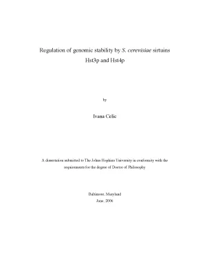
Tải xuống để đọc toàn bộRegulation of genomic stability by S. cerevisiae sirtuins Hst3p and Hst4p by Ivana Celic A dissertation submitted to The Johns Hopkins University in conformity with the requirements for the degree of Doctor of Philosophy Baltimore, Maryland June, 2006 UMI Number: 3243300 UMI Microform 3243300 Copyright 2007 by ProQuest Information and Learning Company. All rights reserved. This microform edition is protected against unauthorized copying under Title 17, United States Code.
ProQuest Information and Learning Company 300 North Zeeb Road P. Box 1346 Ann Arbor, MI 48106-1346 Abstract The Sir2 proteins, also known as sirtuins, represent a large and highly conserved family of NAD+-dependent protein deacetylases that control various fundamental biological processes. The baker’s yeast, Saccharomyces cerevisiae, has five members of this family, Sir2p and Hst1-4p, important for regulation of transcriptional silencing and genomic stability. Hst3p and Hst4p are two redundant sirtuins with the major role in maintenance of genomic stability.
They control genomic stability by regulating the level of acetylation of histone H3 lysine 56. This residue, present in the core of the nucleosome surface, is acetylated during the S phase of the cell cycle and contributes to the repair processes active during DNA replication. At the end of the S phase, K56 of histone H3 is deacetylated in a Hst3p- and Hst4p-dependent manner. Failure to deacetylate K56 leads to a growth defect, sensitivity to DNA-damaging agents and chromosome loss in hst3 hst4 cells.
These phenotypes can be suppressed by mutation of K56 into the nonacetylable residue arginine. Failure to deacetylate K56 also leads to activation of the DNA-damage response and renders hst3 hst4 cells sensitive to perturbations in DNA replication, repair and checkpoint function as is evident from numerous synthetic lethality interactions that hst3 hst4 cells display with mutations in the genes that regulate these processes. The growth defect of hst3 hst4 cells can be suppressed by overexpression of RFC1, the large subunit of RFC, a “clamp loader” that loads PCNA, the “sliding clamp” onto DNA during DNA replication. Interestingly, the growth defect of hst3 hst4 cells can also be suppressed by deletion of CTF18 and, somewhat less efficiently, RAD24 and ELG1.
CTF18, RAD24 and ELG1 all encode large subunits of alternative RFCs, each of ii which shares four smaller subunits (Rfc2p-Rfc5p) with Rfc1p. We propose that ongoing cycles of K56 acetylation and deacetylation contribute to the regulation of the functional equilibrium between different RFC complexes in the cell. (Thesis Advisor and Reader) Professor Department of Molecular Biology and Genetics Johns Hopkins University School of Medicine Brendan Cormack, Ph. (Thesis Reader) Associate Professor Department of Molecular Biology and Genetics Johns Hopkins University School of Medicine iii Acknowledgements This has been a very, very long ride and many people have helped me during this time in different ways.
I would like to thank my advisor, Jef Boeke, for giving me opportunity to work in his lab, for his patience and support over all these years. I am grateful to my committee members, Carol Greider, Cynthia Wolberger and Brendan Cormack for words of encouragement and letting me graduate after all. I am very grateful to Alain Verreault and his postdoctoral fellow Hiroshi Masumoto, who originally discovered that Hst3p and Hst4p regulated histone K56 acetylation, for their generosity in sharing results and willingness to collaborate. Also, many thanks go to Wendell Griffith, who has done some great MS analysis of K56 acetylation.
I thank the Boeke lab members for making the Boeke lab nice place to work. I had a great opportunity to overlap with some exceptional graduate students, Eric Bolton, Jeffrey Han and Siew-Loon Ooi, who thought me lot of little tricks of the trade. I thank to my friends, some close, some far away, who made these years more bearable. Special thanks goes to Jeff Han, for his support and many, many moments of happiness he brought in my life.
My deepest gratitude goes to my family, my parents, my sister and my grandmothers for being always there for me no matter what. My parents have sacrificed a lot to put me through school and ultimately to get me here and I will be always indebted for that. My sister and my grandmothers helped as much as they could. I am, also, very grateful to my sister for taking good care of my parents.
It has given me great peace of mind over these years. iv Table of Contents Page Title Page i Abstract ii Acknowledgments iv Table of Contents v List of Figures and Tables viii Chapter 1: Introduction 1 Chapter 2: The sirtuins Hst3p and Hst4p control histone H3 lysine 56 acetylation 23 Introduction 24 Results 26 Hst3 and Hst4 control histone H3 K56 acetylation during the cell cycle 26 The nicotinamide binding pocket of Hst3 is necessary for regulation of histone H3 K56 acetylation 27 The phenotypes of hst3 hst4 mutant cells are caused by high levels of H3 K56 acetylation 30 The histone chaperone Asf1p is needed for H3 K56 acetylation 32 Hst3p can trigger K56Ac deacetylation in mature chromatin 33 Discussion 35 Experimental Procedures 51 v Chapter 3: Cells lacking Hst3p and Hst4p activate the DNA damage response due to the presence of chronic DNA damage and display genetic interaction with genes involved in DNA metabolism 63 Introduction 64 Results 66 hst3 hst4 cells slow down cell-cycle progression in response to replication inhibition and DNA damage 66 hst3 hst4 cells activate DNA damage checkpoint in the absence of exogenous DNA damage 67 hst3 hst4 mutant depends on replication checkpoint for viability 69 Increased H2A serine 129 phosphorylation in hst3 hst4 cells 71 hst3 hst4 require subset of DNA repair proteins for viability 72 Overexpression of Rfc1p, large subunit of DNA clamp loader, suppresses phenotypes of hst3 hst4 cells 73 Discussion 76 Experimental procedures 96 References 107 Appendices 130 Appendix A: A phylogenetically conserved NAD+-dependent protein deacetylase activity in the Sir2 protein family 131 Appendix B: Chemistry of gene silencing: the mechanism of NAD+-dependent deacetylation reactions 138 Appendix C: Telomeric and rDNA silencing in Saccharomyces cerevisiae vi are dependent on a nuclear NAD+ salvage pathway 147 Appendix D: SIRT3, a human SIR2 homologue, is an NAD+-dependent deacetylase localized to mitochondria 161 Appendix E: Structure of a Sir2 enzyme bound to an acetylated p53 peptide 168 Appendix F: Sir2-dependent activation of acetyl-CoA synthetase by deacetylation of active lysine 182 Curriculum Vitae 186 vii List of Figures Page Figure 1. Phylogenetic tree of sirtuins. Proposed catalytic mechanism of Sir2 proteins.
Schematic alignment of S. Alignment of Hst3p, Hst4p and selected sirtuins. Hst3p and Hst4p control histone H3 K56 acetylation during the cell cycle. Effect of nicotinamide on the acetylation of histone H3 K56.
Mutation in invariant residues of Hst3p affects K56 acetylation level and function of Hst3p in vivo. The phenotypes of hst3 hst4 double mutants are substantially suppressed by mutation of histone H3 K56 into arginine. The hht1-K56R mutation suppresses the mitotic chromosome loss phenotype of hst3 hst4 mutant cells. The Asf1p histone chaperone is needed for H3 K56Ac.
Effects of rapid inactivation of Hst3p on H3 K56 deacetylation. Induction of Hst3p synthesis leads to H3 K56 deacetylation in mature chromatin. Abnormal distribution of histone H3 K56 acetylation with respect to replication forks is likely responsible for the DNA damage sensitivity of hst3 hst4 mutant cells. Drugs sensitivities of hst3 hst4 mutants.
Like wild-type cells, hst3 hst4 cells slow down DNA replication when exposed to MMS. Like wild-type cells, hst3 hst4 cells do not elongate their mitotic spindles when exposed to HU. Induction of RNR3 and HUG1 in hst3 hst4 cells. Hyperphosphorylation of Rad53p in hst3 hst4 cells.
hst3 hst4 cells required functional DNA replication checkpoint for viability. hst3 hst4 cells display increased level of histone H2A Ser128 phosphorylation. Synthetic lethality analysis with hst3 hst4 and suppression with H3 K56R. Overexpression of RFC1 suppresses growth defect, Ts phenotype and HU sensitivity of hst3 hst4 cells.
Overexpression of Rfc1p does not affect histone H3 K56 acetylation level in hst3 hst4 cells. Suppression of hst3 hst4 growth defect and Ts phenotype by inactivation of RAD24, but not RAD9. Suppression of hst3 hst4 growth defect and Ts phenotype by inactivation of alternative RFC complexes. List of the genes whose expression was changed in hst3 hst4 cells.
Synthetic lethal analysis with hst3 hst4. 95 ix Chapter 1 Introduction 1 Protein acetylation and deacetylation have emerged over the past decade or two as important posttranslational mechanisms regulating various aspects of cellular biology. This regulation is carried out through the action of protein acetylases, which modify the ε-NH2 of lysine residues and their counterparts, the protein deacetylases, which remove acetyl groups (Kouzarides, 2000; Kurdistani and Grunstein, 2003). The Sir2 family of proteins is an integral part of this regulatory network.
Sir2 proteins deacetylate lysine residues in histones and other proteins in a very unique enzymatic reaction that requires nicotinamide adenine dinucleotide (NAD+) (Imai et al., 2000; Landry et al., 2000b; Smith et al. The deacetylation reaction catalyzed by Sir2 proteins is absolutely dependent on NAD+. This is what differentiates Sir2 proteins as class III deacetylases, distinct from class I and class II deacetylases that don’t require NAD+ and deacetylate proteins through a more simple hydrolysis mechanism (Finnin et al. Presently, it is not known why sirtuins deacetylate proteins in such an energetically costly mechanism, but it has been suggested that sirtuins may represent a cellular NAD+ sensor linking metabolism to other aspects of cellular physiology, like genome stability, aging, apoptosis and differentiation.
Sir2 proteins or sirtuins form a large and highly conserved protein family. All three kingdoms of life, archea, bacteria and eukaryotes, are represented within the sirtuin family (Figure 1. Some organisms have only one sirtuin, while others have multiple paralogs, for example yeast with five (Brachmann et al., 1995) and mammals with 7 paralogs (Frye, 1999; Frye, 2000). All sirtuins share a conserved catalytic core domain necessary for the NAD+-dependent deacetylation reaction.
Sir2 proteins differ significantly in their N- and C-terminal extensions, which range from non- 2 existent, for example in archeal sirtuins, to very long ones, like in S. These extensions mediate member-specific functions like protein localization, as in the case of human SirT3 which is targeted to mitochondria by its N-terminal extension (Onyango et al., 2002; Schwer et al., 2002), formation of multiprotein complexes with specialized function, as in the case of S.cerevisiae Sir2p, which exists in the form of at least two functionally different protein complexes within a cell (Ghidelli et al., 2001) or they have an autoregulatory function, as in the case of S. cerevisiae Hst2p in which N and C-terminal domain have inhibitory activity by binding to the catalytic core intermolecularly and intramolecularly, respectively (Zhao et al. Phylogenetic tree of sirtuins (published with permission by William Hawse and Cynthia Wolberger).
4 Sirtuins catalyze NAD+-dependent deacetylation of lysine residues in proteins (Imai et al., 2000; Landry et al., 2000b; Smith et al. During this reaction, the glycosidic bond between the C1’ atom of ribose and nicotinamide moiety in NAD+ is cleaved to generate, in addition to the deacetylated lysine residue, free nicotinamide and acetyl-ADP ribose (APDR) (Sauve et al., 2001; Tanner et al., 2000; Tanny and Moazed, 2001). NMR and mass-spectrophotometric analysis of the Sir2 deacetylation product revealed that actual product is 2’- and 3’-acetyl-ADP ribose (Sauve et al. 2’-acetyl-ADP ribose is the first product of the reaction followed by non-enzymatic conversion to 3’- acetyl-ADP ribose to a final equilibrium ratio of 47:67 for the 2’ and 3’ stereoisomers.
Labeling experiments with H218O, MS and NMR analysis led Sauve et al. (Sauve et al., 2001) to propose the mechanism (Figure 1.2), which involves the nucleophilic attack by the carbonyl oxygen of the acetyl-lysine on the C1’ carbon of the nicotinamide ribose (N- ribose) in NAD+. This step involves formation of the highly reactive riboxacarbenium ion that captures the acyl oxygen of acetyl-lysine to generate a 1’-O-alkyl-amidate, followed by the attack of the N-ribose 2’hydroxyl (2’OH) to form a 1’, 2’-acyloxonium structure, which is the precursor for 2’-acetyl-ADP ribose formation.
Nội dung được bảo vệ bản quyền — Tải xuống đầy đủ
Trích dẫn luận án này
Ivana Celic (2006). Sirtuins Hst3p và Hst4p điều hòa ổn định bộ gen [Luận án tiến sĩ, The Johns Hopkins University]. LuanAn.net. https://luanan.net/sinh-hoc/cong-nghe-sinh-hoc/sirtuins-hst3p-hst4p-dieu-hoa-on-dinh-bo-gen-saccharomyces-cerevisiae
Từ khóa và chủ đề nghiên cứu
Từ khóa liên quan
Xem thêm luận án cùng lĩnh vực
Chủ đề nghiên cứu
Câu hỏi thường gặp
Luận án "Sirtuins Hst3p và Hst4p điều hòa ổn định bộ gen" nghiên cứu về vấn đề gì?
Luận án tiến sĩ khám phá vai trò sirtuins Hst3p và Hst4p trong điều hòa ổn định bộ gen. Nghiên cứu cơ chế deacetylase histone H3 lysine 56 và ảnh hưởng đến sửa chữa DNA.
Luận án "Sirtuins Hst3p và Hst4p điều hòa ổn định bộ gen" được bảo vệ tại trường nào?
Luận án này được bảo vệ tại The Johns Hopkins University. Năm bảo vệ: 2006.
Luận án "Sirtuins Hst3p và Hst4p điều hòa ổn định bộ gen" thuộc chuyên ngành gì?
Luận án "Sirtuins Hst3p và Hst4p điều hòa ổn định bộ gen" thuộc chuyên ngành Molecular Biology and Genetics. Danh mục: Công Nghệ Sinh Học.
Luận án "Sirtuins Hst3p và Hst4p điều hòa ổn định bộ gen" có bao nhiêu trang?
Luận án "Sirtuins Hst3p và Hst4p điều hòa ổn định bộ gen" có 197 trang. Bạn có thể xem trước một phần tài liệu ngay trên trang web trước khi tải về.
Cách tải luận án "Sirtuins Hst3p và Hst4p điều hòa ổn định bộ gen" về máy như thế nào?
Để tải luận án về máy, bạn nhấn nút "Tải xuống ngay" trên trang này, sau đó hoàn tất thanh toán phí lưu trữ. File sẽ được tải xuống ngay sau khi thanh toán thành công. Hỗ trợ qua Zalo: 0559 297 239.