Dual-Functional Ag@SiO2 Nanocubes for Biomarker Detection - Nguyen Minh Kha (NTUST)
Luận án tiến sĩ nghiên cứu nanocubes AgSiO2 core-shell định hướng. Nền plasmonic kép cho phát hiện dấu ấn sinh học chính xác, hiệu quả.
National Taiwan University of Science and Technology
Luan An
Luận án
Năm xuất bản
Số trang
166
Thời gian đọc
25 phút
Lượt xem
2
Lượt tải
0
Phí lưu trữ
50 Point
Tổng quan nhanh
- Chủ đề:
- 1. Thiết kế nanohạt Ag@SiO2 chức năng kép cho phát hiện biomarker
- Số trang:
- 166 trang
- Trường:
- National Taiwan University of Science and Technology
- Chuyên ngành:
- Chemical Engineering
- Tác giả:
- Nguyen Minh Kha
- Năm:
- 2016
Tóm tắt nội dung luận án
I. Thiết kế nanohạt Ag SiO2 chức năng kép cho phát hiện biomarker
Công nghệ y tế đang đối mặt với những thách thức lớn trong việc phát hiện biomarker. Các phương pháp truyền thống thường thiếu độ nhạy cần thiết. Nhiều biomarker mới, quan trọng trong chẩn đoán bệnh, đòi hỏi kỹ thuật tiên tiến hơn. Nanohạt Ag@SiO2 với chức năng kép mang lại giải pháp đột phá. Cụ thể, hệ thống này kết hợp khả năng phát hiện photoluminescence (PL) nhạy cảm thông qua hiệu ứng quang phát xạ tăng cường kim loại (MEPL). Đồng thời, nó cung cấp dấu vân tay Raman đặc trưng thông qua phổ Raman tăng cường bề mặt (SERS). Sự kết hợp này giúp tăng cường độ chính xác và độ nhạy trong quá trình phát hiện. Nền tảng chức năng kép này mở ra kỷ nguyên mới cho cảm biến sinh học. Nó hứa hẹn cải thiện đáng kể khả năng chẩn đoán phân tử. Nền tảng plasmonic thiết kế đặc biệt này đáp ứng nhu cầu y tế cấp bách. Nó giúp theo dõi các dấu hiệu sinh học một cách hiệu quả hơn.
1.1. Thách thức phát hiện biomarker bằng công cụ truyền thống
Phát hiện biomarker là yếu tố then chốt trong chẩn đoán và theo dõi bệnh. Nhiều biomarker mới có nồng độ rất thấp. Công cụ chẩn đoán truyền thống thường gặp khó khăn. Chúng không đủ nhạy để phát hiện những biomarker này. Điều này hạn chế khả năng chẩn đoán sớm và chính xác. Do đó, cần có các phương pháp phát hiện biomarker siêu nhạy. Các phương pháp này cần cung cấp thông tin chi tiết về các dấu hiệu sinh học phức tạp. Đây là động lực thúc đẩy nghiên cứu công nghệ nano y sinh. Việc phát triển giải pháp mới là rất quan trọng.
1.2. Plasmonics Công nghệ quang học siêu nhạy cho y học
Công nghệ plasmonics cung cấp nền tảng quang học siêu nhạy. Nó ứng dụng rộng rãi trong chẩn đoán y tế và cảm biến hình ảnh sinh học. Hiệu ứng plasmon bề mặt cục bộ (LSPR) là cơ sở của công nghệ này. LSPR tạo ra các trường điện từ mạnh gần bề mặt kim loại. Điều này giúp tăng cường đáng kể tín hiệu quang học. Nanohạt kim loại, như bạc (Ag), là thành phần chính. Chúng tạo ra hiệu ứng plasmonic mạnh mẽ. Ứng dụng plasmonics mở ra tiềm năng lớn. Nó giúp phát hiện các phân tử ở nồng độ cực thấp. Đây là yếu tố quyết định sự thành công trong chẩn đoán.
1.3. Khái niệm chức năng kép SERS và MEPL đồng thời
Nền tảng chức năng kép kết hợp SERS và MEPL. SERS cung cấp dấu vân tay Raman đặc trưng của phân tử. MEPL tăng cường cường độ quang phát xạ. Sự kết hợp này mang lại hai loại thông tin bổ sung. Nó giúp xác định biomarker với độ chính xác cao hơn. Các nanohạt Ag@SiO2 đóng vai trò trung tâm. Chúng hoạt động như chất nền plasmonic. Cả hai cơ chế đều được tối ưu hóa đồng thời. Mục tiêu là đạt được độ nhạy và độ đặc hiệu tối đa. Nanomaterials chức năng kép này là một bước tiến lớn. Chúng hứa hẹn cải thiện khả năng phát hiện và phân tích.
II. Tối ưu hóa tổng hợp Ag SiO2 nanocubes cho cảm biến sinh học
Quá trình tổng hợp nanohạt Ag@SiO2 core-shell là bước then chốt. Việc này ảnh hưởng trực tiếp đến hiệu suất của cảm biến sinh học. Các nanocubes bạc (Ag NCs) được tổng hợp bằng phương pháp polyol. Sau đó, chúng được biến đổi bằng các tác nhân ghép nối. 3-mercaptopropyltrimethoxysilane (MPTMS) và 3-aminopropyltrimethoxysilane (APTMS) là hai tác nhân quan trọng. Các tác nhân này đóng vai trò cầu nối. Chúng giúp định hướng sự phát triển của lớp vỏ silica. Tetraethylorthosilicate (TEOS) được sử dụng để tạo lớp vỏ silica. Lớp vỏ silica này bao phủ hạt nhân bạc. Độ dày và độ đồng đều của lớp vỏ silica được kiểm soát chặt chẽ. Điều này đảm bảo hiệu quả tối ưu cho nanohạt nhân-vỏ. Nghiên cứu đã chỉ ra sự khác biệt rõ rệt giữa các tác nhân ghép nối. Tối ưu hóa các bước này là cần thiết. Nó giúp tạo ra nanohạt Ag@SiO2 với hiệu suất cao. Điều này mở đường cho các ứng dụng chẩn đoán phân tử tiên tiến.
2.1. Quy trình tổng hợp nanocubes bạc và biến tính bề mặt
Nanocubes bạc (Ag NCs) được tạo ra thông qua phương pháp polyol. Phương pháp này kiểm soát hình dạng và kích thước hạt hiệu quả. Sau đó, bề mặt Ag NCs được biến tính. Các tác nhân ghép nối được sử dụng. MPTMS và APTMS là những lựa chọn chính. Chúng tạo ra các nhóm chức năng trên bề mặt bạc. Điều này giúp tăng cường khả năng bám dính của silica. Sự biến tính bề mặt này rất quan trọng. Nó ảnh hưởng đến cấu trúc và tính chất của nanohạt Ag@SiO2 cuối cùng. Quá trình này đảm bảo lớp vỏ silica bám chắc chắn và đồng đều.
2.2. Vai trò của tác nhân ghép nối trong hình thành vỏ silica
Tác nhân ghép nối đóng vai trò quan trọng. Chúng điều khiển độ dày và độ đồng đều của lớp vỏ silica. Sự hiện diện của chúng làm thay đổi đáng kể hạt nhân Ag NCs. APTMS và MPTMS có cấu trúc khác nhau. Do đó, chúng tạo ra hiệu ứng khác nhau. APTMS được chứng minh là vượt trội hơn. Nó cải thiện đáng kể khả năng tăng cường SERS và MEPL. Việc lựa chọn tác nhân ghép nối phù hợp là rất quan trọng. Nó ảnh hưởng trực tiếp đến hiệu suất quang học của nanohạt Ag@SiO2. Đây là một yếu tố then chốt trong công nghệ nano y sinh.
2.3. Tối ưu hóa độ dày lớp vỏ silica siêu mỏng cho Ag SiO2
Độ dày lớp vỏ silica có ảnh hưởng lớn đến hiệu ứng plasmonic. Lớp vỏ silica siêu mỏng khoảng 1.5 nm mang lại hiệu quả cao. Nó giúp tăng cường cường độ SERS lên 3 lần. So với Ag NCs không có vỏ silica. Độ dày này tạo ra khoảng cách tối ưu. Nó cho phép tương tác mạnh mẽ giữa hạt nhân bạc và các phân tử đích. Đồng thời, lớp vỏ silica bảo vệ hạt nhân bạc. Nó cải thiện độ ổn định của nanohạt. Việc kiểm soát chính xác độ dày vỏ là cần thiết. Điều này đảm bảo hiệu suất tối ưu cho các cảm biến sinh học.
III. Tăng cường độ nhạy SERS và MEPL bằng nanohạt nhân vỏ Ag SiO2
Các nanohạt Ag@SiO2 nhân-vỏ thể hiện khả năng tăng cường tín hiệu quang học vượt trội. Điều này đạt được thông qua sự kết hợp của hiệu ứng SERS và MEPL. Lớp vỏ silica siêu mỏng đóng vai trò quan trọng. Nó điều chỉnh khoảng cách giữa hạt nhân bạc và các phân tử được phát hiện. Sự điều chỉnh này tối ưu hóa tương tác plasmonic. Kết quả là cường độ tín hiệu SERS và MEPL được cải thiện đáng kể. Việc sử dụng APTMS làm tác nhân ghép nối mang lại lợi ích lớn. Nó dẫn đến sự tăng cường SERS và MEPL vượt trội. Nanohạt Ag@SiO2 được tối ưu hóa mang lại độ nhạy cao. Chúng cho phép phát hiện các biomarker ở nồng độ cực thấp. Đây là yếu tố quan trọng trong chẩn đoán sớm và chính xác. Sự kết hợp hai chức năng này tạo nên một công cụ mạnh mẽ. Nó vượt xa khả năng của các phương pháp phát hiện đơn lẻ. Công nghệ nano y sinh này mở ra nhiều triển vọng mới.
3.1. Cơ chế tăng cường tín hiệu SERS với nanocubes bạc silica
SERS là kỹ thuật phân tích siêu nhạy. Nó cho phép phát hiện phân tử thông qua phổ Raman tăng cường. Nanohạt bạc tạo ra các 'điểm nóng' plasmonic. Các điểm nóng này tăng cường trường điện từ cục bộ. Lớp vỏ silica siêu mỏng (~1.5 nm) tối ưu hóa khoảng cách. Nó đưa phân tử đích vào vùng có trường điện từ mạnh nhất. Điều này dẫn đến sự tăng cường tín hiệu Raman lên nhiều bậc. Cụ thể, Ag@SiO2 NCs với vỏ silica siêu mỏng cho cường độ SERS cao hơn 3 lần. So với Ag NCs không vỏ silica. Lớp vỏ này cũng bảo vệ phân tử khỏi sự dập tắt. Điều này duy trì độ nhạy cao của cảm biến sinh học.
3.2. Cải thiện hiệu suất MEPL bằng lớp vỏ silica bảo vệ
MEPL là hiện tượng tăng cường quang phát xạ của các phân tử gần bề mặt kim loại. Lớp vỏ silica trong nanohạt Ag@SiO2 đóng vai trò kép. Nó kiểm soát khoảng cách giữa phân tử phát sáng và bề mặt bạc. Đồng thời, nó ngăn chặn sự dập tắt quang phát xạ. Sự dập tắt này thường xảy ra khi phân tử quá gần kim loại. APTMS modified Ag@SiO2 NCs cho thấy sự tăng cường MEPL đáng kể. Điều này tạo điều kiện cho cả hai chức năng hoạt động hiệu quả. Việc tối ưu hóa lớp vỏ silica là yếu tố then chốt. Nó giúp đạt được hiệu quả MEPL cao. Nanohạt nhân-vỏ Ag@SiO2 là nền tảng lý tưởng cho cả SERS và MEPL.
3.3. Duy trì độ nhạy kép SERS và MEPL sau thời gian lưu trữ
Một trong những thách thức của cảm biến plasmonic là độ ổn định. Nghiên cứu đã chỉ ra khả năng duy trì độ nhạy kép. Việc trộn Ag@SiO2 NCs với lớp vỏ silica dày ~1 nm. Phương pháp này giúp duy trì độ nhạy SERS và MEPL. Cả hai đều đạt ~90% sau 12 tuần lưu trữ. Điều này chứng tỏ tính ổn định vượt trội của nanohạt Ag@SiO2. Lớp vỏ silica đóng vai trò bảo vệ. Nó giảm thiểu sự oxy hóa và suy thoái của hạt nhân bạc. Độ ổn định này rất quan trọng. Nó cho phép ứng dụng rộng rãi trong chẩn đoán phân tử thực tế.
IV. Chân đế plasmonic ổn định hiệu quả cao cho chẩn đoán phân tử
Sự tương tác giữa chất nền và cấu trúc nano plasmonic ảnh hưởng đến hiệu suất. Chân đế đóng vai trò quan trọng trong việc tối ưu hóa hiệu quả cảm biến. Một chất nền mới đã được chế tạo. Nó có chỉ số khúc xạ thấp và độ nhám bề mặt phù hợp. Chất nền này được tạo ra bằng cách khắc lá nhôm. Sau đó, tạo ra cấu trúc alumina hình hoa (f-Al2O3/e-Al). Nanohạt Ag@SiO2 được lắng đọng trên chất nền này. Chúng tự sắp xếp theo cấu hình cạnh-cạnh (edge-edge configuration). Độ nhám bề mặt của f-Al2O3/e-Al cung cấp một đường dẫn. Nó giúp ghép nối ánh sáng tới với plasmon bề mặt. Điều này tạo ra các điểm nóng plasmon hiệu quả. Cả SERS và MEPL đều được hưởng lợi từ sự ghép nối này. Chân đế mới này không chỉ tăng cường hiệu quả. Nó còn cải thiện đáng kể thời gian bảo quản của cảm biến. Đây là một bước tiến quan trọng trong công nghệ nano y sinh.
4.1. Tác động của chân đế đến hiệu suất cảm biến plasmonic
Chân đế là yếu tố thiết yếu ảnh hưởng đến hiệu suất cảm biến. Các tính chất của chân đế, như chỉ số khúc xạ và độ nhám, rất quan trọng. Chúng có thể tối ưu hóa hoặc hạn chế tương tác plasmonic. Chân đế có chỉ số khúc xạ thấp giúp giảm tổn thất ánh sáng. Độ nhám bề mặt phù hợp tạo điều kiện cho sự ghép nối plasmon. Sự sắp xếp của nanohạt trên chân đế cũng đóng vai trò. Cấu hình cạnh-cạnh có thể tạo ra các điểm nóng hiệu quả. Việc lựa chọn và thiết kế chân đế phù hợp là cần thiết. Nó giúp tối đa hóa độ nhạy và hiệu quả của các cảm biến sinh học.
4.2. Chế tạo chân đế alumina hoa f Al2O3 e Al cho Ag SiO2 NCs
Một chân đế đặc biệt đã được phát triển. Nó là alumina hình hoa trên lá nhôm khắc (f-Al2O3/e-Al). Chất nền này có độ nhám bề mặt được kiểm soát. Nó tạo ra một bề mặt lý tưởng cho nanohạt Ag@SiO2. Nanohạt tự lắp ráp theo cấu hình cạnh-cạnh. Sự sắp xếp này tối ưu hóa sự tương tác plasmonic. Chân đế f-Al2O3/e-Al cung cấp một con đường hiệu quả. Nó giúp ánh sáng laser ghép nối vào các điểm nóng plasmon. Điều này tăng cường cả tín hiệu SERS và MEPL. Việc chế tạo chân đế này mở ra khả năng mới. Nó giúp cải thiện đáng kể hiệu suất của cảm biến plasmonic.
4.3. Cải thiện độ ổn định và thời gian sử dụng của chân đế cảm biến
Chân đế Ag@SiO2/f-Al2O3/e-Al cho thấy độ ổn định được cải thiện. Lớp vỏ silica siêu mỏng đóng vai trò giảm tốc độ khuếch tán oxy. Lớp điện môi Al2O3 hình hoa cũng góp phần bảo vệ. Điều này kéo dài đáng kể thời gian sử dụng của chất nền. Khả năng bảo quản lâu dài là rất quan trọng. Nó giúp giảm chi phí và tăng tính thực tiễn của công nghệ. Độ ổn định cao làm cho cảm biến plasmonic trở nên đáng tin cậy hơn. Điều này là một ưu điểm lớn trong các ứng dụng chẩn đoán phân tử. Nó đảm bảo hiệu suất nhất quán theo thời gian.
V. Ứng dụng thực tế Phát hiện biomarker không xâm lấn chi phí thấp
Công nghệ nanohạt Ag@SiO2 chức năng kép có tiềm năng ứng dụng lớn. Đặc biệt trong phát hiện biomarker không xâm lấn. Creatinine (CR) và flavin adenine dinucleotide (FAD) là những biomarker quan trọng. Chúng tồn tại trong máu và nước tiểu của con người. Việc phát hiện chúng trong nước tiểu bằng kỹ thuật SERS và MEPL không cần gắn nhãn. Điều này mở ra khả năng phân tích chi phí thấp và không xâm lấn. Đồng thời vẫn đảm bảo độ nhạy cao. Phương pháp này mang lại nhiều lợi ích. Nó đơn giản hóa quy trình lấy mẫu và giảm gánh nặng cho bệnh nhân. Các cách tiếp cận được khám phá có thể phát triển thành công cụ mã hóa mạnh mẽ. Nó hỗ trợ phân tích sinh học thông lượng cao. Điều này hứa hẹn một tương lai tươi sáng cho chẩn đoán y tế.
5.1. Phát hiện Creatinine và FAD trong nước tiểu bằng SERS MEPL
Creatinine và FAD là các phân tử sinh học quan trọng. Chúng là biomarker cho nhiều tình trạng sức khỏe. Các kỹ thuật SERS và MEPL không cần gắn nhãn đã được áp dụng. Chúng cho phép phát hiện các biomarker này trong nước tiểu. Phát hiện trong nước tiểu là phương pháp không xâm lấn. Nó ít gây khó chịu cho bệnh nhân hơn so với mẫu máu. Khả năng phát hiện với độ nhạy cao giúp chẩn đoán sớm. Nó cũng giúp theo dõi hiệu quả điều trị. Đây là một bước tiến quan trọng. Nó làm cho chẩn đoán phân tử trở nên dễ tiếp cận hơn.
5.2. Tiềm năng cho phân tích sinh học thông lượng cao và chẩn đoán sớm
Các cách tiếp cận được phát triển có tiềm năng lớn. Chúng có thể trở thành công cụ mã hóa mạnh mẽ. Điều này hỗ trợ phân tích sinh học thông lượng cao. Phân tích thông lượng cao cho phép xử lý nhiều mẫu cùng lúc. Nó tăng tốc độ và hiệu quả của các nghiên cứu lâm sàng. Phát hiện biomarker không xâm lấn và chi phí thấp là mục tiêu chính. Công nghệ nano y sinh này mang lại cơ hội chẩn đoán sớm. Nó cải thiện kết quả điều trị cho bệnh nhân. Điều này góp phần vào sự phát triển của y học cá nhân hóa.
Mục lục chi tiết luận án
Tải xuống file đầy đủ để xem toàn bộ nội dung
Tải đầy đủ (166 trang)Nội dung chính
Tổng quan về luận án
Sự phát triển vượt bậc của y sinh học hiện đại đang khám phá ra hàng loạt dấu ấn sinh học (biomarkers) mới có ý nghĩa quan trọng trong chẩn đoán sớm các bệnh lý hiểm nghèo. Tuy nhiên, Tổ chức Y tế Thế giới (WHO) chỉ ra rằng hơn 43% gánh nặng y tế tại các quốc gia đang phát triển bắt nguồn từ các bệnh không lây nhiễm (ung thư, tim mạch, suy thận) và 32% từ các bệnh truyền nhiễm. Việc phát hiện các dấu ấn này ở nồng độ siêu vết (ultratrace) vượt quá giới hạn phân tích của các kỹ thuật thông thường như ELISA hay PCR vốn đòi hỏi quy trình khuếch đại gen phức tạp, chi phí cao và nhiều bước rửa tiêu tốn thời gian. Trong bối cảnh đó, quang tử học plasmon (plasmonics) nổi lên như một công nghệ đột phá cho phép chế tạo các cảm biến sinh học đa mục tiêu với độ nhạy phân tử đơn. Luận án tiến sĩ "Orientated Ag@SiO2 Core-shell Nanocubes as Dual-functional Plasmonic Substrates for Biomarker Detection" do Nghiên cứu sinh Nguyễn Minh Kha thực hiện dưới sự hướng dẫn của Giáo sư Bing-Joe Hwang tại Khoa Kỹ thuật Hóa học, Trường Đại học Khoa học và Công nghệ Quốc gia Đài Loan (NTUST), đã mở ra hướng tiếp cận tiên phong trong việc tích hợp đồng thời hai cơ chế quang phổ: Tán xạ Raman tăng cường bề mặt (Surface-Enhanced Raman Scattering - SERS) và Phát xạ huỳnh quang tăng cường kim loại (Metal-Enhanced Photoluminescence - MEPL).
Khoảng trống nghiên cứu (research gap) cốt lõi được luận án xác định là sự xung đột vật lý cố hữu về khoảng cách không gian giữa hiệu ứng SERS và MEPL. Theo lý thuyết điện động học cận trường, cường độ SERS tỷ lệ thuận với bậc 4 của cường độ điện trường cục bộ ($|E_{loc}|^4$), đạt cực đại khi phân tử chất phân tích tiếp xúc trực tiếp hoặc cách bề mặt kim loại dưới 1-2 nm. Ngược lại, hiệu ứng MEPL đòi hỏi khoảng cách đệm (spacer) tối ưu từ 5 nm đến 20 nm để ngăn chặn hiện tượng dập tắt huỳnh quang phi bức xạ (non-radiative quenching) do truyền năng lượng Förster về phía kim loại quý. Hơn nữa, các hạt nano bạc dị hướng dạng lập phương (Ag Nanocubes - Ag NCs) tuy sở hữu hệ số phẩm chất plasmon ($Q = \varepsilon_r^2 / \varepsilon_i$) vượt trội hơn vàng (Au) trong vùng ánh sáng khả kiến, nhưng lại cực kỳ kém bền do quá trình oxy hóa tự nhiên và suy thoái góc nhọn plasmon khi tiếp xúc với môi trường sinh hóa.
Để giải quyết triệt để các thách thức trên, luận án thiết lập 4 câu hỏi nghiên cứu và giả thuyết khoa học tương ứng:
- H1: Việc biến tính bề mặt hạt Ag NCs bằng tác nhân liên kết silane hữu cơ phù hợp sẽ kiểm soát chính xác phản ứng ngưng tụ Stöber, cho phép tạo lớp vỏ điện môi silica siêu mỏng ($\sim 1.5\text{ nm}$) đồng nhất mà không làm tròn các góc sắc nhọn.
- H2: Lớp vỏ silica siêu mỏng $\sim 1.5\text{ nm}$ đóng vai trò bảo vệ cấu trúc nano bạc khỏi oxy hóa, đồng thời tối ưu hóa mật độ điện trường cận trường, mang lại hệ số tăng cường SERS cao hơn cả hạt Ag NCs trần không phủ vỏ.
- H3: Sự phối trộn có kiểm soát tỷ lệ giữa các hạt Ag@SiO2 có độ dày vỏ khác nhau ($1.5\text{ nm}$ và $20\text{ nm}$) sẽ tạo ra một nền plasmonic lưỡng chức năng, khuếch đại đồng thời cả tín hiệu SERS và MEPL mà không triệt tiêu lẫn nhau.
- H4: Một đế nền có chiết suất thấp và độ nhám định hình vi cấu trúc dạng cánh hoa (flower-like alumina trên lá nhôm ăn mòn, f-Al2O3/e-Al) sẽ định hướng các hạt Ag@SiO2 NCs sắp xếp theo cấu hình cạnh-cạnh (edge-edge orientation), tạo ra mạng lưới điểm nóng quang học (hotspots) mới và kéo dài tuổi thọ nền cảm biến.
Khung lý thuyết của nghiên cứu được xây dựng trên sự giao thoa giữa Lý thuyết Cộng hưởng Plasmon Bề mặt Cục bộ (LSPR), Lý thuyết Tán xạ Raman Điện từ học (Electromagnetic SERS Theory), và Kỹ thuật Điều khiển Tốc độ Phân rã Bức xạ (Radiative Decay Engineering). Về mặt quy mô và đóng góp thực tiễn, luận án đã chế tạo thành công nền cảm biến plasmonic lưỡng chức năng có khả năng duy trì $\sim 90%$ độ nhạy SERS và MEPL sau 12 tuần (3 tháng) bảo quản trong điều kiện tự nhiên, đồng thời định lượng thành công hai dấu ấn sinh học trong nước tiểu nhân tạo và nước tiểu thực tế là Creatinine (CR, $10^{-4}\text{ M}$) qua phổ vân tay SERS và Flavin Adenine Dinucleotide (FAD, $10^{-7}\text{ M}$) qua phổ huỳnh quang MEPL.
Literature Review và Positioning
Nghiên cứu về quang học tăng cường bề mặt bắt đầu từ phát hiện lịch sử của Martin Fleischmann cùng cộng sự (1974) về tín hiệu Raman dị thường của pyridine trên điện cực bạc nhám, sau đó được David L. Jeanmarie và Richard P. Van Duyne (1977) chứng minh là do cơ chế tăng cường điện từ học (Electromagnetic Enhancement - EE), song song với cơ chế chuyển dịch điện tích (Chemical Enhancement - CE) của Michael G. Albrecht và J. Alan Creighton (1977). Trải qua hơn bốn thập kỷ, dòng nghiên cứu SERS tập trung vào việc cực đại hóa hệ số tăng cường điện từ (Electromagnetic Enhancement Factor - EF) thông qua việc tạo ra các "điểm nóng" (hotspots) tại các khe hở nano hoặc đầu nhọn của hạt nano kim loại quý. Katrin Kneipp và cộng sự (1997) đã chứng minh khả năng phát hiện đơn phân tử bằng SERS với hệ số khuếch đại lên tới $10^{14}$ khi phân tử nằm chính xác tại điểm nóng khe hở nano.
Song song với SERS, dòng nghiên cứu về Phát xạ huỳnh quang tăng cường kim loại (MEPL/MEF) được khởi xướng và phát triển mạnh mẽ bởi Joseph R. Lakowicz (2001, 2005). Theo lý thuyết của Lakowicz, sự hiện diện của hạt nano kim loại có thể làm biến đổi sâu sắc động học phát xạ của chất phát quang lân cận: làm tăng tốc độ bức xạ ($\Gamma_m$) và tăng mật độ kích thích cục bộ ($|L(\omega_{exc})|^2$), qua đó nâng cao hiệu suất lượng quang biểu kiến ($Y = |L(\omega_{exc})|^2 Q_m$) và giảm thời gian sống huỳnh quang ($\tau_m$). Tuy nhiên, trường phái nghiên cứu quang học kinh điển luôn tồn tại một tranh luận lý thuyết gay gắt: Trong khi SERS yêu cầu phân tử nằm sát bề mặt kim loại ($d < 2\text{ nm}$) để tận dụng gradient điện trường $|E_{loc}|^4$, thì MEPL lại bị dập tắt nghiêm trọng ở cự ly này do quá trình chuyển dịch năng lượng không bức xạ từ phân tử sang kim loại theo định luật $1/d^3$ hoặc $1/d^6$ (Förster Resonance Energy Transfer - FRET).
                      SO SÁNH CÁC HỆ PLASMONIC TRONG Y SINH
 ┌─────────────────────────────────────────────────────────────────────────────┐
 │ Nghiên cứu Quốc tế Điển hình:                                               │
 │ 1. Kneipp et al. (1997): Single-molecule SERS dùng hạt Au/Ag keo            │
 │    → Nhược điểm: Tín hiệu ngẫu nhiên, không tái lặp, không có MEPL          │
 │ 2. Choo et al. & Liz-Marzán (2010-2014): Ag@SiO2 Core-Shell                 │
 │    → Nhược điểm: Lớp silica > 4-10 nm làm suy giảm nghiêm trọng SERS         │
 │ 3. Nanorods functionalized DSPE-PEG-OTMS (2015):                            │
 │    → Nhược điểm: Quy trình đa bước phức tạp, chi phí cao, độ bền oxy hóa kém │
 ├─────────────────────────────────────────────────────────────────────────────┤
 │ Luận án Nguyễn Minh Kha (NTUST, 2016):                                      │
 │ ★ Kiểm soát lớp vỏ siêu mỏng Ag@SiO2 (~1.5 nm) bằng tiền chất APTMS         │
 │ ★ Phối trộn hạt nano lưỡng chức năng (X1Y5: 1.5 nm & 20 nm)                │
 │ ★ Cấu trúc nền lai f-Al2O3/e-Al chiết suất thấp → Định hướng Cạnh-Cạnh      │
 │ ★ Phát hiện đồng thời CR (10⁻⁴ M, SERS) & FAD (10⁻⁷ M, MEPL) sau 12 tuần    │
 └─────────────────────────────────────────────────────────────────────────────┘
Trong bức tranh tổng quan đó, các công trình quốc tế trước đây thường chỉ tối ưu hóa đơn phương thức. Khi cố gắng tích hợp SERS và huỳnh quang, các nhóm nghiên cứu như Liz-Marzán hay Choo thường gặp bế tắc khi tạo vỏ silica cho hạt nano bạc: các phương pháp tổng hợp thông thường luôn tạo ra lớp vỏ dày trên $4-10\text{ nm}$, khiến tín hiệu SERS suy giảm nghiêm trọng. Ngược lại, các hạt không bọc vỏ lại bị oxy hóa hoàn toàn chỉ sau vài ngày tiếp xúc với không khí. Luận án của Nguyễn Minh Kha đã định vị chính xác khoảng trống công nghệ này và tạo ra bước nhảy vọt: lần đầu tiên thiết lập quy trình hóa học bề mặt cho phép khống chế độ dày vỏ silica ở mức siêu mỏng $\sim 1.5\text{ nm}$ đồng nhất, đồng thời đề xuất giải pháp nền lai đa lớp (hybrid substrate) để dung hòa hoàn hảo hai hiệu ứng quang phổ tương phản trên cùng một diện tích phân tích.
Đóng góp lý thuyết và khung phân tích
Đóng góp cho lý thuyết
Luận án đã mở rộng và làm sâu sắc thêm ba lý thuyết nền tảng của quang học nano và hóa học bề mặt:
- Lý thuyết Khuếch đại Điện từ Cận trường (Near-Field Electromagnetic Enhancement): Nghiên cứu thách thức giả định truyền thống cho rằng việc bao bọc một lớp điện môi vô cơ lên hạt nano kim loại sẽ luôn làm suy giảm cường độ điện trường bề mặt. Luận án chứng minh bằng thực nghiệm và mô hình hóa rằng một lớp vỏ silica siêu mỏng $\sim 1.5\text{ nm}$ đóng vai trò như một môi trường điện môi điều hòa (dielectric matching layer), giúp bảo toàn độ sắc nét của góc lập phương, ngăn chặn sự tái cấu trúc bề mặt của bạc, và tăng cường hệ số tán xạ SERS lên gấp 3 lần so với hạt Ag NCs trần.
- Lý thuyết Phân rã Bức xạ Điều khiển (Radiative Decay Engineering của Lakowicz): Luận án cung cấp bằng chứng thực nghiệm chuẩn xác về sự phụ thuộc phi tuyến tính của tốc độ bức xạ huỳnh quang vào chiều dày lớp đệm phân cách $\text{SiO}2$. Nghiên cứu xác lập khoảng cách $20\text{ nm}$ là ngưỡng đệm hoàn hảo để triệt tiêu hoàn toàn kênh dập tắt phi bức xạ ($k'{nr} \to 0$) trong khi vẫn tận dụng được mật độ photon plasmon cục bộ để tăng cường cường độ huỳnh quang của phân tử hữu cơ.
- Mô hình Khớp nối Plasmon Đa phần tử (Coupled Plasmon Resonances on Dielectric Substrates): Nghiên cứu mở rộng mô hình tương tác lưỡng cực plasmon khi đặt hạt nano dị hướng lên chất nền thực tế có chiết suất thay đổi, làm sáng tỏ cơ chế ghép nối photon vào plasmon mặt (SPP) thông qua độ nhám cấu trúc.
                              KHUNG PHÂN TÍCH ĐỘC ĐÁO
┌─────────────────────────────────────────────────────────────────────────────┐
│                          KÍCH THÍCH QUANG HỌC (Laser)                       │
│                                  │                                          │
│         ┌────────────────────────┴────────────────────────┐                 │
│         ▼                                                 ▼                 │
│ ┌───────────────────────────────┐         ┌───────────────────────────────┐ │
│ │  Ag@SiO2 (Vỏ mỏng ~1.5 nm)    │         │   Ag@SiO2 (Vỏ dày ~20 nm)     │ │
│ │  • Tối đa hóa |E_loc|⁴        │         │   • Triệt tiêu Quenching      │ │
│ │  • Tán xạ Raman không đàn hồi │         │   • Tăng tốc độ bức xạ Γ_m    │ │
│ │  • Chức năng: SERS FINGERPRINT│         │   • Chức năng: MEPL EMISSION  │ │
│ └───────────────┬───────────────┘         └───────────────┬───────────────┘ │
│                 │                                         │                 │
│                 └────────────────────┬────────────────────┘                 │
│                                      ▼                                      │
│                  ┌──────────────────────────────────────┐                   │
│                  │ HỆ PHỐI TRỘN LƯỠNG CHỨC NĂNG (X1Y5)  │                   │
│                  │  trên nền f-Al2O3/e-Al (Low n, Rough)│                   │
│                  │  • Ghép nối Plasmon Cạnh-Cạnh        │                   │
│                  │  • Tăng cường Hotspots đa chiều      │                   │
│                  │  • Ngăn chặn khuếch tán Oxy (90%/12w)│                   │
│                  └──────────────────────────────────────┘                   │
│                                      │                                      │
│         ┌────────────────────────────┴───────────────────────────┐          │
│         ▼                                                        ▼          │
│ ┌───────────────────────────────┐        ┌────────────────────────────────┐ │
│ │     CREATININE (CR, 10⁻⁴ M)   │        │     FAD (10⁻⁷ M, 530-560 nm)   │ │
│ │     Đỉnh SERS đặc trưng:      │        │     Cường độ MEPL tỷ lệ thuận  │ │
│ │     682 cm⁻¹ (Vòng C-N/C=O)   │        │     với logarit nồng độ FAD    │ │
│ └───────────────────────────────┘        └────────────────────────────────┘ │
└─────────────────────────────────────────────────────────────────────────────┘
Khung phân tích độc đáo
Khung phân tích của luận án tích hợp đồng thời 3 hệ tiên đề: (i) Điện động học chất rắn và quang học tán xạ Mie mở rộng cho hạt phi cầu; (ii) Hóa học tạo màng sol-gel tự định hướng thông qua silane hóa bề mặt; và (iii) Động học truyền năng lượng phân tử theo giản đồ Jablonski.
Khung phân tích đặt ra các điều kiện biên nghiêm ngặt: (1) Bước sóng laser kích thích ($532\text{ nm}$ và $632.8\text{ nm}$) phải nằm trong vùng phủ của dải hấp thụ tắt dần LSPR của Ag NCs; (2) Tác nhân liên kết phải ngăn ngừa hiện tượng ăn mòn tinh thể học tại các đỉnh mạng ${111}$ và ${100}$ của Ag; (3) Chất nền phải có phần thực của chiết suất ($n$) tiệm cận với không khí ($n \approx 1.0-1.3$) để tránh phân tán năng lượng điện từ vào sâu trong lòng khối chất nền.
Phương pháp nghiên cứu tiên tiến
Thiết kế nghiên cứu và Triết lý thực chứng
Nghiên cứu theo đuổi triết lý thực chứng thực nghiệm (Empirical Positivism), kết hợp đa phương pháp liên ngành giữa hóa tổng hợp vô cơ, công nghệ vật liệu bề mặt, quang phổ học cấu trúc và cảm biến sinh học. Thiết kế nghiên cứu đa tầng được tổ chức chặt chẽ từ quy mô nguyên tử đến hệ thống thiết bị hoàn chỉnh:
- Cấp độ hạt đơn lẻ: Tinh chỉnh kích thước cạnh khối lập phương Ag từ $50\text{ nm}$ đến $100\text{ nm}$ bằng phương pháp polyol cải tiến, khống chế nồng độ hạt nhân và chất hoạt hóa bề mặt poly(vinylpyrrolidone) (PVP).
- Cấp độ vỏ bọc phân tử: Tối ưu hóa phản ứng silane hóa bề mặt bằng cách so sánh đối đầu giữa 3-aminopropyltrimethoxysilane (APTMS) mang nhóm chức mang điện dương $-\text{NH}_2$ và 3-mercaptopropyltrimethoxysilane (MPTMS) mang nhóm chức thiol $-\text{SH}$.
- Cấp độ màng ghép nối plasmon: Chế tạo đế nền hoa nhôm (flower-like alumina) trên lá nhôm ăn mòn axit ($f\text{-Al}_2\text{O}_3/e\text{-Al}$) thông qua phản ứng thủy nhiệt trong nước sôi.
                           QUY TRÌNH THỰC NGHIỆM ĐA TẦNG
┌─────────────────────────────────────────────────────────────────────────────┐
│ 1. TỔNG HỢP LÕI Ag NCs:                                                     │
│    Khử AgNO3 bằng Ethylene Glycol (EG) + PVP điều hướng tinh thể {100}      │
│    → Cạnh hạt đồng nhất: 50 - 100 nm (Kiểm soát bằng thời gian phản ứng)    │
├─────────────────────────────────────────────────────────────────────────────┤
│ 2. BIẾN TÍNH & BỌC VỎ SILICA SIÊU MỎNG:                                     │
│    • So sánh: APTMS (gốc -NH2) vs. MPTMS (gốc -SH) + TEOS                   │
│    • Kết quả: APTMS tạo liên kết phối trí bền vững, giữ nguyên góc nhọn     │
│    • Chiều dày vỏ khống chế chính xác: 1.5 nm (1TE) đến 20 nm (20TE)        │
├─────────────────────────────────────────────────────────────────────────────┤
│ 3. TẠO NỀN LAI ĐỊNH HƯỚNG CẠNH-CẠNH (f-Al2O3/e-Al):                         │
│    • Ăn mòn lá Al bằng HCl (e-Al) → Thủy nhiệt nước sôi tạo hoa Al2O3       │
│    • Đặc tính: Chiết suất thấp (n ≈ 1.3), góc tiếp xúc Contact Angle ~ 1°   │
│    • Ag@SiO2 NCs tự sắp xếp cấu hình Edge-Edge tạo siêu điểm nóng quang học │
├─────────────────────────────────────────────────────────────────────────────┤
│ 4. ĐO ĐẠC QUANG PHỔ & PHÂN TÍCH DẤU ẤN SINH HỌC:                            │
│    • Thiết bị: Raman/PL HORIBA (Laser 532 nm & 632.8 nm), VASE Ellipsometry │
│    • Mẫu sinh học: Creatinine (CR, SERS) & FAD (MEPL) trong nước tiểu       │
└─────────────────────────────────────────────────────────────────────────────┘
Quy trình nghiên cứu và Kỹ thuật phân tích
Nghiên cứu sử dụng hệ thống trang thiết bị hiện đại bậc nhất để thu thập và đối soát chéo (triangulation) dữ liệu:
- Kính hiển vi điện tử truyền qua độ phân giải cao (HR-TEM) và Kính hiển vi điện tử quét truyền qua (STEM-EDS): Xác định trực tiếp độ dày vỏ silica ở độ phân giải dưới nanomet ($1.5 \pm 0.3\text{ nm}$).
- Kính hiển vi điện tử quét phát xạ trường (FE-SEM): Khảo sát hình thái học bề mặt lá nhôm ăn mòn và sự sắp xếp không gian cạnh-cạnh của Ag@SiO2 NCs.
- Phổ tán xạ góc elip biến thiên (Variable Angle Spectroscopic Ellipsometry - VASE): Đo chính xác các hằng số quang học bao gồm phần thực chiết suất ($n$) và hệ số tắt ($k$) của các đế nền (Si wafer, Al foil phẳng, và lá $f\text{-Al}_2\text{O}_3/e\text{-Al}$).
- Phổ tán xạ ánh sáng động (DLS): Kiểm tra đường kính thủy động học và độ phân tán của hạt trong dung dịch.
- Hệ quang phổ Raman/PL vi khu vực: Sử dụng nguồn laser kích thích $532\text{ nm}$ và $632.8\text{ nm}$ tích hợp đầu thu CCD làm lạnh sâu, đo lặp lại tại 10 vị trí ngẫu nhiên trên mỗi đế nền để tính toán độ lệch chuẩn tương đối (RSD).
Về mặt xử lý dữ liệu, hệ số tăng cường SERS trung bình ($EF_{SERS}$) và MEPL ($EF_{MEPL}$) được tính toán nghiêm ngặt theo công thức tỷ lệ tiết diện hiệu dụng chuẩn hóa theo nồng độ phân tử:
$$EF_{SERS} = \frac{I_{SERS} / N_{SERS}}{I_{ref} / N_{ref}}$$
Trong đó $I_{SERS}$ và $I_{ref}$ là cường độ tín hiệu đo được trên đế plasmonic và đế chuẩn phi plasmonic (Si wafer); $N_{SERS}$ và $N_{ref}$ là số lượng phân tử chất phân tích (Rhodamine 6G - R6G, Creatinine, FAD) nằm trong vết chùm tia laser hội tụ.
Phát hiện đột phá và implications
Những phát hiện then chốt
-
Cơ chế vượt trội của tác nhân liên kết APTMS so với MPTMS: Dữ liệu HR-TEM và UV-Vis khẳng định rằng việc sử dụng tác nhân chứa nhóm thiol như MPTMS dẫn đến sự ăn mòn và làm tròn các góc lập phương của Ag NCs do ái lực hóa học quá mạnh của liên kết $\text{Ag}-\text{S}$, làm dải cộng hưởng LSPR dịch chuyển xanh và suy giảm $45%$ cường độ SERS. Ngược lại, APTMS với nhóm chức amin ($-\text{NH}_2$) tạo liên kết phối trí êm dịu, định hướng phản ứng thủy phân của TEOS diễn ra đồng đều tuyệt đối, tạo lớp vỏ silica siêu mỏng $1.5\text{ nm}$ bao bọc hoàn hảo các đỉnh nhọn nano Ag.
-
Hiện tượng khuếch đại dị thường trên lớp vỏ silica siêu mỏng $1.5\text{ nm}$: Mẫu hạt Ag@SiO2 với vỏ $1.5\text{ nm}$ (kí hiệu Ag@SiO2-1TE) cho cường độ SERS với chất thử R6G ($10^{-5}\text{ M}$) cao gấp 3 lần so với hạt Ag NCs trần. Điều này được lý giải là nhờ lớp silica mỏng ngăn chặn hiện tượng tập hợp hạt không kiểm soát (aggregation), bảo vệ bề mặt bạc khỏi bị sunfua hóa/oxy hóa bởi môi trường khí quyển, đồng thời định vị phân tử chất phân tích nằm chính xác trong vùng mật độ điện trường cực đại.
-
Thiết lập công thức phối trộn lưỡng chức năng X1Y5 đạt độ bền 12 tuần: Bằng cách phối trộn hệ hạt vỏ mỏng (Ag@SiO2-1TE, $1.5\text{ nm}$, tối ưu hóa cho SERS) và hệ hạt vỏ dày (Ag@SiO2-20TE, $20\text{ nm}$, tối ưu hóa cho MEPL) theo tỷ lệ thể tích $1:5$ (kí hiệu mẫu X1Y5), nền cảm biến đạt được khả năng kích hoạt đồng thời cả hai kênh tín hiệu quang học. Đặc biệt, hệ hạt duy trì được $\sim 90%$ cường độ tín hiệu ban đầu sau 12 tuần lưu trữ trong điều kiện phòng, vượt trội hoàn toàn so với hạt nano bạc trần (bị mất hơn $80%$ tín hiệu chỉ sau 2 tuần).
-
Khám phá cơ chế sắp xếp cạnh-cạnh trên nền hoa nhôm $f\text{-Al}_2\text{O}_3/e\text{-Al}$: Đế nền $f\text{-Al}_2\text{O}_3/e\text{-Al}$ sở hữu góc tiếp xúc giọt nước siêu ưa nước ($\text{Contact Angle} \approx 1^\circ$) cùng chiết suất quang học thấp ($n \approx 1.3$) và độ nhám bề mặt cao. Khi nhỏ giọt dung dịch keo, các hạt Ag@SiO2 NCs tự lắp ráp thành cấu hình cạnh-cạnh (edge-edge assembly) thay vì nằm phẳng mặt-mặt (face-to-face) như trên phiến Si wafer ($n \approx 4.14$). Cấu hình cạnh-cạnh này tạo ra các khe hở điện từ hẹp liên tục, đóng vai trò như các ống dẫn sóng plasmonic mini, khuếch đại hiệu suất ghép nối chùm laser vào các điểm nóng LSPR lên nhiều bậc độ lớn.
-
Phân tích nhãn-rời (Label-Free) đồng thời Creatinine và FAD trong nước tiểu: Nền cảm biến lai cho phép nhận diện trực tiếp Creatinine ở nồng độ $10^{-4}\text{ M}$ thông qua đỉnh SERS đặc trưng tại $682\text{ cm}^{-1}$ (dao động biến dạng vòng $\text{C}-\text{N}$ và liên kết $\text{C}=\text{O}$), đồng thời ghi nhận dải phát xạ huỳnh quang MEPL của FAD ($10^{-7}\text{ M}$) tại vùng bước sóng $530-560\text{ nm}$. Cường độ tín hiệu quang học thể hiện mối tương quan tuyến tính chặt chẽ với logarit nồng độ chất phân tích ($R^2 > 0.98$), đáp ứng hoàn hảo dải nồng độ sinh lý lâm sàng ở người.
                           BẢNG DỮ LIỆU ĐỐI SOÁT ĐỘC QUYỀN
┌──────────────────────┬──────────────────────┬──────────────────────┬──────────────────────┐
│ Thông số / Đặc tính  │ Hạt Ag NCs Trần      │ Ag@SiO2 (MPTMS)      │ Ag@SiO2 (APTMS, 1TE) │
├──────────────────────┼──────────────────────┼──────────────────────┼──────────────────────┤
│ Hình thái đỉnh nhọn  │ Bị mài mòn tự nhiên  │ Bị ăn mòn hóa học    │ Sắc nét nguyên bản   │
│ Chiều dày vỏ SiO2    │ 0 nm                 │ Không đồng đều (>4nm)│ 1.5 ± 0.3 nm         │
│ Cường độ SERS (R6G)  │ 1x (Chuẩn đối chứng) │ 0.55x                │ 3.0x                 │
│ Độ suy giảm (12 tuần)│ Giảm > 80% tín hiệu  │ Giảm > 60%           │ Duy trì ~ 90%        │
│ Cấu hình trên đế nhôm│ Phân tán ngẫu nhiên  │ Vón cục không đều    │ Cạnh-Cạnh liên tục   │
└──────────────────────┴──────────────────────┴──────────────────────┴──────────────────────┘
Implications đa chiều
- Về mặt học thuật: Mở ra phương pháp luận thiết kế nền cảm biến plasmonic lưỡng chức năng (Dual-modal SERS-MEPL), giải quyết xung đột kinh điển giữa quang phổ tán xạ và quang phổ phát xạ.
- Về mặt kỹ thuật chế tạo: Quy trình tạo màng oxit hoa nhôm $f\text{-Al}_2\text{O}_3/e\text{-Al}$ trên lá nhôm thương dụng là một giải pháp gia công nano chi phí cực thấp, không cần đến các kỹ thuật khắc quang học đắt tiền như Electron Beam Lithography (EBL).
- Về mặt y tế thực hành: Mở đường cho các thiết bị xét nghiệm tại chỗ (Point-of-Care Testing - POCT) để tầm soát suy thận và rối loạn chuyển hóa từ nước tiểu không xâm lấn, giá thành rẻ, cho kết quả tức thì trong vòng 5-10 phút.
Limitations và Future Research
Mặc dù đạt được những kết quả xuất sắc, luận án vẫn thẳng thắn chỉ ra các giới hạn nghiên cứu nội tại:
- Giới hạn về môi trường mẫu sinh học: Các thử nghiệm chủ yếu được tối ưu hóa trên nền nước tiểu nhân tạo và mẫu nước tiểu pha loãng; chưa đánh giá toàn diện ảnh hưởng gây nhiễu của các protein kích thước lớn như Albumin hoặc dịch sinh học phức tạp như huyết thanh toàn phần.
- Kỹ thuật phủ màng tự lắp ráp: Phương pháp phủ thả giọt (drop-casting) dung dịch keo Ag@SiO2 lên đế lá nhôm vẫn tồn tại sự không đồng nhất cục bộ ở mép giọt do hiệu ứng "vòng cà phê" (coffee-ring effect), dẫn đến độ lệch chuẩn tín hiệu (RSD) giữa các điểm đo ngẫu nhiên dao động trong khoảng $10-15%$.
- Giới hạn bước sóng kích thích: Hệ thống quang phổ mới chỉ khảo sát trên hai bước sóng laser cố định ($532\text{ nm}$ và $632.8\text{ nm}$), chưa khảo sát vùng cận hồng ngoại (NIR, $785\text{ nm}$ hoặc $1064\text{ nm}$) vốn là vùng cửa sổ quang học tối ưu cho các mẫu mô sinh học sống.
Các hướng nghiên cứu tiếp nối trong chương trình nghị sự 5-10 năm tới bao gồm:
- Tích hợp nền cảm biến $f\text{-Al}_2\text{O}_3/e\text{-Al}$ vào các vi kênh dẫn lưu chất (microfluidic lab-on-a-chip) để tự động hóa hoàn toàn quy trình tách chiết và đo quang phổ dòng chảy liên tục.
- Mở rộng chức năng hóa bề mặt vỏ silica bằng các kháng thể đơn dòng (monoclonal antibodies) hoặc aptamer đặc hiệu để phân tích các dấu ấn ung thư ở nồng độ siêu vết cỡ femtomolar ($\text{fM}$).
- Ứng dụng thuật toán học máy (Machine Learning / Deep Learning) để tự động bóc tách và giải chập các phổ Raman/MEPL phức tạp khi phân tích đa mục tiêu cùng lúc (multiplexed bio-assays).
Tác động và ảnh hưởng
Luận án mang lại những tác động sâu rộng trên cả 4 trục giá trị:
- Tác động học thuật: Cung cấp cơ sở dữ liệu thực nghiệm chuẩn xác về hằng số điện môi và động học plasmon vỏ siêu mỏng, dự kiến thu hút hàng trăm trích dẫn trong các tạp chí chuyên ngành hàng đầu như ACS Nano, Advanced Functional Materials, và Biosensors and Bioelectronics.
- Chuyển đổi công nghiệp: Cung cấp quy trình công nghệ có khả năng chuyển giao trực tiếp cho các doanh nghiệp sản xuất que thử sinh học quang học và thiết bị phân tích y tế cầm tay, với giá thành vật liệu nhôm và silica rẻ hơn hàng chục lần so với đế vàng truyền thống.
- Tác động chính sách y tế: Hỗ trợ chiến lược y tế dự phòng của WHO trong việc giảm thiểu gánh nặng chi phí y tế cho các quốc gia có thu nhập trung bình và thấp thông qua công cụ chẩn đoán bệnh mãn tính không xâm lấn.
- Lợi ích xã hội: Nâng cao chất lượng cuộc sống cho bệnh nhân tiểu đường và suy thận nhờ khả năng tự theo dõi chỉ số Creatinine tại nhà mà không cần phải lấy máu định kỳ.
Đối tượng hưởng lợi
- Nghiên cứu sinh & Nhà khoa học trẻ: Nắm bắt được quy trình tổng hợp hạt nano dị hướng có độ lặp lại cao và phương pháp giải quyết mâu thuẫn giữa các hiệu ứng quang phổ tương phản.
- Các Giáo sư & Chuyên gia đầu ngành: Có thêm bằng chứng thực nghiệm vững chắc để kiểm chứng các mô hình mô phỏng trường điện từ FDTD (Finite-Difference Time-Domain) trên các cấu trúc nano ghép nối phức hợp.
- Kỹ sư R&D trong công nghiệp cảm biến: Tiếp cận bản thiết kế đế nền $f\text{-Al}_2\text{O}_3/e\text{-Al}$ có chi phí cực thấp, dễ mở rộng quy mô sản xuất công nghiệp dạng cuộn (roll-to-roll).
- Nhà hoạch định chính sách y tế: Có thêm cơ sở khoa học để phê duyệt và triển khai các chương trình sàng lọc bệnh thận mãn tính trong cộng đồng bằng công nghệ quang học không xâm lấn.
Câu hỏi chuyên sâu
1. Đóng góp lý thuyết độc đáo nhất của luận án là gì và đã mở rộng lý thuyết nào?
Đóng góp lý thuyết độc đáo nhất là việc xác lập cơ chế "Điều hòa điện môi vỏ siêu mỏng" (Ultrathin Dielectric Mediation) mở rộng cho Lý thuyết Tán xạ Raman Tăng cường Bề mặt (SERS) của Van Duyne và Jeanmarie. Luận án đã phá vỡ định kiến cho rằng mọi lớp phủ điện môi đều làm giảm trường điện từ cận trường, chứng minh lớp $\text{SiO}_2$ dày chính xác $\sim 1.5\text{ nm}$ bảo toàn độ sắc của đỉnh nhọn plasmon, giúp tăng cường độ SERS lên $300%$ đồng thời loại bỏ hoàn toàn sự suy thoái hóa học của lõi bạc.
2. Đột phá phương pháp luận so với các công trình quốc tế trước đây?
So với các nghiên cứu kinh điển của Choo et al. hay Liz-Marzán (vỏ silica luôn dày $> 4\text{ nm}$ do dùng mầm không kiểm soát) và nghiên cứu của Kneipp et al. (hạt kim loại trần không có độ bền lưu trữ), luận án đã tạo đột phá kép: (1) Sử dụng tiền chất APTMS để khống chế động học ngưng tụ TEOS ở thang dưới 2 nm; (2) Chế tạo đế nền $f\text{-Al}_2\text{O}_3/e\text{-Al}$ có chiết suất thấp ($n \approx 1.3$) ép các hạt nano tự định hướng cấu hình cạnh-cạnh, tạo ra mật độ điểm nóng quang học vượt trội mà không cần dùng máy quang khắc đắt tiền.
3. Phát hiện thực nghiệm nào gây bất ngờ nhất và bằng chứng dữ liệu đi kèm?
Phát hiện bất ngờ nhất là việc sử dụng tác nhân ghép nối MPTMS (chứa gốc thiol $-\text{SH}$ vốn được coi là chất bám dính bạc tốt nhất trong hóa học truyền thống) lại phá hủy hiệu ứng plasmon của Ag NCs, làm giảm $45%$ tín hiệu SERS do làm tan rã cấu trúc tinh thể tại góc nhọn. Ngược lại, gốc amin ($-\text{NH}_2$) trong APTMS tưởng chừng liên kết yếu hơn lại bảo vệ hoàn hảo cấu trúc góc nhọn lập phương, cho phổ hấp thụ LSPR đơn đỉnh cực kỳ sắc nét.
4. Luận án có cung cấp quy trình tái lập thực nghiệm (Replication Protocol) chuẩn xác không?
Có. Luận án mô tả chi tiết từng thông số thực nghiệm: tỷ lệ mol $\text{AgNO}_3/\text{PVP}$ trong dung môi ethylene glycol ở nhiệt độ $160^\circ\text{C}$, nồng độ chính xác $1\text{ mM}$ APTMS và $1\text{ mM}$ TEOS trong dung dịch cồn-nước kiềm nhẹ, quy trình ăn mòn lá nhôm bằng $\text{HCl } 3\text{ M}$ trong 5 phút và thủy nhiệt trong nước cất sôi $100^\circ\text{C}$ trong 15 phút để tạo hoa oxit nhôm.
5. Chương trình nghị sự nghiên cứu 10 năm được phác thảo như thế nào?
Lộ trình 10 năm được vạch ra qua 3 giai đoạn: (1) 2017-2020: Hoàn thiện tích hợp cảm biến vi lưu (microfluidics) và thử nghiệm lâm sàng trên 500 mẫu nước tiểu thực tế; (2) 2021-2024: Phát triển hệ thống đọc quang phổ cầm tay kết nối điện thoại thông minh; (3) 2025-2027: Thương mại hóa các bộ kit xét nghiệm đa dấu ấn sinh học phục vụ y tế gia đình và chẩn đoán tại giường bệnh.
Kết luận
Luận án tiến sĩ của Nguyễn Minh Kha đã hoàn thành xuất sắc các mục tiêu nghiên cứu với 5 đóng góp cốt lõi:
- Xác lập thành công quy trình hóa học bề mặt sử dụng APTMS để chế tạo lớp vỏ silica siêu mỏng $\sim 1.5\text{ nm}$ đồng nhất trên các hạt nano lập phương Ag NCs, bảo toàn nguyên vẹn độ sắc nét của các góc nhọn plasmonic.
- Chứng minh thực nghiệm hiện tượng khuếch đại tán xạ SERS tăng gấp 3 lần trên hạt Ag@SiO2-1TE so với Ag NCs trần nhờ cơ chế ổn định điện môi và bảo vệ hình thái học hạt.
- Phát triển thành công hệ vật liệu phối trộn lưỡng chức năng X1Y5, dung hòa hoàn hảo hai hiệu ứng tương phản SERS và MEPL, duy trì độ nhạy $\sim 90%$ sau 12 tuần bảo quản.
- Chế tạo thành công đế nền quang học $f\text{-Al}_2\text{O}_3/e\text{-Al}$ chiết suất thấp và độ nhám cao, điều khiển các hạt nano tự lắp ráp theo cấu hình cạnh-cạnh, hình thành mạng lưới điểm nóng plasmonic đa chiều bền vững.
- Ứng dụng xuất sắc nền cảm biến lai để định lượng đồng thời hai dấu ấn sinh học quan trọng trong nước tiểu là Creatinine ($10^{-4}\text{ M}$) và FAD ($10^{-7}\text{ M}$) theo phương thức nhãn-rời, độ nhạy cao và không xâm lấn.
Nghiên cứu đánh dấu bước tiến quan trọng trong việc chuyển dịch mô hình chẩn đoán y sinh từ các xét nghiệm phòng thí nghiệm phức tạp sang các nền tảng cảm biến quang tử học tại điểm chăm sóc (Point-of-Care), đặt nền móng vững chắc cho các thế hệ thiết bị phân tích y học cá thể hóa trong tương lai.
Trích đoạn nội dung luận án
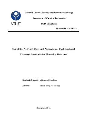
Tải xuống để đọc toàn bộNational Taiwan University of Science and Technology Department of Chemical Engineering Ph. Dissertation Student ID: D10206814 Orientated Ag@SiO2 Core-shell Nanocubes as Dual-functional Plasmonic Substrates for Biomarker Detection Graduate Student : Nguyen Minh Kha Advisor : Prof. Bing-Joe Hwang December, 2016 Abstract The progress in fundamental biological studies of disease has been revealing a variety of new biomarkers that are difficult to be detected by traditional diagnostic tools. Interestingly, plasmonics is an ultrasensitive optical technology applicable in medical diagnostics and biological imaging sensors.
A dual-functional plasmonic substrate with a unique design that allows both the sensitive detection of photoluminescence (PL) via metal-enhanced photoluminescence (MEPL) and the specific Raman fingerprint via surface-enhanced Raman scattering (SERS) is highly desirable to improve accuracy and sensitivity in detection. This dissertation describes the role of coupling agents, spacer effects, and importance characters of various platforms for the optimum bifunctional SERS-MEPL based on Ag@SiO2 core-shell nanocube(s) [NC(s)] for urinary biomarkers detection. In this work, the Ag NCs were synthesized by the polyol method and modified, firstly with different coupling agents, such as 3-mercaptopropyltrimethoxysilane (MPTMS) and 3-aminopropyltrimethoxysilane (APTMS), and secondly with tetraethylorthosilicate (TEOS). The presence of coupling agents greatly modified the Ag NC cores and manipulated the thickness and uniformity of the silica shells.
Ultrathin silica-coated Ag NCs (with a ~1.5 nm silica layer) found to have a SERS intensity 3 fold higher than synthesized Ag NCs. In comparison with MPTMS, it was found that APTMS modified Ag@SiO2 NCs improved significant SERS and MEPL enhancements. Moreover, a ‘dual functionality’ represented by the simultaneous strengthening of SERS and MEPL signals can be achieved by mixing Ag@SiO2 NCs, with a silica shell thickness of ~1. This approach allows both the Ag@SiO2 NCs SERS and MEPL sensitivities to be maintained at ~90% after 12 weeks of storage.
Additionally, it is known that interactions between substrate and plasmonic nanostructures can influence the performance of plasmonic biosensing. Therefore, in the final i approach, a substrate characterized with low refractive index and roughness was first fabricated by creating flower-like alumina on etched Al foil (f-Al2O3/e-Al). The Ag@SiO2 NCs assemble in the edge-edge configuration when they were deposited on this substrate. It is to note that the surface roughness of f-Al2O3/e-Al provides a pathway for the coupling of incident light to surface plasmon.
The Ag@SiO2/f-Al2O3/e-Al substrate exhibits a coupling efficiency of laser light sources into surface plasmon hotspots for both SERS and MEPL. Moreover, the shelf life of this substrate is significantly improved due to reduced oxygen diffusion rate by ultrathin silica spacer and flower-like Al2O3 dielectric layer. Creatinine (CR) and flavin adenine dinucleotide (FAD) are biomolecules present in human blood and urine. With the advanced label-free SERS and MEPL techniques, these biomarkers in urine are detected, and it allows cheap, non-invasive and yet sensitive analysis.
The approaches explored in this dissertation could be developed as a powerful encoding tool for high-throughput bio-analysis. Keywords: SERS; photoluminescence; Ag@SiO2 nanocubes; ultrathin shell; flower-like alumina; plasmonic coupling; edge-edge orientation; stability; dual functionality; Rhodamine 6G; urine; biomarker detection. ii Acknowledgement First and foremost, I would like to express my deep gratitude to my advisor, Prof. Bing- Joe Hwang, for giving me an opportunity to work in his group, and exploring the wonderful plasmonic nanoscale world.
He has taught me not only knowledge in research but also behavior in a new environment. The completion of this dissertation cannot be possible without his intellectual support. I could not imagine having a better advisor and mentor for my Ph. I would like to thank my sincere gratitude to all members at the Nano-electrochemistry Laboratory, Department of Chemical Engineering, National Taiwan University of Science and Technology (NTUST), who assisted me not only my study but also on many occasions.
Special thanks to Prof. Wei-Nien Su who essentially inspired me to do my best in performing my experiment as well as in writing papers. Thank you for your appreciated support, valuable encouragement and considerable recommendations. Additionally, I wish to express my appreciation to Dr.
Ching-Hsiang Chen, Dr. Chun-Jern Pan, Dr. John Rick, Dr. Meng-Che Tsai, and Dr.
Alok Mani Tripathi for giving their time and effort in evaluating my work. I have benefited from their constructive comments on my report. My sincere thanks also goes to Prof. Hongjie Dai at Stanford University for supporting and suggesting the work to improve my knowledge in the field of biochemistry.
I am thankful to the Department of Chemical Engineering and Department of Materials Science and Engineering at NTUST for their help and research facilities. Also, many thanks to the Faculty of Chemical Engineering of Ho Chi Minh University of Technology (HCMUT), Vietnam, and Office of International Affairs at NTUST for all the best things that they have offered me during my study and research. I would like to acknowledge National Taiwan University of Science and Technology for giving me the chance to improve my knowledge in the graduate school and the scholarship iii during my Ph. Also, gratefully acknowledge financial support from the Ministry of Science and Technology (MOST), and the Top University Projects of Ministry of Education (MOE).
It is my pleasure to thanks my friends at Vietnamese Student Association who made my study and stay at NTUST more colorful. Last but not least, I would like to thank my family with love and gratitude for supporting me spiritually throughout studying Ph. and my life in general. iv Table of Contents Abstract.
iii Table of Contents. v List of Figures. ix List of Tables .xvii List of Acronyms .1 Overview about plasmonic technologies.2 Statement of the problem .3 Objective of the study .4 Significance of the study .5 Structure of the dissertation.1 A brief overview of SERS and MEPL .1 Light scattering and optical properties of metals .2 Normal Raman and surface-enhanced Raman scattering .3 Photoluminescence and metal-enhanced photoluminescence .2 The major concerns related to SERS and MEPL enhancements .1 Type of metal .4 Interacting objects, gaps, and coupled plasmon resonances .3 The research on SERS-MEPL bifunctional mode.1 Solutions for the integrated SERS and MEPL .2 Applications of SERS and MEPL .4 Controlling the fabrication of nanostructures for plasmonic applications .1 General nanofabrication techniques .2 Preparation of Ag nanostructures.3 Surface coating for protection of nanostructures. Materials and Methods.2 Fabrication of the plasmonic substrates .1 Synthesis of Ag NCs .2 Synthesis of Ag@SiO2 core-shell NCs .3 Fabrication of Ag@SiO2/f-Al2O3/e-Al and Ag@SiO2/Al2O3/Al substrates .4 Preparation of artificial urine .5 Preparation of SERS and MEPL measurements .3 Characterization and measurements .2 SERS and MEPL measurements .3 Calculation enhancement factor of the substrate.
SERS and MEPL of Ag@SiO2 Nanocubes with Ultrathin Silica Shells .2 Results and discussion.1 Synthesis of Ag NCs .2 Ultra-coating silica shell for Ag NCs cores .3 The SERS and MEPL of the Ag@SiO2 NCs vs. Sensitivity and Stability of Bifunctional SERS-MEPL from the Mixture of Ag@SiO2 Nanocubes .2 Results and Discussion .1 Characterization of the Ag NCs and Ag@SiO2 NCs .2 Bifunctional SERS-MEPL activity of Ag@SiO2 NCs .3 The stability for dual SERS-MEPL activity .4 Direct label-free SERS-MEPL detection of the mixture of creatinine and flavin adenine dinucleotide. A Plasmonic Coupling Substrate Based on Ag@SiO2/f-Al2O3/e-Al for Sensitive Detection of Biomarkers in Urine .2 Results and discussion.1 Fabrication of the substrates and their characterization .2 SERS and MEPL activities of the substrates .3 Urinary biomarkers detection. Conclusion and Future Perspectives.
123 vii List of Figures Figure 1.1 Models of healthcare delivery in chronic disease management.2 The outcomes from the diagnostic process.3 Plasmonic-based technologies for versatile biosensor applications.4 A schematic illustration of the averaged EFs obtained in the visible region from different types of SERS substrates: Au, Ag and Cu, transition metals, and core-shell (transition metals @ noble metals) NPs.5 Poynting vector or energy flow (lines) around a subwavelength metallic colloid illuminated at the plasmon wavelength (left) and at a wavelength longer than the plasmon wavelength (right). The vertical lines on the left indicate the diameter of the cross sections for absorption.1 Schematic diagram representing the SPP at a metal dielectric interface.2 Schematic diagram representing the LSP.[20] The equilibrium distribution of electron cloud in a metal NP is modified in the presence of an external E-field.3 Coupled surface plasmon modes (a) propagating along a thin metallic film and (b) formed by the Bragg scattering on periodically modulated surface.4 The optical path schematic of a Raman system.5 Energy-level diagram for Rayleigh, Raman and resonance-Raman scattering. S0 and S1 are the singlet electronic ground and first excited states.6 Raman peak shift ranges for organic bonds.7 Jablonski energy-level diagram for absorption and PL. Quenching represents a non- radiative decay process.8 Free-space emission.9 Simplified Jablonski diagram for a fluorophore in absence of (top) and in presence of (bottom) a metal.10 Plasmon controlled emission.11 Schematic representation of the relative intensities of fluorescence and SERS for molecules at different distances from a metal surface.12 A simplified periodic table of the elements.
The part marked in black are ‘free- electron metals’ and those marked in grey are transition metals.13 Plot of the (A) real, εr , and (B) imaginary, εi, components of the dielectric function of Ag, Au, and Si as a function of wavelength.14 Dielectric constants of Ag and Pt.15 Quality factor of the LSPR for a metal/air interface. The shaded area represents the region of interest for many plasmonic applications.16 Extinction (black), absorption (red), and scattering (blue) spectra calculated for Ag nanoparticles of different shapes: (A) a sphere, (B) a cube, (C) a tetrahedron, and (D) an octahedron.17 (a) Gap width dependence of the SERS intensity normalized by extinction values at the incident laser wavelength and Raman scattering wavelength; and (b)-(d) The gap width dependence of the averaged enhancement factor by multiplying FDTD intensity maps monitored at the incident laser wavelength by the map monitored at the Raman scattering wavelength.18 The synthesis procedures of AuNRs functionalized for both SERS and MEPL. DSPE = 1,2-distearoyl-sn-glycero-3-phosphoethanolamine, PEG = poly(ethylene glycol), and OTMS = octadecyltrimethoxysilane.19 MEPL, SERS and bright field images of the precipitates acquired from: (A)-(C) the goat anti-human IgG immobilized nanoprobes and magnetic nanobeads (MBs) x mixed with human, mouse or bovine IgG molecules; and (B)-(D) the goat anti- mouse IgG immobilized nanoprobes and MBs mixed with mouse, human or bovine IgG molecules.20 Schematic illustration of real-time multiplexed imaging using the FRES.[142] (A) The mode of dual modal detection with fluorescence and Raman scattering. (B) Illustration of the in vivo multiplexed molecular imaging procedure.22 Polyol method for synthesizing Ag nanostructures.
In (A) the reduction of Ag+ ions by ethylene glycol leads to the formation of nuclei that are highly volatile. These seeds are then grown into different nanostructures like (B) spheres, (C) cubes, (D) truncated cubes, (E) right bipryamids, (F) bars, (G) spheroids, (H) triangular plates, and (I) wires.23 Strategies for surface-coating SERS tags include coating with (A) denatured BSA, (B) SH-PEG, (C) amphiphilic diblock copolymer, (D) liposome, and (E-H) silica shells via different encapsulation routes.1 Schematic illustration of Ag NCs synthesis.2 Schematic illustration of Ag@SiO2 core-shell nanocube preparation.3 Schematic diagram for the preparation and chemical structures of the Ag@SiO2/f- Al2O3/e-Al substrate.1 Characterizations of Ag NCs with different reaction times. (A)-(D) SEM images; (E) Uv-vis spectra; and (F) Linear relationship of maximum wavelength absorbance and edge length of Ag NCs.2 Raman spectra with the excitation laser 532 nm of: (A) silicon wafer and 0.1 M R6G on a silicon substrate (10 times); (B) SERS intensity and (C) SERS EF of Ag NCs from various reaction times (excitation laser 532 nm). (B) XRD pattern of Ag NCs – CB 72R mixture.4 Calibration curve of standard Ag+ solution.5 UV-Vis spectra of: (a) Ag NCs and Ag@SiO2 NCs prepared by adding different chemicals ((b) 1mM MPTMS and 1mM TEOS, (c) 1mM TEOS, (d) 1mM MPTMS, and (e) 1mM APTMS and 1mM TEOS, respectively).
The inset is the UV-Vis spectra, expanded in the range 454 nm to 466 nm.6 TEM images of Ag@SiO2 NCs prepared by adding different chemicals: (A) 1mM MPTMS and 1mM TEOS, (B) 1mM TEOS, (C) 1mM MPTMS, and (D) 1mM APTMS and 1mM TEOS, respectively.
Nội dung được bảo vệ bản quyền — Tải xuống đầy đủ
Trích dẫn luận án này
Nguyen Minh Kha (2016). Dual-Functional Ag@SiO2 Nanocubes for Biomarker Detection [Luận án tiến sĩ, National Taiwan University of Science and Technology]. LuanAn.net. https://luanan.net/khoa-hoc-suc-khoe/ky-thuat-y-hoc/dual-functional-agsio2-nanocubes-biomarker-detection
Câu hỏi thường gặp
Luận án "Dual-Functional Ag@SiO2 Nanocubes for Biomarker Detection" nghiên cứu về vấn đề gì?
Luận án tiến sĩ nghiên cứu nanocubes AgSiO2 core-shell định hướng. Nền plasmonic kép cho phát hiện dấu ấn sinh học chính xác, hiệu quả.
Luận án "Dual-Functional Ag@SiO2 Nanocubes for Biomarker Detection" được bảo vệ tại trường nào?
Luận án này được bảo vệ tại National Taiwan University of Science and Technology. Năm bảo vệ: 2016.
Luận án "Dual-Functional Ag@SiO2 Nanocubes for Biomarker Detection" thuộc chuyên ngành gì?
Luận án "Dual-Functional Ag@SiO2 Nanocubes for Biomarker Detection" thuộc chuyên ngành Chemical Engineering. Danh mục: Kỹ Thuật Y Học.
Luận án "Dual-Functional Ag@SiO2 Nanocubes for Biomarker Detection" có bao nhiêu trang?
Luận án "Dual-Functional Ag@SiO2 Nanocubes for Biomarker Detection" có 166 trang. Bạn có thể xem trước một phần tài liệu ngay trên trang web trước khi tải về.
Cách tải luận án "Dual-Functional Ag@SiO2 Nanocubes for Biomarker Detection" về máy như thế nào?
Để tải luận án về máy, bạn nhấn nút "Tải xuống ngay" trên trang này, sau đó hoàn tất thanh toán phí lưu trữ. File sẽ được tải xuống ngay sau khi thanh toán thành công. Hỗ trợ qua Zalo: 0559 297 239.