Luận án Tiến sĩ: Tối ưu hóa tái tạo dây thần kinh ngoại vi bằng tín hiệu cấu trúc và sinh hóa
Tái tạo dây thần kinh ngoại vi: Khám phá giải pháp đột phá bằng tín hiệu cấu trúc & sinh hóa, mở đường cho phục hồi chức năng hiệu quả.
Anatomy & Neurobiology
Luan An
Luận án tiến sĩ
Năm xuất bản
Số trang
249
Thời gian đọc
38 phút
Lượt xem
0
Lượt tải
0
Phí lưu trữ
50 Point
Tổng quan nhanh
- Chủ đề:
- 1. Tái tạo dây thần kinh ngoại vi: Vai trò tín hiệu cấu trúc
- Số trang:
- 249 trang
- Trường:
- Virginia Commonwealth University
- Chuyên ngành:
- Anatomy & Neurobiology
- Tác giả:
- Balendu Shekhar Jha
- Năm:
- 2011
Tóm tắt nội dung luận án
I. Tái tạo dây thần kinh ngoại vi Vai trò tín hiệu cấu trúc
Phục hồi dây thần kinh ngoại vi sau chấn thương là một thách thức y khoa lớn. Sự tái tạo thần kinh thường không đạt được hiệu quả hoàn chỉnh. Cần có những phương pháp hỗ trợ mạnh mẽ từ môi trường xung quanh tế bào. Tín hiệu cấu trúc đóng vai trò then chốt trong quá trình này. Chúng cung cấp định hướng vật lý, dẫn đường cho sự phát triển của sợi trục. Điều này là thiết yếu để khôi phục kết nối chức năng.
1.1. Tầm quan trọng của tín hiệu cấu trúc
Tái tạo thần kinh ngoại vi đòi hỏi một môi trường hỗ trợ tối ưu. Tín hiệu cấu trúc từ môi trường vi mô hướng dẫn sự tăng trưởng sợi trục. Chúng là yếu tố quyết định sự tái kết nối của dây thần kinh. Việc thiếu các tín hiệu này cản trở sự phục hồi chức năng.
1.2. Ứng dụng giá đỡ sinh học tái tạo
Giá đỡ sinh học (biomaterial scaffolds) cung cấp một khung vật lý. Chúng mô phỏng chất nền ngoại bào (ECM) tự nhiên. ECM chứa các yếu tố cấu trúc quan trọng. Các giá đỡ này hướng dẫn tế bào Schwann và sợi trục. Điều này thúc đẩy quá trình tái tạo thần kinh. Chúng tạo ra môi trường thuận lợi cho phục hồi dây thần kinh.
1.3. Cơ chế tái tạo thần kinh ngoại vi
Cơ chế tái tạo thần kinh liên quan đến nhiều yếu tố phức tạp. Tín hiệu cấu trúc từ giá đỡ định hướng sự phát triển. Các sợi trục cần một con đường rõ ràng để vượt qua khoảng trống tổn thương. Giá đỡ cung cấp các kênh dẫn hướng vật lý. Điều này tối ưu hóa quá trình phục hồi dây thần kinh.
II. Cải thiện phục hồi thần kinh Tín hiệu sinh hóa then chốt
Bên cạnh tín hiệu cấu trúc, tín hiệu sinh hóa cũng đóng vai trò cực kỳ quan trọng trong tái tạo thần kinh. Chúng điều khiển các quá trình tế bào ở cấp độ phân tử. Sự hiện diện và phân phối đúng đắn của các yếu tố này có thể đẩy nhanh và cải thiện đáng kể quá trình phục hồi. Tín hiệu sinh hóa hoạt động như chất xúc tác, khuyến khích sự phát triển khỏe mạnh của dây thần kinh.
2.1. Yếu tố tăng trưởng thần kinh và Neurotrophin
Tín hiệu sinh hóa bao gồm các yếu tố tăng trưởng thần kinh. Neurotrophin là nhóm protein thiết yếu. Chúng thúc đẩy sự sống sót và phát triển của tế bào thần kinh. Yếu tố tăng trưởng thần kinh hỗ trợ sự nảy chồi sợi trục. Chúng là thành phần cốt lõi cho phục hồi dây thần kinh.
2.2. Vai trò tín hiệu sinh hóa trong tái tạo
Tín hiệu sinh hóa cung cấp các thông điệp hóa học quan trọng. Chúng điều chỉnh hành vi tế bào, bao gồm di chuyển, tăng sinh, và biệt hóa. Tái tạo thần kinh ngoại vi phụ thuộc vào các tín hiệu này. Sự kết hợp hiệu quả với tín hiệu cấu trúc mang lại kết quả vượt trội.
2.3. Phát triển nền tảng phân phối thuốc
Phân phối các yếu tố sinh hóa một cách có kiểm soát là cực kỳ quan trọng. Nền tảng phân phối thuốc đảm bảo nồng độ tối ưu tại vị trí tổn thương. Chúng giải phóng yếu tố tăng trưởng thần kinh một cách chính xác. Điều này tăng cường khả năng phục hồi dây thần kinh. Nó giúp cải thiện đáng kể hiệu quả tái tạo.
III. Công nghệ sợi điện Giá đỡ sinh học tái tạo thần kinh
Công nghệ electrospinning (sợi điện) mang lại giải pháp tiên tiến cho việc chế tạo giá đỡ sinh học. Kỹ thuật này tạo ra các cấu trúc sợi nano có khả năng định hướng cao, mô phỏng chính xác cấu trúc chất nền ngoại bào của dây thần kinh tự nhiên. Điều này tạo ra một môi trường lý tưởng để thúc đẩy sự tái tạo thần kinh ngoại vi, cung cấp các kênh dẫn hướng cho sự phát triển của sợi trục.
3.1. Giới thiệu kỹ thuật electrospinning
Electrospinning là một phương pháp chế tạo sợi nano. Kỹ thuật này sử dụng điện trường mạnh để kéo giãn dung dịch polyme. Nó tạo ra các sợi polyme cực mỏng và liên tục. Sợi này có thể sắp xếp theo một hướng cụ thể. Điều này tạo ra cấu trúc giống như chất nền ngoại bào (ECM).
3.2. Chế tạo giá đỡ 3D định hướng
Công nghệ electrospinning cho phép tạo ra giá đỡ 3D. Các giá đỡ này có cấu trúc định hướng cao. Chúng bắt chước cấu trúc sợi của dây thần kinh tự nhiên. Điều này cung cấp các kênh dẫn hướng cho sợi trục. Nó hỗ trợ tái tạo dây thần kinh ngoại vi hiệu quả.
3.3. Tối ưu hóa biến số sợi điện
Hiệu quả của giá đỡ phụ thuộc vào các biến số của quá trình electrospinning. Cần tối ưu hóa điện áp, tốc độ bơm, khoảng cách thu thập. Điều này đảm bảo sợi có chất lượng cao và đồng đều. Các đặc tính này ảnh hưởng trực tiếp đến khả năng tái tạo thần kinh.
IV. Giá đỡ cấu trúc 3D Hướng dẫn tái tạo dây thần kinh
Việc thiết kế và ứng dụng các giá đỡ cấu trúc 3D là một bước tiến quan trọng trong tái tạo thần kinh. Những giá đỡ này không chỉ cung cấp hỗ trợ vật lý mà còn có thể được thiết kế để tích hợp các tín hiệu sinh hóa. Điều này tạo ra một môi trường phức tạp và được kiểm soát, tối đa hóa tiềm năng phục hồi chức năng của dây thần kinh bị tổn thương. Nghiên cứu so sánh hiệu quả của chúng rất cần thiết.
4.1. Thiết kế giá đỡ 3D cho tái tạo
Giá đỡ cấu trúc 3D được thiết kế đặc biệt. Chúng định hướng sự phát triển của tế bào thần kinh. Cấu trúc này tối ưu cho việc tái tạo thần kinh ngoại vi. Thiết kế có thể tích hợp tín hiệu cấu trúc và sinh hóa. Mục tiêu là tạo ra môi trường thuận lợi nhất cho sự phát triển.
4.2. So sánh hiệu quả ống dẫn thần kinh
Các ống dẫn thần kinh làm từ sợi điện được nghiên cứu sâu rộng. Hiệu quả của chúng được so sánh với phương pháp truyền thống. Ống dẫn này cung cấp một môi trường được kiểm soát chặt chẽ. Chúng có thể cải thiện phục hồi dây thần kinh đáng kể. So sánh giúp đánh giá ưu điểm của công nghệ mới này.
4.3. Đánh giá chức năng phục hồi thần kinh
Việc đánh giá chức năng rất quan trọng. Nó xác định mức độ thành công của tái tạo thần kinh. Các chỉ số như chỉ số chức năng thần kinh hông (SFI) được sử dụng rộng rãi. Phản xạ rút chi cũng được kiểm tra cẩn thận. Dữ liệu này chứng minh hiệu quả của giá đỡ sinh học.
V. Phục hồi chức năng thần kinh Phương pháp nghiên cứu
Để đánh giá hiệu quả của các giá đỡ sinh học và tín hiệu tái tạo, cần áp dụng các phương pháp nghiên cứu khoa học nghiêm ngặt. Việc này bao gồm quy trình chế tạo vật liệu chính xác và các thử nghiệm chức năng chuyên biệt. Những phương pháp này cung cấp dữ liệu định lượng, giúp hiểu rõ hơn về cơ chế phục hồi và tiềm năng ứng dụng lâm sàng.
5.1. Quy trình sản xuất sợi điện PCL
PCL (Polycaprolactone) là một polyme sinh học phổ biến. PCL được sử dụng để sản xuất sợi điện trong nghiên cứu. Quy trình này tạo ra các sợi nano có độ bền cao. Sợi PCL có khả năng tương thích sinh học xuất sắc. Đây là lựa chọn tốt cho giá đỡ sinh học tái tạo thần kinh.
5.2. Phân tích chỉ số chức năng thần kinh
Chỉ số chức năng thần kinh hông (SFI) đánh giá khả năng vận động. Đây là phương pháp định lượng sự phục hồi sau chấn thương dây thần kinh. SFI cung cấp dữ liệu về chức năng tái tạo thần kinh. Nó giúp so sánh hiệu quả của các phương pháp điều trị khác nhau một cách khách quan.
5.3. Thử nghiệm phản xạ rút chi
Thử nghiệm phản xạ rút chi đo lường cảm giác. Nó đánh giá mức độ phục hồi cảm giác ở chi bị ảnh hưởng. Đây là một chỉ số quan trọng khác trong đánh giá tái tạo thần kinh. Kết quả từ thử nghiệm này bổ sung cho SFI, cung cấp cái nhìn toàn diện về phục hồi dây thần kinh.
VI. Tương lai tái tạo dây thần kinh Kết luận và hướng đi
Nghiên cứu về tái tạo dây thần kinh ngoại vi đã đạt được những tiến bộ đáng kể. Sự kết hợp của tín hiệu cấu trúc và sinh hóa thông qua giá đỡ sinh học electrospun mở ra nhiều hướng đi mới. Những thành tựu này không chỉ nâng cao hiểu biết về cơ chế phục hồi mà còn hứa hẹn những ứng dụng lâm sàng đột phá, cải thiện chất lượng cuộc sống cho bệnh nhân chấn thương thần kinh.
6.1. Thành tựu trong tái tạo thần kinh
Nghiên cứu chứng minh tiềm năng của tín hiệu cấu trúc và sinh hóa. Việc sử dụng giá đỡ sinh học (biomaterial scaffolds) làm tăng khả năng tái tạo thần kinh ngoại vi. Các phương pháp kỹ thuật như electrospinning đã tạo ra bước đột phá. Chúng mang lại hy vọng mới cho phục hồi dây thần kinh bị tổn thương.
6.2. Hướng nghiên cứu tiếp theo
Cần tiếp tục khám phá các yếu tố tăng trưởng thần kinh mới. Nghiên cứu cần tập trung vào việc tối ưu hóa thiết kế giá đỡ. Kết hợp nhiều loại tín hiệu là một hướng đi hứa hẹn. Mục tiêu là đạt được sự phục hồi chức năng hoàn hảo và toàn diện hơn.
6.3. Tiềm năng ứng dụng lâm sàng
Những phát hiện này có tiềm năng lớn cho ứng dụng lâm sàng. Chúng có thể dẫn đến các phương pháp điều trị chấn thương thần kinh ngoại vi hiệu quả hơn. Mục tiêu cuối cùng là cải thiện chất lượng cuộc sống cho bệnh nhân. Công nghệ này hứa hẹn một tương lai tốt đẹp hơn trong y học tái tạo.
Mục lục chi tiết luận án
Tải xuống file đầy đủ để xem toàn bộ nội dung
Tải đầy đủ (249 trang)Trích đoạn nội dung luận án
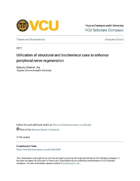
Tải xuống để đọc toàn bộVirginia Commonwealth University VCU Scholars Compass Theses and Dissertations Graduate School 2011 Utilization of structural and biochemical cues to enhance peripheral nerve regeneration Balendu Shekhar Jha Virginia Commonwealth University Follow this and additional works at: https://scholarscompass.edu/etd Part of the Nervous System Commons © The Author Downloaded from https://scholarscompass.edu/etd/2650 This Dissertation is brought to you for free and open access by the Graduate School at VCU Scholars Compass. It has been accepted for inclusion in Theses and Dissertations by an authorized administrator of VCU Scholars Compass. For more information, please contact libcompass@vcu. © Balendu Shekhar Jha 2011 All Rights Reserved UTILIZATION OF STRUCTURAL & BIOCHEMICAL CUES TO ENHANCE PERIPHERAL NERVE REGENERATION A dissertation submitted in partial fulfillment of the requirements for the degree of Doctor of Philosophy at Virginia Commonwealth University.
by BALENDU SHEKHAR JHA B.) Physical Therapy, Delhi University, 2003. Simpson, PhD Associate Professor Department of Anatomy & Neurobiology Virginia Commonwealth University Richmond, Virginia August, 2011. ii Acknowledgement Earning a PhD degree is truly a marathon event, and I would not have been able to complete this journey without the aid and support of countless people over these years. I must first express my gratitude towards my advisor, Dr.
David Simpson for his help and guidance. With his enthusiasm, inspiration, and great new ideas, he helped to make research work fun for me. I always considered him as Mr. He has a solution to each and every problem, and can make sense out anything (literally any data).
His way of seeing things and handling situations have set an example I hope to match someday. I would like to express my appreciation to my committee members: Dr. Raymond Colello, Dr. Scott Henderson, Dr.
Babette Fuss, Dr. Bob Diegelmann, and Dr. Gary Bowlin for their guidance towards completion of my bench work, and for the taking time for careful reading and commenting of my dissertation. Your expectations and concerns have always been right to the point.
This work would not have been possible without the constant assistance, guidance, and inputs provided by Dr. John Bigbee and Dr. Both of them have been my regular consultants, training me how to interpret science. I would like to thank the past and present Simpson lab fellows.
Rusty Bowman has always been a second mentor after my advisor. I am sure he has a big brain with more than 50% hippocampus where he has a huge knowledge database stored. He has an answer to any question with statistical and demographic figures. I huge thanks goes out to Thomas Turner for being the fun guy in the lab, keeping the lab alive with his jokes and funny online videos; you kept things light and smiling.
I would also like to thank Chantal Ayres for making me realize every now and then, that I should work in an organized fashion, keep the lab clean (glutaraldehyde-free), eat healthy and exercise regularly. A special thanks to Casey Grey for dealing with me every day now, and who has been always there for editing and proof-reading my work. Thank you for your encouragement, support, and most of all your humor. I would like to thank all my friends; thank you for being the surrogate family during my years at the VCU and for your continued moral support.
Most importantly, I am forever indebted to my parents and my wife, Vandana for their understanding, endless patience and encouragement when it was most required. I would also like to thank my younger sister, Pragya for being there with my parents, and taking care of them when needed, in my absence during the course of my PhD training. iii Table of Contents Page Acknowledgement……………………………………………………………………. ii List of tables……………………………………………………………………….
v List of figures……………………………………………………………………….… vi List of abbreviations…………………………………………………………………. Introduction to electrospinning…………. Regulating electrospinning – tweaking its variables……………… 12 3. Electrospun collagen: A tissue engineering scaffold with unique functional properties in a wide variety of applications……….
Materials and methods……. Two pole air gap electrospinning: Fabrication of highly aligned, three- dimensional scaffolds for nerve reconstruction…. Designing of a drug delivery platform for sustained release of gradients of growth factors at precise locations…. Electrospun 3D nerve guides: A comparative study….
Conclusions and future research directions …. Future research directions ………………………………………. 235 v List of Tables Page Table 4.1: Summary of specific electrospinning conditions for PCL in two pole electrospinning system…………………………………………………….1: Statistical analysis for sciatic functional index (SFI) assay……………….2: Statistical analysis for withdrawal reflex assay…………………………… 176 vi List of Figures Page Figure 2.1: Schematic of the process of electrospinning………………………….2A: Effect of Coulombic repulsion forces………………………….2B: Coiling of the electrospun jet………………………….1: Endothelial interactions with electrospun collagen and gelatin……….2: Osteoblast interactions with electrospun collagen & electrospun gelatin. Rates of wound closure in lesions treated with electrospun collagen or electrospun gelatin………………………….
Healing response to electrospun collagen and electrospun gelatin as a function of fiber diameter and pore dimension….5: Muscle fabrication: 3 weeks………………………….6: Muscle fabrication: 8 weeks………………………….7: Analysis of Type I collagen α chain content: Analysis of Type I collagen α chain content………………………….8: Ultrastructural and functional characteristics of collagen.1: Schematic representation of the mechanism of two pole air gap electrospinning. Schematic of the ground target used in a two pole air gap electrospinning system. Representative scanning electron micrographs (SEM).4: Average fiber diameter………………………….5: Analysis of fiber alignment by 2D FFT………………………….7: Cell culture experimentation………………………….8: Nerve reconstruction – frozen sections………………………….9: Nerve reconstruction – semi-thin sections. 107 vii Page Figure 4.10: Transmission electron microscopy………………………….1: Structure of alginic acid residues.2: Schematic of the characteristic egg-box structure……………………….3: Schematic of the electrospraying apparatus for preparing alginate microbeads………………………….4: Fabrication of alginate thread with concentration gradients…………….5: SEM images of alginaate microbeads, macrobeads, threads…………….6: NGF capture efficiency of different forms of alginate delivery platforms.7A: NGF capture efficiency of alginate threads and total NGF release in 7 days from different concentration alginate threads ….7B: NGF release profile from varying concentration alginate threads…….8 (A,B): NGF capture efficiency of alginate threads loaded with varying concentration of NGF.9: % NGF loss in the calcium chloride bath during the process of alginate thread polymerization………………………….10: NGF release profile from alginate threads………………………….11: NGF release and capture from alginate thread inside the electrospun 3D nerve guide………………………….12: DRG culture in scaffold with NGF in alginate delivery platform…….13: NGF gradient in the alginate thread…………………………………….1: Sciatic Functional Index……………………………………………….2: Gastrocnemius muscle atrophy comparison……………………………… 173 Figure 6.3: Sensory testing using the withdrawal reflex……………………………… 176 Figure 6.4: Lumbrical motor end plates…………………………………………….
178 viii Page Figure 6.5: Signal amplitudes across the implants at post-operative day 45………….6: Tangential semi-thin sections 45 days post-surgery ….8: Electron microscopy……………………………………………………… 191 ix List of Abbreviations 3D Three-dimensional ANOVA Analysis of variance BDNF Brain-derived neurotrophic factor BSA Bovine serum albumin CNTF Ciliary neurotrophic factor DRG Dorsal root ganglion ECM Extracellular matrix ELISA Enzyme-linked immunosorbent assay esC Electrospun collagen esG Electrospun gelatin FBS Fetal bovine serum FFT Fast fourier transform GAPDH Glyceraldehyde 3-phosphate dehydrogenase GDNF Glial cell line derived neurotrophic factor HDF Human dermal fibroblasts HFP 1,1,1,3,3,3-hexafluoro-2-propanol MEM Minimum essential media N-CAM Neural cell adhesion molecule NGF Nerve growth factor PBS Phosphate buffered saline PCL Poly-ε-caprolactone PGA/PLA Polylactic acid / Polyglycolic acid PNS Peripheral nervous system rEC Recovered electrospun collagen rEG Recovered electrospun gelatin RGD Arginine-glycine-aspartate SDS Sodium dodecyl sulfate SEM Scanning electron microscopy SFI Sciatic functional index TEM Transmission electron microscopy TFE 1,1,1-trifluoroethanol TGF Transforming growth factor x Abstract UTILIZATION OF STRUCTURAL & BIOCHEMICAL CUES TO ENHANCE PERIPHERAL NERVE REGENERATION. By Balendu Shekhar Jha, PMP, PT A dissertation submitted in partial fulfillment of the requirements for the degree of Doctor of Philosophy at Virginia Commonwealth University. Virginia Commonwealth University, 2011 Major Director: David G. Associate Professor, Department of Anatomy and Neurobiology This study examines the prospects of using the electrospinning process to fabricate tissue engineering scaffolds targeting a variety of regenerative applications, with a primary focus on the production of nerve guides for the treatment of long-defect nerve injuries in the peripheral nervous system.
A basic overview of the conventional electrospinning process is provided, and the utility of this fabrication scheme in the production of collagen-based tissue engineering scaffolds is demonstrated. Next, a novel modification of the basic electrospinning process is xi presented. This process, called two pole air gap electrospinning, was developed to produce nerve guides that exhibit an anisotropic structure that mimics the extracellular matrix of native peripheral nerve tissue. This electrospinning process makes it possible to produce macroscopic nerve guides that are cylindrical in shape and composed of dense arrays of nano- to micron-scale diameter fibers.
Unlike, conventional hollow core nerve guides, these electrospun constructs lack a central lumen, hence the designation 3D (for three-dimensional) nerve guide. The fibers are nearly exclusively arrayed in parallel with the long axis of the construct. This architectural feature provides thousands of individual channels, and aligned fibers that provide guidance cues that are designed to drive regenerating axons to grow in a highly directed fashion down the longitudinal axis of the guide. To supplement the structural cues provided by the fibrillar arrays of the electrospun 3D nerve guides, an alginate-based platform designed to deliver therapeutic reagents was developed and characterized.
This platform makes it possible to fabricate gradients of therapeutic reagents within the fibrillar arrays of an electrospun nerve guide. Functional and structural analyses of these constructs supplemented with or without a gradient of NGF, in a long-defect nerve injury in the rodent sciatic nerve indicate that the 3D design is superior to the gold standard treatment, the autologous nerve graft. Animals treated with the 3D grafts recovered motor and sensory function faster and exhibited far higher nerve-to-nerve and nerve- to-muscle signal amplitudes in electrophysiological studies than animals treated with autologous grafts or conventional hollow core cylindrical grafts. Overview The central hypothesis of this study states that tissue regeneration after injury can be maximized by identifying and recapitulating key features of the native extracellular matrix (ECM) [1].
In this study the central role that scaffold structure and composition play in the tissue engineering paradigm is explored. Tissue engineering is an evolving multidisciplinary field that has the potential to revolutionize medical practice and improve the health and quality of life for millions of people worldwide by restoring the structure and function to diseased or damaged tissues and organs. As a science, tissue engineering encompasses a broad range of potential applications including the repair, augmentation, or replacement of body tissues such as bone, muscle, skin, blood vessels, nerve, cartilage, and other connective tissues such as ligaments and tendons. Fundamental to nearly all tissue engineering processes is the scaffold used to establish the three-dimensional space necessary for cell attachment and growth at the injury site [1].
Typically, these scaffolds biodegrade or integrate themselves into the host tissue as the nascent ECM regenerates at the injury site. In effect, the scaffolds represent a template that act to guide the regenerative process and in most applications these structures are designed to be remodeled and completely replaced by native tissues. These scaffolds may or may not be supplemented with various types of cells designed to promote the reconstitution of functional tissue. A primary assumption of the tissue engineering paradigm is the notion that functional tissue will develop if the proper biological, guidance and or positional cues are provided by the tissue engineering scaffold [1].
It is becoming increasingly clear that each specific tissue requires its own unique set of these signals. The cues to be used in any specific application may be driven by biological, clinical, commercial and / or regulatory considerations. In the example of 1 peripheral nervous tissue, it may be guidance and / or positional cues that are paramount in design of the regenerative template. Superimposed on these basic considerations are the processing limitations that limit the ability to fabricate different materials into scaffolds with the features suitable to function as a regenerative template for the reconstruction of organs and tissue.
Nội dung được bảo vệ bản quyền — Tải xuống đầy đủ
Trích dẫn luận án này
Balendu Shekhar Jha (2011). Tái tạo dây thần kinh ngoại vi: Cải thiện bằng tín hiệu cấu trúc và sinh hóa [Luận án tiến sĩ, Virginia Commonwealth University]. LuanAn.net. https://luanan.net/y-hoc/tai-tao-day-than-kinh-ngoai-vi-cai-thien-tin-hieu-cau-truc-sinh-hoa
Câu hỏi thường gặp
Luận án "Tái tạo dây thần kinh ngoại vi: Cải thiện bằng tín hiệu cấu trúc và sinh hóa" nghiên cứu về vấn đề gì?
Tái tạo dây thần kinh ngoại vi: Khám phá giải pháp đột phá bằng tín hiệu cấu trúc & sinh hóa, mở đường cho phục hồi chức năng hiệu quả.
Luận án "Tái tạo dây thần kinh ngoại vi: Cải thiện bằng tín hiệu cấu trúc và sinh hóa" được bảo vệ tại trường nào?
Luận án này được bảo vệ tại Virginia Commonwealth University. Năm bảo vệ: 2011.
Luận án "Tái tạo dây thần kinh ngoại vi: Cải thiện bằng tín hiệu cấu trúc và sinh hóa" thuộc chuyên ngành gì?
Luận án "Tái tạo dây thần kinh ngoại vi: Cải thiện bằng tín hiệu cấu trúc và sinh hóa" thuộc chuyên ngành Anatomy & Neurobiology. Danh mục: Y Học.
Luận án "Tái tạo dây thần kinh ngoại vi: Cải thiện bằng tín hiệu cấu trúc và sinh hóa" có bao nhiêu trang?
Luận án "Tái tạo dây thần kinh ngoại vi: Cải thiện bằng tín hiệu cấu trúc và sinh hóa" có 249 trang. Bạn có thể xem trước một phần tài liệu ngay trên trang web trước khi tải về.
Cách tải luận án "Tái tạo dây thần kinh ngoại vi: Cải thiện bằng tín hiệu cấu trúc và sinh hóa" về máy như thế nào?
Để tải luận án về máy, bạn nhấn nút "Tải xuống ngay" trên trang này, sau đó hoàn tất thanh toán phí lưu trữ. File sẽ được tải xuống ngay sau khi thanh toán thành công. Hỗ trợ qua Zalo: 0559 297 239.