Luận án: Điều hòa vận chuyển chủ động ở ống thận người bởi MT-3: Vai trò của miền C
Nghiên cứu về cơ chế điều chỉnh vận chuyển tích cực theo hướng vector trong ống thận gần của con người.
University of North Dakota School of Medicine and Health Sciences
Biochemistry and Molecular Biology
Luan An
Luận án tiến sĩ
Năm xuất bản
Số trang
117
Thời gian đọc
18 phút
Lượt xem
0
Lượt tải
0
Phí lưu trữ
40 Point
Tổng quan nhanh
- Chủ đề:
- 1. Vai trò MT-3: Điều hòa vận chuyển chủ động ống thận
- Số trang:
- 117 trang
- Trường:
- University of North Dakota School of Medicine and Health Sciences
- Chuyên ngành:
- Biochemistry and Molecular Biology
- Tác giả:
- Andrea Marie Nore
- Năm:
- 2015
Tóm tắt nội dung luận án
I. Vai trò MT 3 Điều hòa vận chuyển chủ động ống thận
Nghiên cứu khám phá cơ chế điều hòa vận chuyển chủ động tại các tế bào ống lượn gần (ống thận người). Protein Metallothionein-3 (MT-3) đóng vai trò trung tâm. MT-3 kiểm soát quá trình vận chuyển vector, thiết yếu cho chức năng thận. Việc hiểu rõ chức năng MT-3 giúp làm sáng tỏ sinh lý học thận. Nó cũng cung cấp thông tin về quá trình tái hấp thu chất điện giải. Cân bằng nội môi trong cơ thể phụ thuộc vào hoạt động này. Nghiên cứu này tập trung vào cách MT-3 tác động lên các protein liên quan đến vận chuyển.
1.1. Tổng quan về Protein Metallothionein 3
Metallothionein-3 (MT-3) là một protein đặc biệt. MT-3 khác biệt so với các metallothionein khác. Nó có một miền C-terminus độc đáo. Miền này có khả năng ảnh hưởng đến tương tác protein-protein. MT-3 được tìm thấy nhiều trong não. Sự hiện diện của nó trong thận cũng rất quan trọng. MT-3 liên kết với các ion kim loại, đặc biệt là kẽm. Kẽm trong thận có vai trò điều hòa nhiều quá trình sinh học.
1.2. Chức năng MT 3 trong sinh lý học thận
MT-3 điều hòa nhiều chức năng tế bào ở thận. Nó ảnh hưởng đến sự gắn kết tế bào biểu mô và tính phân cực. Các quá trình này là nền tảng cho vận chuyển chủ động. Vận chuyển chủ động giúp tái hấp thu các chất quan trọng. Các chất này bao gồm glucose, axit amin và chất điện giải. Bơm ion hoạt động hiệu quả nhờ sự điều hòa này. MT-3 cũng có thể bảo vệ thận khỏi tác nhân gây độc. Ví dụ về tác nhân gây độc như Cadmium, gây độc cho thận.
1.3. Cơ chế điều hòa vận chuyển qua màng
Vận chuyển qua màng là một quá trình phức tạp. Nó đòi hỏi sự phối hợp của nhiều protein. Các kênh ion và bơm ion hoạt động đồng bộ. MT-3 can thiệp vào các tín hiệu tế bào. Điều này ảnh hưởng đến biểu hiện của các protein màng. Sự kiểm soát này đảm bảo hiệu quả của tái hấp thu. Nồng độ ion và nước trong cơ thể được duy trì ổn định. Đây là yếu tố sống còn đối với cân bằng nội môi tổng thể.
II. Cơ chế tác động Protein MT 3 ở ống thận người
Nghiên cứu làm rõ cơ chế phân tử MT-3 điều hòa vận chuyển. Miền C-terminus của Protein MT-3 giữ vai trò then chốt. Miền này tương tác với các protein liên kết tế bào. Cụ thể là E-cadherin và N-cadherin. Sự điều hòa các cadherin này ảnh hưởng đến cấu trúc tế bào. Nó cũng thay đổi tính phân cực của tế bào ống thận. Điều này có tác động trực tiếp đến khả năng vận chuyển chủ động. Việc nghiên cứu các tương tác protein-protein cung cấp thông tin chi tiết. Nó giúp hiểu rõ về con đường tín hiệu của MT-3.
2.1. Ảnh hưởng của MT 3 lên cadherin E và N cadherin
Cadherin là các protein quan trọng. Chúng tham gia vào sự gắn kết giữa các tế bào. E-cadherin duy trì đặc tính biểu mô. N-cadherin thường liên quan đến tế bào trung mô. MT-3 có khả năng điều hòa biểu hiện của cả hai cadherin này. Sự thay đổi biểu hiện cadherin tác động đến sự phát triển lớp đơn tế bào chặt chẽ. Điều này là thiết yếu cho chức năng vận chuyển vector ở ống thận. Tỷ lệ E/N-cadherin ảnh hưởng đến hình thái và chức năng tế bào.
2.2. Vai trò miền C terminus trong tương tác Protein MT 3
Miền C-terminus của MT-3 là duy nhất. Nó không có trong các metallothionein khác. Nghiên cứu tập trung vào vai trò của miền này. Miền C-terminus được chứng minh là liên quan đến các tương tác protein-protein cụ thể. Nó có thể là yếu tố quyết định sự gắn kết với E- và N-cadherin. Hiểu rõ miền này giúp giải thích các chức năng riêng biệt của MT-3. Điều này có ý nghĩa quan trọng trong việc thiết kế các liệu pháp điều trị.
2.3. Quá trình chuyển đổi biểu mô trung mô EMT
EMT là một quá trình sinh học. Tế bào biểu mô mất đặc tính của chúng. Chúng chuyển sang trạng thái trung mô. Quá trình này có thể xảy ra trong bệnh lý thận. MT-3 được phát hiện có vai trò trong chuyển đổi biểu mô-trung mô ngược (MET). MT-3 thúc đẩy tế bào trở lại trạng thái biểu mô. Điều này có thể khôi phục chức năng của ống thận. Việc điều hòa EMT/MET bởi MT-3 có ý nghĩa lớn đối với bệnh lý thận.
III. Tương tác MT 3 và cadherin Ảnh hưởng sinh lý thận
Mối quan hệ giữa MT-3 và cadherin có ý nghĩa sâu sắc. Nó ảnh hưởng đến khả năng tái hấp thu và tiết chất của ống thận. Sự điều hòa E-cadherin và N-cadherin bởi MT-3 định hình môi trường tế bào. Môi trường này hỗ trợ các bơm ion. Các bơm này thực hiện vận chuyển chủ động. Cân bằng nội môi của cơ thể phụ thuộc vào hoạt động này. Nghiên cứu cũng khảo sát biểu hiện cadherin trong các loại tế bào khác nhau. Điều này giúp hiểu rõ hơn về tính đặc hiệu mô của MT-3.
3.1. Phân tích biểu hiện E và N cadherin ở ống thận
Nghiên cứu đã phân tích biểu hiện của E-cadherin và N-cadherin. Phân tích được thực hiện trên tế bào ống lượn gần người. Nó cũng được thực hiện trên dòng tế bào HK-2. Các điều kiện nuôi cấy khác nhau ảnh hưởng đến biểu hiện này. MT-3 có thể thay đổi tỷ lệ E/N-cadherin. Điều này ảnh hưởng đến sự duy trì tính toàn vẹn của hàng rào biểu mô. Hàng rào này rất quan trọng cho chức năng lọc của thận.
3.2. Liên kết Protein MT 3 với protein liên quan biểu mô
Nghiên cứu xác nhận các tương tác protein-protein của MT-3. MT-3 liên kết với các protein quan trọng. Các protein này thúc đẩy phái sinh biểu mô. Sự liên kết này góp phần vào việc duy trì hình thái và chức năng tế bào. Điều này đảm bảo hiệu quả của vận chuyển chủ động ở ống thận. Các kỹ thuật như pulldowns và miễn dịch kết tủa đã được sử dụng. Chúng giúp xác định các đối tác tương tác của MT-3.
3.3. Tái hấp thu chất điện giải và cân bằng nội môi
Khả năng của MT-3 điều hòa vận chuyển chủ động là rất quan trọng. Nó ảnh hưởng trực tiếp đến tái hấp thu chất điện giải. Natri, kali, clorua được tái hấp thu. Quá trình này giữ cho nồng độ các chất trong máu ổn định. Nếu vận chuyển bị rối loạn, cân bằng nội môi bị ảnh hưởng. Điều này có thể dẫn đến các bệnh lý nghiêm trọng. Việc điều hòa của MT-3 đảm bảo rằng các bơm ion hoạt động tối ưu.
IV. Ứng dụng nghiên cứu MT 3 trong sức khỏe thận
Những phát hiện về Protein MT-3 mở ra hướng mới. Nó giúp hiểu biết về sinh lý học thận và các bệnh lý liên quan. Việc làm rõ vai trò của MT-3 trong điều hòa vận chuyển chủ động là một bước tiến. Nó có thể dẫn đến phát triển các chiến lược điều trị mới. Đặc biệt là đối với các tình trạng rối loạn chức năng ống thận. Kẽm trong thận cũng là một yếu tố cần được xem xét. Tương tác của MT-3 với các protein khác mang lại cái nhìn toàn diện hơn. Nó về mạng lưới điều hòa phức tạp trong tế bào thận.
4.1. Khám phá tương tác protein protein của MT 3
Nghiên cứu đã xác nhận các tương tác trực tiếp của MT-3. Nó tương tác với các protein liên quan đến việc duy trì kiểu hình biểu mô. Các tương tác này giải thích cách MT-3 điều hòa sự gắn kết tế bào. Chúng cũng giải thích cách MT-3 ảnh hưởng đến vận chuyển qua màng. Việc hiểu rõ các đối tác tương tác này là cơ sở. Nó để nghiên cứu sâu hơn về con đường tín hiệu của MT-3. Điều này có thể dẫn đến việc xác định các mục tiêu thuốc mới.
4.2. Vai trò kẽm trong thận và chức năng MT 3
MT-3 là một metallothionein. Nó có khả năng liên kết với các ion kim loại, đặc biệt là kẽm. Nồng độ kẽm trong thận có thể ảnh hưởng đến hoạt động của MT-3. Sự điều hòa kẽm bởi MT-3 có thể gián tiếp ảnh hưởng đến các quá trình tế bào khác. Điều này bao gồm hoạt động của bơm ion và tái hấp thu chất điện giải. Kẽm đóng vai trò quan trọng trong nhiều enzyme và protein. Sự cân bằng kẽm là cần thiết cho chức năng thận khỏe mạnh.
4.3. Tiềm năng điều trị bệnh lý ống thận
Những hiểu biết về MT-3 có thể mở ra tiềm năng điều trị. Đặc biệt là đối với các bệnh lý ống thận. Các bệnh này bao gồm tổn thương thận cấp và mạn tính. Việc điều hòa chức năng MT-3 có thể giúp khôi phục chức năng thận. Nó có thể giảm nhẹ tác động của các yếu tố gây hại. Nghiên cứu tiếp theo cần tập trung vào việc phát triển các hợp chất. Các hợp chất này có thể điều biến hoạt động của MT-3 một cách chọn lọc. Điều này nhằm mục đích cải thiện sức khỏe thận.
Tải xuống file đầy đủ để xem toàn bộ nội dung
Tải đầy đủ (117 trang)Trích đoạn nội dung luận án
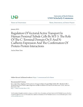
Tải xuống để đọc toàn bộUniversity of North Dakota UND Scholarly Commons Theses and Dissertations Theses, Dissertations, and Senior Projects January 2015 Regulation Of Vectorial Active Transport In Human Proximal Tubule Cells By MT-3: The Role Of The C-Terminal Domain On E-And N- Cadherin Expression And The Confirmation Of Protein-Protein Interactions Andrea Marie Nore Follow this and additional works at: https://commons.edu/theses Recommended Citation Nore, Andrea Marie, "Regulation Of Vectorial Active Transport In Human Proximal Tubule Cells By MT-3: The Role Of The C- Terminal Domain On E-And N-Cadherin Expression And The Confirmation Of Protein-Protein Interactions" (2015). Theses and Dissertations.edu/theses/1938 This Dissertation is brought to you for free and open access by the Theses, Dissertations, and Senior Projects at UND Scholarly Commons. It has been accepted for inclusion in Theses and Dissertations by an authorized administrator of UND Scholarly Commons. For more information, please contact zeineb.
REGULATION OF VECTORIAL ACTIVE TRANSPORT IN HUMAN PROXIMAL TUBULE CELLS BY MT-3: THE ROLE OF THE C-TERMINAL DOMAIN ON E-AND N-CADHERIN EXPRESSION AND THE CONFIRMATION OF PROTEIN-PROTEIN INTERACTIONS by Andrea Marie Nore Bachelor of Science, Minnesota State University Moorhead, 2010 A Dissertation Submitted to the Graduate Faculty of the University of North Dakota School of Medicine and Health Sciences in partial fulfillment of the requirements for the degree of Doctor of Philosophy Grand Forks, North Dakota December 2015 Copyright 2015 Andrea Nore ii This dissertation, submitted by Andrea Nore in partial fulfillment of the requirements for the Degree of Doctor of Philosophy from the University of North Dakota, has been read by the Faculty Advisory Committee under whom the work has been done and is hereby approved. Garrett __________________________________________ Seema Somji __________________________________________ Donald A. Dunlevy This dissertation is being submitted by the appointed advisory committee as having met all of the requirement of the School of Graduate Studies at the University of North Dakota and is hereby approved. __________________________________ Wayne Swisher Dean of the School of Graduate Studies _________________________________ Date iii PERMISSION Title REGULATION OF VECTORIAL ACTIVE TRANSPORT IN HUMAN PROXIMAL TUBULE CELLS BY MT-3: THE ROLE OF THE C- TERMINAL DOMAIN ON E-AND N-CADHERIN EXPRESSION AND THE CONFIRMATION OF PROTEIN-PROTEIN INTERACTIONS Department Biochemistry and Molecular Biology Degree Doctor of Philosophy In presenting this dissertation in partial fulfillment of the requirements for a graduate degree from the University of North Dakota, I agree that the library of this University shall make it freely available for inspection.
I further agree that permission for extensive copying for scholarly purposes may be granted by the professor who supervised my dissertation work or, in his absence, by the Chairperson of the department or the dean of the School of Graduate Studies. It is understood that any copying or publication or other use of this dissertation or part thereof for financial gain shall not be allowed without my written permission. It is also understood that due recognition shall be given to me and to the University of North Dakota in any scholarly use which may be made of any material in my dissertation. Andrea Nore November 5, 2015 iv TABLE OF CONTENTS LIST OF FIGURES.
vii LIST OF TABLES. 1 Hypothesis and Rationale. 3 Epithelial Cell Adhesion and Polarity. 6 Epithelial-Mesenchymal Transition.
8 Cadmium is Nephrotoxic. 18 Metallothionein-3 Protein-Protein Interactions. 22 Laser-Capture Microdissection. 26 RNA isolation and qRT-PCR.
34 Expression of E- and N-Cadherin in Human Kidney Proximal Tubules. 34 Expression of E- and N-Cadherin in HK-2 Cells as a Function of Growth Medium Composition. 38 Comparison of E- and N-Cadherin Expression in HK-2 and HPT Cell Cultures. 39 Alterations in Tight Monolayer Development.
44 Association of the Unique C-Terminal Domain of MT-3 With MT-3 Induced MET in HK-2 Cells. 46 Effect of Forced E-cadherin Expression on HK-2 Vectorial Active Transport, N-Cadherin Expression, and Cell Morphology. 52 Zn7MT-3 mediated pulldowns in HK-2 lysates. 55 V5-mediated immunoprecipitations in MT-3 expressing HK-2 lysates.
60 HK-2 Cells have Features Associated with the Initial Stages of EMT. 60 HK-2 Cells Undergo MET Mediated by MT-3. 65 MT-3 Interacts with Proteins that Promote an Epithelial Phenotype. 82 vi LIST OF FIGURES Figure Page III-1.
Immunohistochemical staining of E-cadherin and N- Cadherin in the human kidney. Expression of E-and N-cadherin in microdissected proximal tubules. Influence of growth medium on E-and N-cadherin expression in HK-2 cells. Comparison of E-cadherin expression in HPT and HK-2 cell cultures.
Comparison of N-cadherin expression in HPT and HK-2 cell cultures. The pattern of E- and N-cadherin expression is unaltered when cells are grown on transwell inserts. Transepithelial resistance comparison of HPT and HK-2 cell cultures. Connexin 32 expression in HK-2, HPT, and HK-2 cells expressing MT-3.
Mutated metallothionein constructs. Effect of the altered domains of MT-3 on the formation of domes in stably transfected HK-2 cells. Effect of the altered domains of MT-3 on the expression of E-and N-cadherin in stably transfected HK-3 cells. Effect of the altered domains of MT-3 on the expression of connexin 32 in stably transfected HK-3 cells.
The effect of forced overexpression of E-cadherin on the expression of N-cadherin and on dome formation in HK-2 cells. Schematic representation of the methodology used to pulldown MT-3 interacting proteins. MT-3 interacts with β-actin, Tropomyosin 3, enolase-1, and aldolase A. Zinc supplementation 6 hours post transient transfection of V5-tagged MT-3 into HK-2 cells increases MT-3 protein expression.
MT-3 interacts with several proteins in cultured proximal tubule cells. Co-immunoprecipitation of MT-3 protein complexes in HK-2 cells transfected with V5 tagged-MT-3. Immunostaining of MT-3 in Skin Pigment Lesions. Immunostaining of MT-3 in Nevi and Melanoma Cells.
Expression of MT-3 in primary normal human keratinocytes (NHEK), Immortalized Human Keratinocytes (HaCaT), and normal human melanocytes (HEMa-LP). 94 viii LIST OF TABLES Table Page I-1. MT-3 interacting proteins. Primary antibodies used for western blotting.
Transepithelial resistance and dome formation in HK-2 cells overexpressing MT-3 with altered protein domains. Immunostaining of MT-3 in normal skin, SCC, BCC, Nevi, and Melanoma. 89 ix ACKNOWLEDGMENTS First and foremost I would like to thank Dr. Don Sens, Dr.
Van Doze, Dr. Jim Porter and Dr. Joe Provost for recognizing my potential when I was an undergraduate researcher. Their combined support and mentorship has greatly impacted my scientific success and I am forever grateful.
I would like to recognize the many individuals who made this body of work a possibility. Xudong Zhou, Dr. Yun Zhen, and Dr. Mary Ann Sens procured the tissue and aided in all of immunohistochemistry related data.
I would also like to thank Dr. Chandra Bathula for designing the mutant metallothionein constructs, identifying putative protein interactions with the assistance of Dr. John Shabb, and Wallace Muhonen, and finally for performing the microdissection of proximal tubules from paraffin-embedded blocks of tissue. I would like to thank Dr.
Dunlevy for her assistance with microscopy, expertise in cell biology, and mentorship. Brent Voels performed much of the cloning and purification of the 6.2/V5 vectors prior to kindly giving them to me for linearization and transfection. Seema Somji and Dr. Scott Garrett have been fantastic mentors as well, always available to assist with any struggles I encountered and their independent strengths have made me a well-rounded researcher.
Finally, I would like to thank my family and friends that have supported me as I achieved my goals. My husband (and best friend) John, who has been with me from the beginning of graduate school, is my rock. The many great friends who started and ended this journey with me, in particular Elizabeth Sandquist, Katie Collette, and Dani Rastedt. x These girls hold a special place in my heart, and I will cherish our many “knit nights,” happy hours, potlucks, seminars, practice sessions, comprehensive test review, and cakes.
I couldn’t have done it without any of you. xi ABSTRACT The proximal tubule of the kidney is particularly susceptible to toxicant-induced damage and cell cultures of human proximal tubule cells are widely utilized to study the role of epithelial-mesenchymal transition (EMT) in renal disease. Cadmium is a toxic metal ion that is known to produce renal tubular necrosis and accumulate in the proximal tubule. This metal binds to a family of cysteine rich metal binding proteins known as metallothioneins (MT) that are found in abundance in the kidney.
Previous studies from our laboratory have shown that the third isoform of metallothionein (MT-3) is expressed in the epithelial cells of the human kidney, including those of the proximal tubule. An immortalized proximal tubule cell line does not express MT-3 and does not demonstrate vectorial active transport. Transfection of the MT-3 gene into the HK-2 cells restores vectorial active transport as evidenced by dome formation. This suggests that MT-3 is involved in mesenchymal to epithelial transition (MET), the reverse of EMT, and promotes and epithelial phenotype.
The goals of the present study were to examine the role of growth media composition on classic EMT responses, quantitatively evaluate the expression levels of E- and N-cadherin, define the functional epitope of MT-3 that mediates MET in HK-2 cells, and identify proteins that interact with MT-3 to promote epithelial features in the proximal tubule. It was shown that both E- and N-cadherin mRNA and protein are expressed in the human renal proximal tubule. Based on the pattern of cadherin expression, vectorial active transport, and transepithelial resistance, it seems that the HK-2 cell line has already undergone many of the early features associated xii with EMT. Our data indicates the unique, six amino acid C-terminal sequence of MT-3 is required to induce MET in HK-2 cells.
A combination of co-immunoprecipitation and western blotting indicate that MT-3 interacts with myosin-IIa, β-actin, enolase-1, tropomyosin-3, and aldolase-a in vitro. Together, the data suggests the HK-2 cell line can be an effective model to study later stages in the conversion of the renal epithelial cell to a mesenchymal cell and when transfected with MT-3 it may be an effective model to study the process of MET. MT-3 protein-protein interactions provide insight into the potential mechanism by which MT-3 promotes cytoskeletal organization in non-diseased epithelial proximal tubule cells and offers the opportunity to investigate these interactions under pathological conditions. xiii CHAPTER I INTRODUCTION Significance The incidence of chronic kidney disease (CKD) is steadily rising and has reached epidemic proportions in the western and industrialized world.
Chronic kidney disease is associated with albuminuria, decreased creatinine clearance, altered glomerular morphology, and tubular degeneration (Eddy & Neilson, 2006). Epidemiological evidence and animal models have demonstrated chronic cadmium exposure as a significant contributory factor for developing CKD (Gobe & Crane, 2010; Klaassen, Liu, & Diwan, 2009; Walter C Prozialeck & Edwards, 2012; Thévenod, 2003) Clinicopathological studies have shown tubulo-interstitial fibrosis to be the hallmark of CKD progression (Bohle, Müller, Wehrmann, Mackensen-Haen, & Xiao, 1996; Eddy & Neilson, 2006; Fine, Ong, & Norman, 1993; Zeisberg & Neilson, 2010). This suggests that halting the progression of CKD could be achieved by stopping the progression of or even by inducing remission of fibrosis. Renal fibrosis is defined as the scarring of the tubulo-interstitial space after kidney damage of any type.
It appears to be initiated at random in small areas that are preceded by interstitial inflammation, then expands to become diffuse if drivers of fibrosis persist (Prunotto et al. Accumulation and proliferation of activated fibroblasts (myofibroblasts) in these small areas are linked to the risk of progression of fibrosis (Hinz et al. The exact source 1 of renal myofibroblasts remains to be definitely defined. Several hypotheses exist in the literature including: migration of circulating fibrocytes to the site of the lesion, differentiation of local fibroblasts or pericytes, direct transformation of resident endothelial cells by endothelial-mesenchymal transition (endoMT), or transformation of resident epithelial cells through epithelial mesenchymal transition (EMT).
Studies in experimental models have shown that it is the pericytes that respond to chronic injury and profibrotic signals through proliferation and differentiation into myofibroblasts (S.
Nội dung được bảo vệ bản quyền — Tải xuống đầy đủ
Trích dẫn luận án này
Andrea Marie Nore (2015). Điều hòa vận chuyển chủ động ở ống thận người: Vai trò MT-3 [Luận án tiến sĩ, University of North Dakota School of Medicine and Health Sciences]. LuanAn.net. https://luanan.net/y-hoc/dieu-hoa-van-chuyen-chu-dong-ong-than-nguoi-mt-3
Câu hỏi thường gặp
Luận án "Điều hòa vận chuyển chủ động ở ống thận người: Vai trò MT-3" nghiên cứu về vấn đề gì?
Nghiên cứu về cơ chế điều chỉnh vận chuyển tích cực theo hướng vector trong ống thận gần của con người.
Luận án "Điều hòa vận chuyển chủ động ở ống thận người: Vai trò MT-3" được bảo vệ tại trường nào?
Luận án này được bảo vệ tại University of North Dakota School of Medicine and Health Sciences. Năm bảo vệ: 2015.
Luận án "Điều hòa vận chuyển chủ động ở ống thận người: Vai trò MT-3" thuộc chuyên ngành gì?
Luận án "Điều hòa vận chuyển chủ động ở ống thận người: Vai trò MT-3" thuộc chuyên ngành Biochemistry and Molecular Biology. Danh mục: Y Học.
Luận án "Điều hòa vận chuyển chủ động ở ống thận người: Vai trò MT-3" có bao nhiêu trang?
Luận án "Điều hòa vận chuyển chủ động ở ống thận người: Vai trò MT-3" có 117 trang. Bạn có thể xem trước một phần tài liệu ngay trên trang web trước khi tải về.
Cách tải luận án "Điều hòa vận chuyển chủ động ở ống thận người: Vai trò MT-3" về máy như thế nào?
Để tải luận án về máy, bạn nhấn nút "Tải xuống ngay" trên trang này, sau đó hoàn tất thanh toán phí lưu trữ. File sẽ được tải xuống ngay sau khi thanh toán thành công. Hỗ trợ qua Zalo: 0559 297 239.